Description
CD40/CD40L: A key costimulatory axis in adaptive immunity and its therapeutic intervention
- Gene Information: CD40 is a protein-coding gene located on chromosome 20q13.12, encoding a cell surface receptor that belongs to the tumor necrosis factor receptor (TNFR) superfamily. Its corresponding ligand, CD40L (CD154), is encoded by a gene situated on chromosome Xq26.3 and is a member of the TNF superfamily.
- Protein Expression: CD40 is constitutively expressed primarily by antigen-presenting cells (APCs) including B cells, dendritic cells, and macrophages, as well as endothelial cells. CD40L is predominantly and transiently expressed on the surface of activated T helper cells and platelets during an immune response.
- Signaling Pathway: CD40L exerts its effects by binding as a functional homotrimer to CD40 receptors on target cells, inducing receptor clustering. This interaction recruits intracellular tumor necrosis factor receptor-associated factors (TRAFs), activating downstream canonical/non-canonical NF-κB and MAPK pathways to drive B cell activation, antibody class switching, and macrophage differentiation.
- Therapeutic Intervention: By selectively modulating this costimulatory axis, antagonistic therapeutics (such as dapirolizumab or frexalimab) block CD40/CD40L binding to inhibit pathogenic autoantibody production and systemic inflammation in autoimmune diseases like lupus or multiple sclerosis. Alternatively, agonistic anti-CD40 antibodies (such as selicrelumab) are utilized in immuno-oncology to "license" dendritic cells and unleash cell-mediated anti-tumor immunity.
Targeting strategy
CD40
- The exons 2-7 of mouse Cd40 gene that encodes the extracellular region were replaced by human CD40 exons 2-7 in B-hCD40/hCD40L mice(C).
- The endogenous mouse promoter, 5′ UTR, and 3′ UTR regions are retained, allowing human CD40 expression to be driven by the native mouse Cd40 promoter, while endogenous mouse Cd40 transcription and translation are abolished.
CD40L
- The exons 2-5 of mouse Cd40l gene that encode the extracellular region were replaced by human CD40L exons 2-5 in B-hCD40/hCD40L mice(C).
- The endogenous mouse promoter, 5′ UTR, and 3′ UTR regions are retained, allowing human CD40L expression to be driven by the native mouse Cd40l promoter, while endogenous mouse Cd40l transcription and translation are abolished.
B-hCD40/hCD40L Mice(C) were obtained by mating B-hCD40 Mice(C) (113933) and B-hCD40L Mice(C) (113932)
CD40 Protein Expression in Spleen
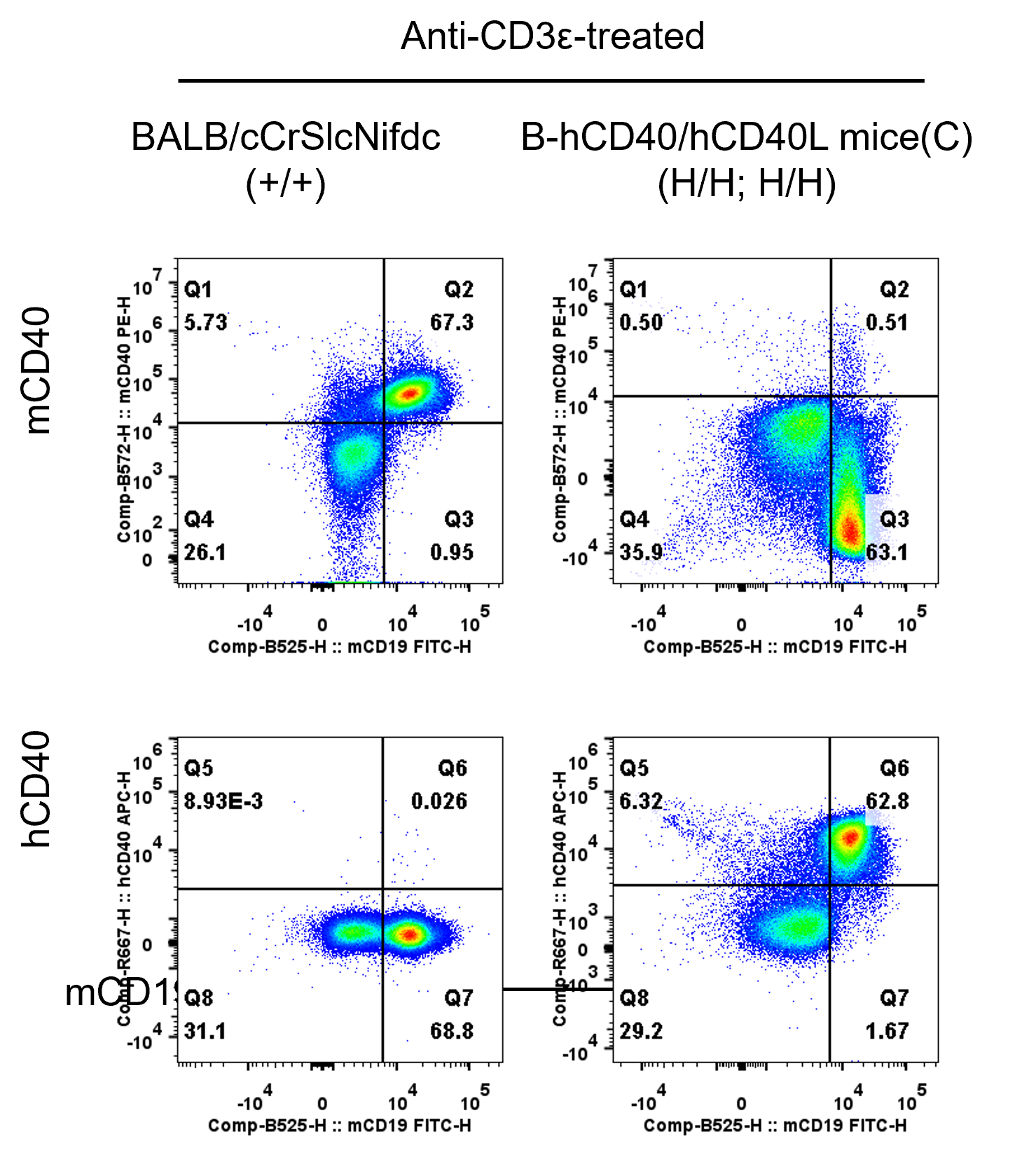
- Mouse CD40 was detected on B cells in wild-type BALB/cCrSlcNifdc mice, but not in B-hCD40/hCD40L mice(C).
- Human CD40 was detected on B cells in B-hCD40/hCD40L mice(C), but not in wild-type BALB/cCrSlcNifdc mice.
Strain specific CD40 expression analysis in homozygous B-hCD40/hCD40L mice(C) by flow cytometry. Splenocytes were collected from wild-type BALB/cCrSlcNifdc mice (+/+) and homozygous B-hCD40/hCD40L mice(C) (H/H; H/H) stimulated with anti-CD3ε in vivo (7.5 μg/mice, stimulation for 24 hours, i.p.), and analyzed by flow cytometry with anti-mouse CD40 antibody (Biolegend, 124609) and anti-human CD40 antibody (Biolegend, 313008). Mouse CD40 was detectable in wild-type BALB/cCrSlcNifdc mice. Human CD40 was exclusively detectable in homozygous B-hCD40/hCD40L mice(C) but not in wild-type BALB/cCrSlcNifdc mice.
CD40L Protein Expression in Thymus
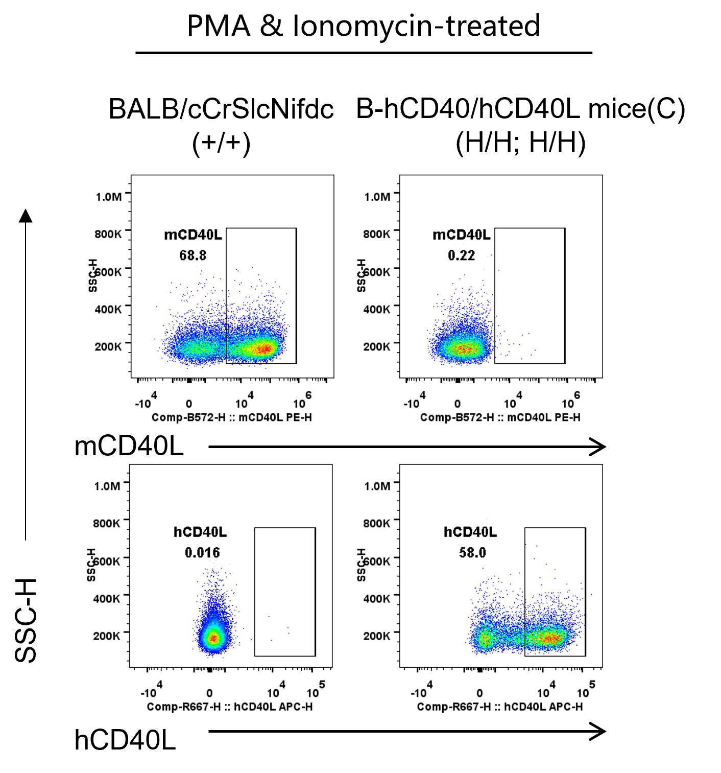
- Mouse CD40L was detected on T cells in wild-type BALB/cCrSlcNifdc mice, but not in B-hCD40/hCD40L mice(C).
- Human CD40L was detected on T cells in B-hCD40/hCD40L mice(C), but not in wild-type BALB/cCrSlcNifdc mice.
Strain specific CD40L expression analysis in homozygous B-hCD40/hCD40L mice(C) by flow cytometry. Thymocytes were collected from wild-type BALB/cCrSlcNifdc mice (+/+) and homozygous B-hCD40/hCD40L mice(C) (H/H; H/H) and stimulated with PMA & Ionomycin, then analyzed by flow cytometry with anti-mouse CD40L antibody (Biolegend, 106509) and anti-human CD40L antibody (Biolegend, 310805). Mouse CD40L was detectable in wild-type BALB/cCrSlcNifdc mice. Human CD40L was exclusively detectable in homozygous B-hCD40/hCD40L mice(C) but not in wild-type BALB/cCrSlcNifdc mice.
In Vivo Efficacy of Anti-Human CD40 Antibody and Anti-Human CD40L in a T cell-Dependent Antibody Response Model
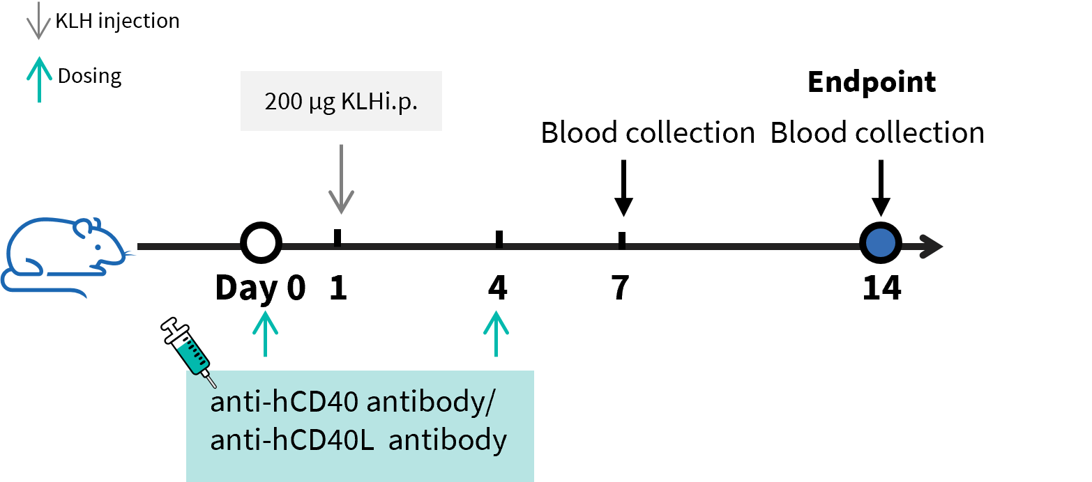
B-hCD40/hCD40L mice(C) were used to establish T cell-Dependent Antibody Response assay (TDAR assay) and evaluate the efficacy of anti-CD40 antibody and anti-CD40L antibody. B-hCD40/hCD40L mice(C) (n=6) were intraperitoneally immunized with 200 μg KLH on day 1 and treated with anti-CD40L antibody dapirolizumab analog (in house) or anti-CD40 antibody bleselumab analog (in house) on day 0 and day 4. Blood was collected on day 7 and day 14 and analyzed by ELISA with KLH specific IgG antibody and KLH specific IgM antibody.
- Concentration of mouse KLH specific IgM and IgG were significantly reduced in the anti–human CD40 antibody–treated group (G3) and anti–human CD40L antibody–treated group (G4) compared with the modelling group (G2).
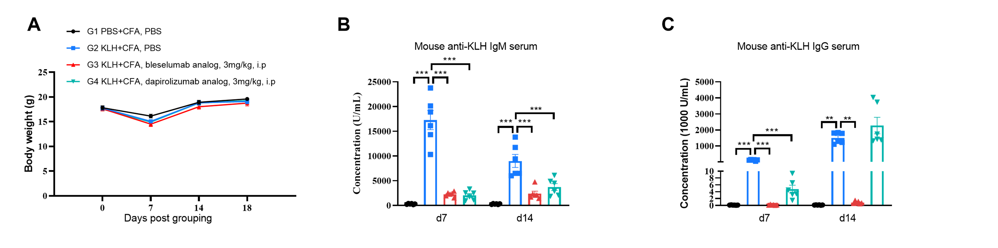
B-hCD40/hCD40L mice(C) were used to establish T cell-Dependent Antibody Response assay (TDAR assay) and evaluate the efficacy of anti-CD40 antibody and anti-CD40L antibody. (A) Body weight of B-hCD40/hCD40L mice(C) increased steadily; (B, C) Concentration of mouse KLH specific IgM and IgG were significantly increased after immunization. But the concentration of KLH specific IgG and IgM in the groups treated with anti-CD40L antibody dapirolizumab analog (in house) or anti-CD40 antibody bleselumab analog (in house) were significantly decreased when compared to that in the control group. The concentration of KLH specific IgG in the groups treated with anti-CD40L antibody dapirolizumab analog (in house) was not decreased on day 14, which may be due to incomplete blockade of B cells by inhibiting the CD40-CD40L pathway. These results demonstrated that the B-hCD40/hCD40L mice(C) provide a powerful preclinical model for in vivo evaluation of anti-CD40 antibody and anti-CD40L antibody. Values are expressed as mean ± SEM. Significance was determined by one-way ANOVA test . * p < 0.5, **p < 0.01, ***p < 0.001.
* When publishing results obtained using this animal model, please acknowledge the source as follows: The animal model [B-hCD40/hCD40L mice(C)] (Cat# 114463) was purchased from Biocytogen.