Description
- Parkinson’s disease (PD) is a complex age-related neurodegenerative disease associated with dopamine deficiency and both motor and nonmotor deficits. The presence of alpha-synuclein positive cytoplasmic inclusions, termed Lewy bodies, in surviving neurons is one of the main hallmark of PD.
- The full coding sequences of human SNCA with A53T mutation that is driven by mouse Thy1 promoter are inserted into mouse Gt(ROSA)26Sor locus in B-hSNCA*A53T mice plus. To achieve the overexpression of human SNCA, we inserted two expression cassettes in which three copies of the human SNCA CDS were driven by mouse Thy1 promoter.
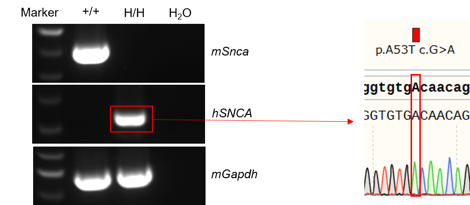
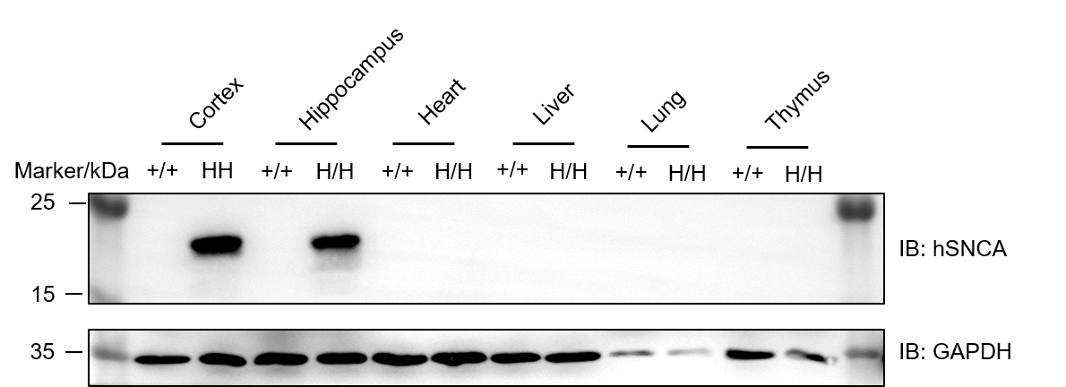
- Human SNCA mRNA was only detectable in B-hSNCA*A53T mice plus, and the A53T point mutation was confirmed via Sanger sequencing. SNCA protein was only detectable in brain from B-hSNCA*A53T mice plus. Abnormal phosphorylated α-Syn accumulations were exclusively detectable in cortex and hippocampus from B-hSNCA*A53T mice plus.
- This model is suitable for drug screening applications, such as knockdown screening for small nucleic acids, but is not intended for efficacy studies.
Targeting strategy
Gene targeting strategy for B-hSNCA*A53T mice plus
The full coding sequences of human SNCA with A53T mutation that is driven by mouse Thy1 promoter are inserted into mouse Gt(ROSA)26Sor locus in B-hSNCA*A53T mice plus. To achieve the overexpression of human SNCA, we inserted two expression cassettes in which three copies of the human SNCA CDS were driven by mouse Thy1 promoter.
Note: The gene editing was on the B-Snca KO mouse model, so there was no endogenous mouse Snca expression in B-hSNCA*A53T mice plus.
mRNA expression analysis
Strain specific analysis of SNCA mRNA expression in wild-type C57BL/6JNifdc mice and B-hSNCA*A53T mice plus by RT-PCR. Brain RNA were isolated from wild-type C57BL/6JNifdc mice (+/+) and homozygous B-hSNCA*A53T mice plus (H/H), then cDNA libraries were synthesized by reverse transcription, followed by PCR with mouse or human SNCA primers. Mouse Snca mRNA was only detectable in wild-type mice. Human SNCA mRNA was exclusively detectable in homozygous B-hSNCA*A53T mice plus, but not in wild-type mice.
Protein expression analysis
Western blot analysis of SNCA protein expression in homozygous B-hSNCA*A53T mice plus. Various tissue lysates were collected from C57BL/6JNifdc wild-type (+/+) mice and homozygous B-hSNCA *A53T mice plus (H/H), and then analyzed by western blot with human-specific anti-SNCA antibody (abcam, ab138501). 40 μg total proteins were loaded for western blotting analysis. SNCA was only detected in hippocampus, cortex from B-hSNCA*A53T mice plus, but not from wild-type mice.
Neuropathology analysis
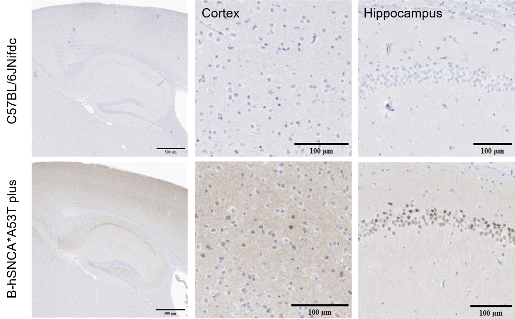
Neuropathology analysis in homozygous B-hSNCA*A53T mice plus. Pathological neuronal accumulation of phosphorylated α-Synuclein in neurons from 4-month-old homozygous male B-hSNCA*A53T mice plus. Brain from both wild-type control mice and homozygous B-hSNCA*A53T mice plus were stained with anti-alpha-synuclein (phosphor S129) antibody (abcam, ab51253). Abnormal phosphorylated α-Syn accumulations were exclusively detectable in cortex and hippocampus from B-hSNCA*A53T mice plus.
* When publishing results obtained using this animal model, please acknowledge the source as follows: The animal model [B-hSNCA*A53T mice plus] (Cat# 114090) was purchased from Biocytogen.