NOD.Cg-Egfrtm1(EGFR)Bcgen/Bcgen • 113309

| Product name | B-NDG hEGFR mice |
|---|---|
| Catalog number | 113309 |
| Strain name | NOD.Cg-Egfrtm1(EGFR)Bcgen/Bcgen |
| Strain background | NOD |
| NCBI gene ID | 13649,16186,19090 (Human) |
| Aliases | Wa5; wa2; Erbb; Errp; wa-2; Errb1; 9030024J15Rik; gc; p64; [g]c; CD132; gamma(c); p460; scid; slip; DNAPK; DNPK1; HYRC1; XRCC7; dxnph; DOXNPH; DNAPDcs; DNA-PKcs |
Gene targeting strategy for B-NDG hEGFR mice. A chimeric CDS that encode mouse EGFR signal peptide, human EGFR extracellular domain, mouse transmembrane and cytoplasmic domain, followed by mouse 3’UTR-STOP was inserted right after mouse Egfr exon 2. The chimeric EGFR protein expression was driven by endogenous mouse Egfr promoter, while mouse Egfr gene transcription and translation will be disrupted.

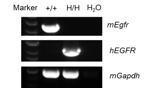
Strain specific analysis of EGFR mRNA expression in B-NDG mice and B-NDG hEGFR mice by RT-PCR. Liver RNA were isolated from B-NDG mice (+/+) (female, 8-week-old), and homozygous B-NDG hEGFR mice (H/H) (female, 9-week-old), then cDNA libraries were synthesized by reverse transcription, followed by PCR with mouse or human EGFR primers. mouse Egfr mRNA was only detectable in B-NDG mice. Human EGFR mRNA was exclusively detectable in homozygous B-NDG hEGFR mice but not in B-NDG mice.

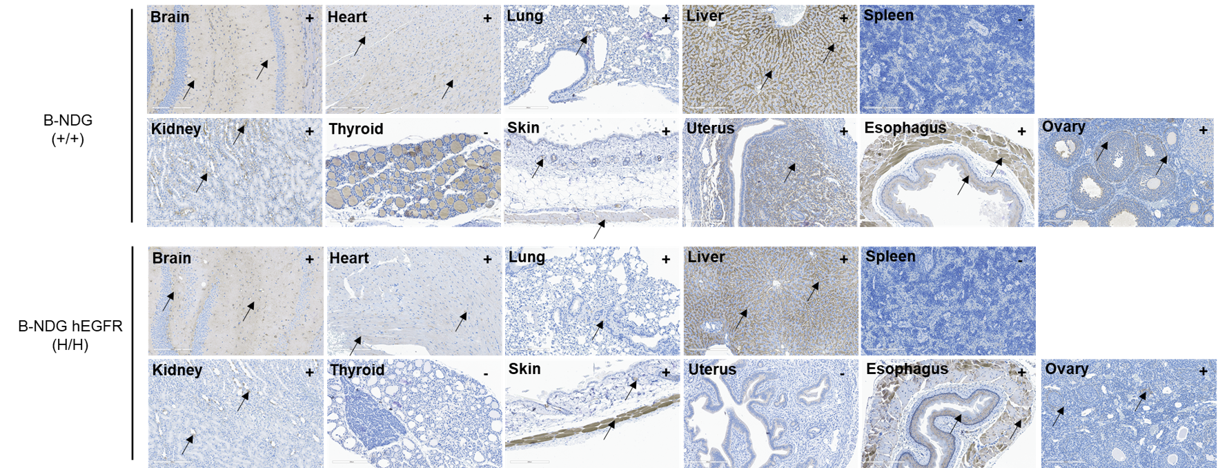
Immunohistochemical (IHC) analysis of EGFR expression in homozygous B-NDG hEGFR mice. The brain, heart, lung, liver, spleen, kidney, thyroid, skin, uterus, esophagus and ovary were collected from B-NDG mice (+/+) (female, 8-week-old) and homozygous B-NDG hEGFR mice (H/H) (female, 9-week-old) , analyzed by IHC with anti-EGFR (abcam, ab32198). EGFR was detectable in both B-NDG mice and homozygous B-NDG hEGFR mice because of antibody crossover. The arrow indicates tissue cells with positive EGFR staining (brown). “+” indicates that the tissue is positive and “-” indicates that the tissue is negative.