B-HLA-A2.1/hPRAME MC38

• 322453

B-HLA-A2.1/hPRAME MC38

Product nameB-HLA-A2.1/hPRAME MC38
Catalog number322453
Strain backgroundC57BL/6
NCBI gene ID567,3105,1489078,1489078 (Human)
Chromosome2
AliasesIMD43, AMYLD6, MHC1D4; HLAA; PRAME
TissueColon
DiseaseColon carcinoma

在此页面上

  • Description
  • Phenotypic analysis
  • Tumorigenicity

海报

查看全部

    发表文章

      Description
      • Origin: The MC38 cell line is derived from C57BL/6 murine colon adenocarcinoma cells. The cell line is a commonly used murine model for colorectal carcinoma.
      • Background Information: HLA-A belongs to the HLA class I heavy chain paralogues. This class I molecule is a heterodimer consisting of a heavy chain and a light chain (beta-2 microglobulin). The heavy chain is anchored in the membrane. Class I molecules play a central role in the immune system by presenting peptides derived from the endoplasmic reticulum lumen so that they can be recognized by cytotoxic T cells. This gene encodes an antigen that is preferentially expressed in human melanomas and that is recognized by cytolytic T lymphocytes. It is not expressed in normal tissues, except testis. The encoded protein acts as a repressor of retinoic acid receptor, and likely confers a growth advantage to cancer cells via this function. Alternative splicing results in multiple transcript variants.
      • Gene targeting strategy: The B2M gene (Exon2 to Exon3) of mouse were replaced by the sequence encompassing the human B2M CDS, HLA-A*0201 gene that included leader sequence, α1 and α2 domains ligated to a fragment of the murine H-2Db gene containing the α3, transmembrane and cytoplasmic domains, PRAME CDS. Human HLA-A2.1 is highly expressed on the surface of B-HLA-A2.1/hPRAME MC38.
      • Application: B-HLA-A2.1/hPRAME MC38 tumor models can be used for preclinical evaluation of cancer vaccines.
      Protein expression analysis

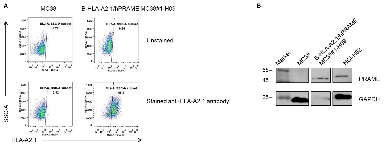
      HLA-A2.1 and human PRAME expression analysis in B-HLA-A2.1/hPRAME MC38 by flow cytometry and western blot, respectively. Single cell suspensions from wild-type MC38 and B-HLA-A2.1/hPRAME MC38 cultures were stained with species-specific anti-HLA-A2.1 antibody (Biolegend, 343306) and anti-human PRAME(Cell Signaling Technology, 56426S). Human HLA-A2.1 was detected on the surface of B-HLA-A2.1/hPRAME MC38 cells but not wild-type MC38 cells(A). Human PRAME was highly expressed in the tumor cells(B). The 1-H09 clone of B-HLA-A2.1/hPRAME MC38 cells was used for in vivo experiments.

      Protein expression analysis of tumor tissue

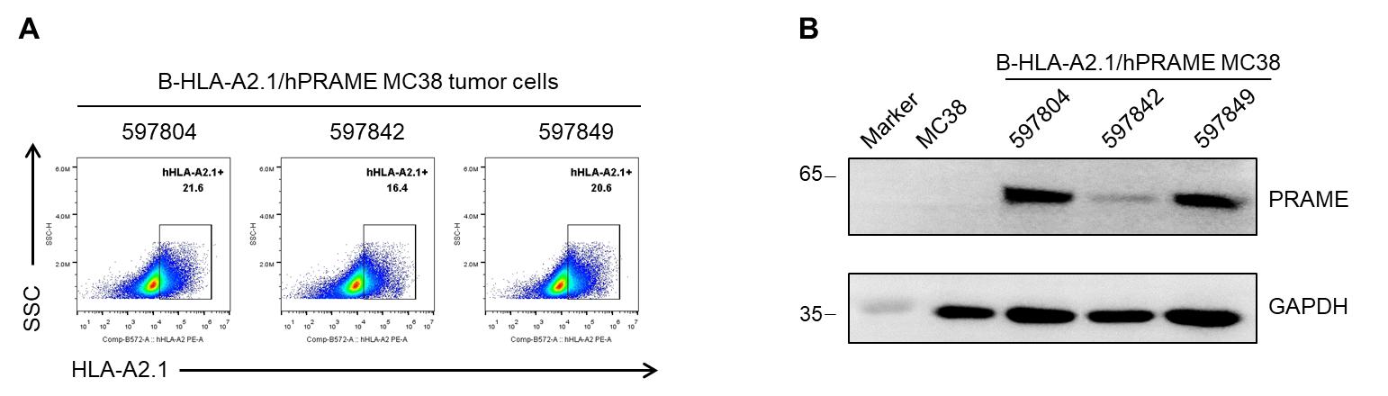
      Human HLA-A2.1 and human PRAME expression evaluated in B-HLA-A2.1/hPRAME MC38 tumor cells by flow cytometry and western blot, respectively. B-HLA-A2.1/hPRAME MC38 cells were subcutaneously transplanted into B-HLA-A2.1 mice (female, 7-week-old, n=6), and on 28 days post inoculation, tumor cells were harvested and assessed for human HLA-A2.1 expression(Biolegend, 343306) by flow cytometry and PRAME expression(Cell Signaling Technology, 56426S) by western blot, respectively. As shown, human HLA-A2.1 was highly expressed on the surface of tumor cells(A). Human PRAME was highly expressed in the tumor cells(B). Therefore, B-HLA-A2.1/hPRAME MC38 cells can be used for in vivo efficacy studies of novel PRAME therapeutics.

      Tumor growth curve & body weight changes

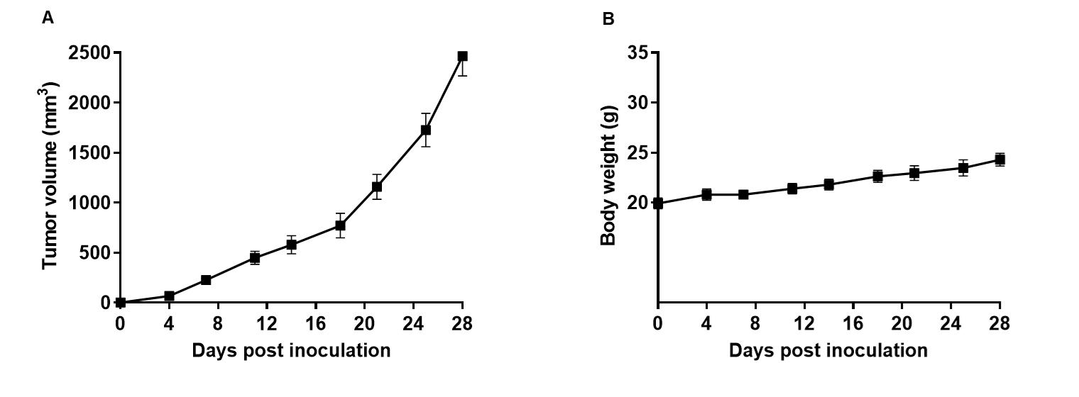
      Subcutaneous homograft tumor growth of B-HLA-A2.1/hPRAME MC38 cells. B-HLA-A2.1/hPRAME MC38 cells (1x10⁶) were subcutaneously implanted into B-HLA-A2.1 mice (male, 7-week-old, n=6). Tumor volume and body weight were measured three times a week. (A) Average tumor volume ± SEM. (B)  Body weight (Mean± SEM). Volume was expressed in mm3 using the formula: V=0.5 X long diameter X short diameter2. As shown in panel AB-HLA-A2.1/hPRAME MC38 cells were able to establish tumors in vivo and can be used for efficacy studies.