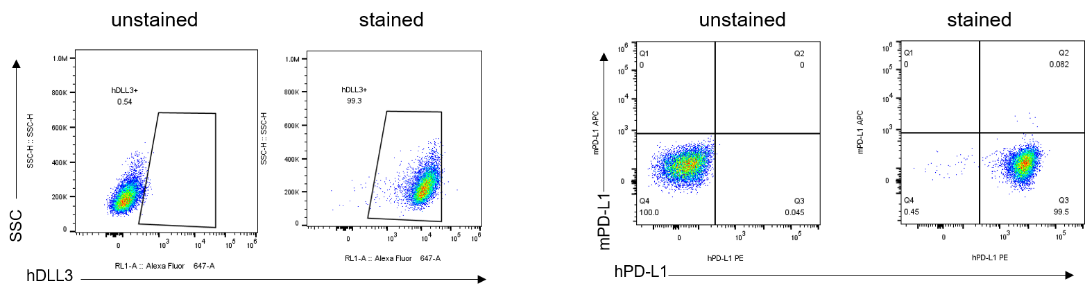
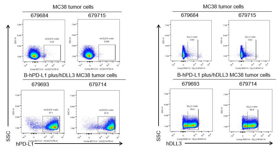
B-hPD-L1/hDLL3 MC38

• 322420

B-hPD-L1/hDLL3 MC38

Product nameB-hPD-L1/hDLL3 MC38
Catalog number322420
Strain backgroundC57BL/6
NCBI gene ID29126,10683 (Human)
Chromosome19, 7
AliasesB7-H; B7H1; PDL1; PD-L1; ADMIO5; hPD-L1; PDCD1L1; PDCD1LG1; SCDO1
TissueColon
DiseaseColon carcinoma

在此页面上

  • Description
  • Phenotypic analysis
  • Tumorigenicity

海报

查看全部

    发表文章

      Description
      • Origin: The MC38 cell line is derived from C57BL6 murine colon adenocarcinoma cells. The cell line is a commonly used murine model for colorectal carcinoma.
      • Background Information: PD-1 (programmed death receptor-1) is an immune checkpoint protein on the surface of T cells. When it binds to its ligands (PD-L1/PD-L2) in the tumor microenvironment, it suppresses T cell activity, leading to immune evasion. DLL3 (Delta-like ligand 3) is a ligand in the Notch signaling pathway. It is highly expressed in various neuroendocrine tumors (such as small cell lung cancer, SCLC) but is nearly absent in normal tissues.PD-1 antibodies rely on the immune system to kill tumor cells, while DLL-3 antibodies independently target and eliminate tumors through direct action or ADC-mediated cytotoxicity. The combination of these two mechanisms can complement each other and overcome resistance to monotherapy.
      • Gene targeting strategy: The exogenous promoter, human PD-L1 and luciferase coding sequences were inserted to replace part of murine exon 3. The exogenous promoter and chimeric DLL3 coding sequence that include human extracellular region and mouse intracellular region were inserted to replace part of murine Dll3 exon 2 and all of exons 3-5. The insertion disrupts the endogenous murine Pdl1 and Dll3 gene, resulting in a non-functional transcript.
      • Tumorigenicity: Confirmed in C57BL/6J mice.
      • Application: The B-hPD-L1/hDLL3 MC38 tumor models can be used for preclinical evaluation.
      Protein expression analysis

      PD-L1 and DLL3 expression analysis in B-hPD-L1/hDLL3 MC38 cells by flow cytometry. Single cell suspensions from B-hPD-L1/hDLL3 MC38 #1-G10 were stained with species-specific anti-PD-L1 antibody (anti-mouse PD-L1: Biolegend, 124312; anti-human PD-L1: Biolegend, 329706) and anti-human DLL3 (AMG-757 analog, in-house). Human PD-L1 and DLL3 was detected on the surface of B-hPD-L1/hDLL3 MC38 cells.

      Protein expression analysis of tumor tissue

      PD-L1 and DLL3 expression were evaluated on B-hPD-L1/hDLL3 MC38 tumor cells by flow cytometry. B-hPD-L1/hDLL3 MC38 cells were subcutaneously transplanted into C57BL/6J mice. Upon conclusion of the experiment, tumor cells were harvested and assessed for human PD-L1 (Biolegend, 329714) and DLL3 (AMG-757 analog, in-house) expression by flow cytometry. As shown, human PD-L1 and DLL3 was highly expressed on the surface of tumor cells. Therefore, B-hPD-L1/hDLL3 MC38 cells can be used for in vivo efficacy studies evaluating novel PD-L1 and DLL3 combination therapeutics.

      Tumor growth curve & body weight changes

      Subcutaneous tumor growth of B-hPD-L1/hDLL3 MC38 cells. B-hPD-L1/hDLL3 MC38 cells (1×10⁶) and wild-type MC38 cells (5×10⁵) were subcutaneously implanted into C57BL/6J mice (female, 8-week-old, n=8). Tumor volume and body weight were measured twice a week. (A) Average tumor volume. (B) Body weight. Volume was expressed in mm3 using the formula: V=0.5 × long diameter × short diameter2. Results indicate that B-hPD-L1/hDLL3 MC38 cells were able to establish tumors in vivo and can be used for efficacy studies. Values are expressed as mean ± SEM.