C57BL/6 • 322416

| Product name | B-hPD-L1 plus/hF3 MC38 |
|---|---|
| Catalog number | 322416 |
| Strain name | C57BL/6 |
| Strain background | C57BL/6 |
| NCBI gene ID | 60533,14066 (Human) |
| Chromosome | 19, 3 |
| Aliases | B7h1; Pdl1; Pdcd1l1; Pdcd1lg1; A530045L16Rik; TF; Cf3; Cf-3; CD142 |
| Tissue | Colon |
| Disease | Colon carcinoma |
Inoculated cell lines can be suspended with DMEM stock solution.
In the experiment, it is necessary to ensure that the number of animals inoculated subcutaneously is at least 1.6 times the actual grouping number.

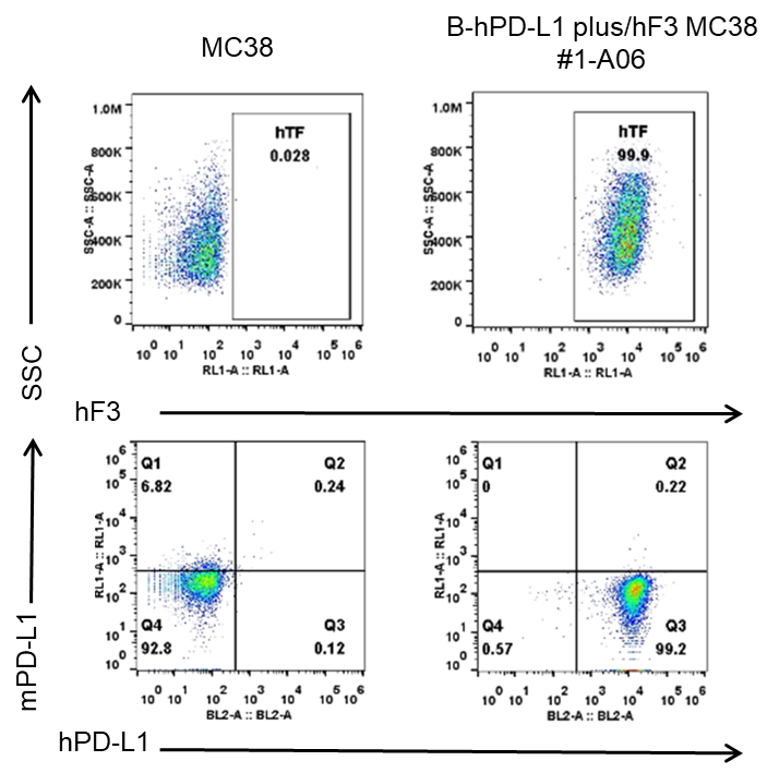
F3 and PD-L1 expression analysis in B-hPD-L1 plus/hF3 MC38 cells by flow cytometry. Single cell suspensions from wild-type MC38 and B-hPD-L1 plus/hF3 MC38 #1-A06 were stained with anti-F3 antibody (R&D, FAB23391A) and anti-PD-L1 antibody (human anti-PD-L1: Biolegend, 329706; mouse anti-PD-L1: Biolegend, 124312). Human F3 and human PD-L1 were detected on the surface of B-hPD-L1 plus/hF3 MC38 cells but not wild-type MC38 cells.

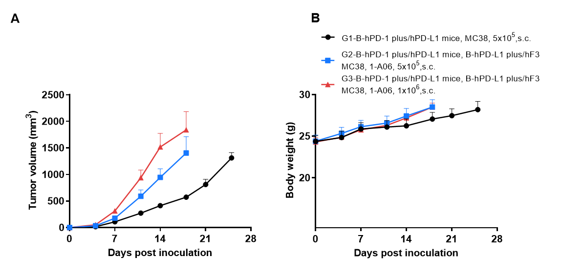
Subcutaneous tumor growth of B-hPD-L1 plus/hF3 MC38 cells. B-hPD-L1 plus/hF3 MC38 cells (5×10⁵, 1×10⁶) and wild-type MC38 cells (5×10⁵) were subcutaneously implanted into B-hPD-1 plus/hPD-L1 mice (Male, 8-week-old, n=6). Tumor volume and body weight were measured twice a week. (A) Average tumor volume. (B) Body weight. Volume was expressed in mm3 using the formula: V=0.5 × long diameter × short diameter2. Results indicate that B-hPD-L1 plus/hF3 MC38 cells were able to establish tumors in vivo and can be used for efficacy studies. Values are expressed as mean ± SEM.

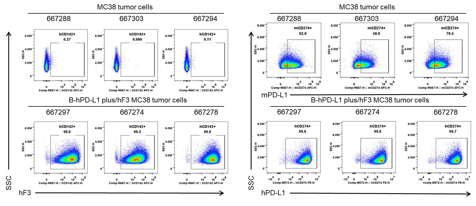
F3 and PD-L1 expression evaluated on B-hPD-L1 plus/hF3 MC38 tumor cells by flow cytometry. B-hPD-L1 plus/hF3 MC38 cells were subcutaneously transplanted into B-hPD-1 plus/hPD-L1 mice (n=6). Upon conclusion of the experiment, tumor cells were harvested and assessed for F3 (Biolegend, 365206) and PD-L1 (human anti-PD-L1: Biolegend, 329706; mouse anti-PD-L1: Biolegend, 124312) expression by flow cytometry. As shown, human F3 and human PD-L1 were highly expressed on the surface of tumor cells. Therefore, B-hPD-L1 plus/hF3 MC38 cells can be used for in vivo efficacy studies evaluating novel F3 therapeutics.