CBA/Ola-Csf1rtm1Bcgen/Bcgen • 113819
Gene targeting strategy for B-Csf1r KO mice(CBA). The Fms-intronic regulatory element (FIRE), a super-enhancer located in intron 2 of the mouse Csf1r locus, is selectively deleted in B-Csf1r KO (CBA) mice.
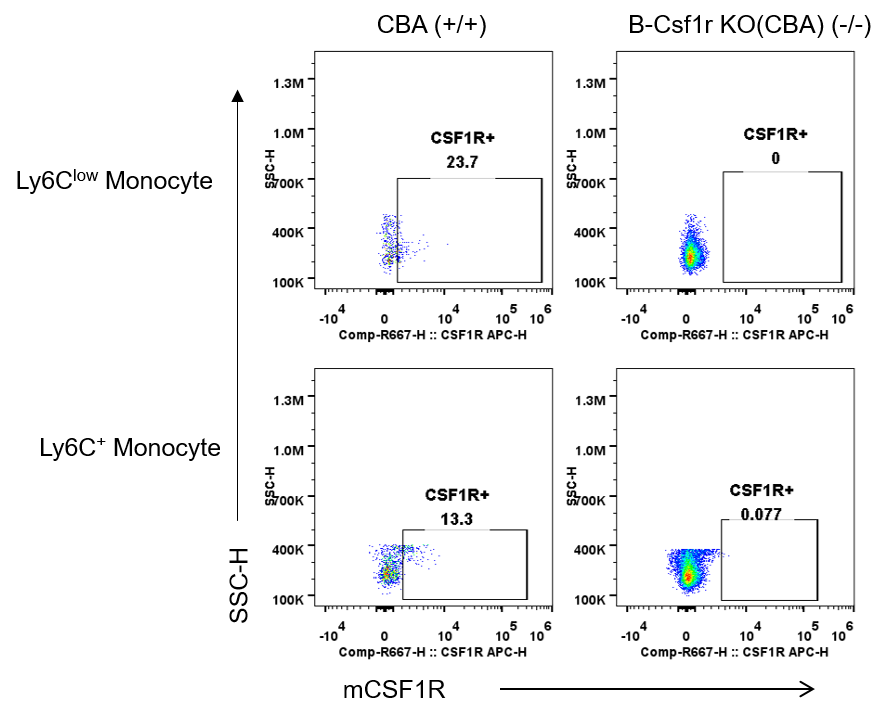
CSF1R expression analysis in wild-type CBA mice and homozygous B-Csf1r KO mice(CBA) by flow cytometry. Blood were collected from wild-type CBA mice (+/+) and homozygous B-Csf1r KO mice(CBA) (-/-). Protein expression was analyzed with anti-mouse CSF1R antibody (Biolegend, 135509) by flow cytometry in Ly6Clow and Ly6C+ monocyte populations. Mouse CSF1R was only detectable in wild-type CBA mice, but not in homozygous B-Csf1r KO mice(CBA).
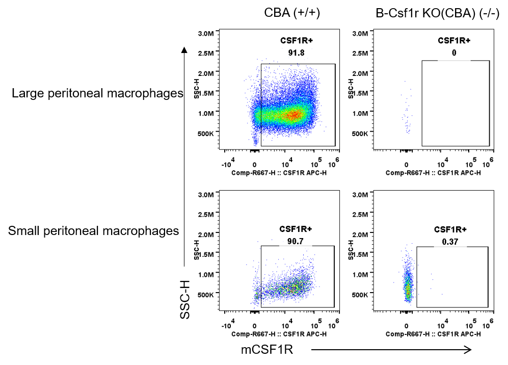
CSF1R expression analysis in wild-type CBA mice and homozygous B-Csf1r KO mice(CBA) by flow cytometry. Peritoneal lavage cells were collected from wild-type CBA mice (+/+) and homozygous B-Csf1r KO mice(CBA) (-/-). Protein expression was analyzed with anti-mouse CSF1R antibody (Biolegend, 135509) by flow cytometry. There was two macrophages populations in the peritoneal lavage cells: small peritoneal macrophages (SPM) identified as F4/80lowCD11b+, and large peritoneal macrophages (LPM) identified as F4/80highCD11b+. Mouse CSF1R was only detectable in wild-type CBA mice, but not in homozygous B-Csf1r KO mice(CBA). Besides, the LPM population was almost lost in B-Csf1r KO mice(CBA).
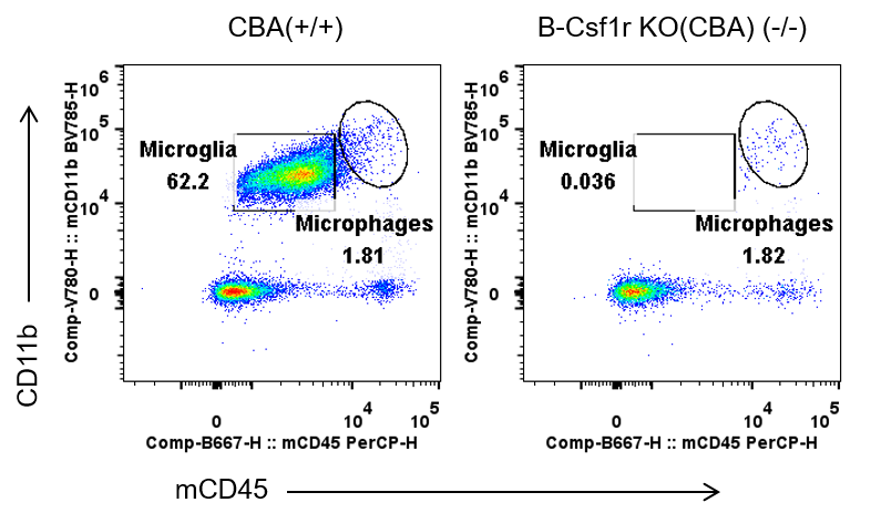
Loss of microglia in homozygous B-Csf1r KO mice(CBA) by flow cytometry. Brains were collected from wild-type CBA mice (+/+) and homozygous B-Csf1r KO mice(CBA) (-/-, 6-7-week-old, female). Myelin-depleted single cells obtained by using Myelin Removal Beads II kit from Miltenyi Biotec (130-096-433) were analyzed by flow cytometry for CD45 and CD11b expression. Microglia=CD45lowCD11b+. Microphages=CD45+CD11b+. CD45lowCD11b+ microglia were almost absent only in the homozygous B-Csf1r KO mice(CBA) but not in wild-type control mice.
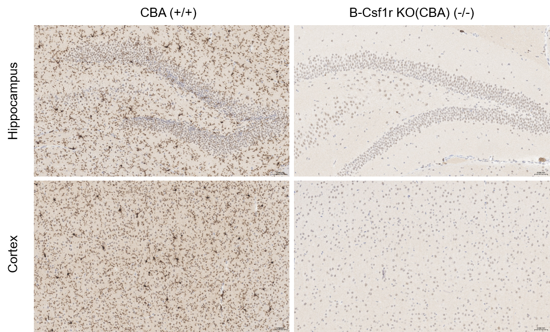
Microglia analysis in brain from wild-type CBA mice and homozygous B-Csf1r KO mice(CBA). Brains were collected from 7-week-old female wild-type mice and homozygous B-Csf1r KO mice(CBA), and then processed for immunohistochemical analysis. Microglia in hippocampus and cortex were detected using the IBA1 antibody (Abcam, ab178846) at 200X magnification. Microglia were absent in the hippocampus and cortex only from homozygous B-Csf1r KO mice(CBA), but not in wild-type mice.