BALB/cCrSlcNifdc-Tnfrsf4tm1(TNFRSF4)Bcgen Tnfsf4tm1(TNFSF4)Bcgen/Bcgen • 114051
Gene targeting strategy for B-hOX40/hOX40L mice(C).
The exons 1-5 of mouse OX40 gene that encode the extracellular domain were replaced by human OX40 exons 1-5 in B-hOX40/hOX40L mice(C).
The exons 2-3 of mouse Ox40l gene that encode the extracellular region were replaced by human OX40L exons 2-3 in B-hOX40/hOX40L mice(C).
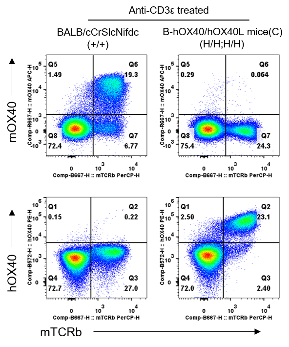
Strain specific OX40 expression analysis in homozygous B-hOX40/hOX40L mice(C) by flow cytometry. Splenocytes were collected from wild-type BALB/cCrSlcNifdc mice(+/+) and homozygous B-hOX40/hOX40L mice(C) (H/H;H/H) stimulated with anti-CD3ε in vivo, and protein expression was analyzed with anti-mouse OX40 antibody (Biolegend, 119414) and anti-human OX40 antibody (Biolegend, 350004) by flow cytometry. mOX40 was only detectable in T cells of wild-type BALB/cCrSlcNifdc mice. hOX40 was only detectable in T cells of homozygous B-hOX40/hOX40L mice(C), but not in wild-type BALB/cCrSlcNifdc mice.
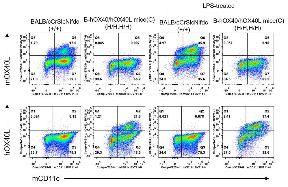
Strain specific OX40L expression analysis in homozygous B-hOX40/hOX40L mice(C) by flow cytometry. Bone marrow cells were collected from wild-type BALB/cCrSlcNifdc mice(+/+) and homozygous B-hOX40/hOX40L mice(C)(H/H;H/H). BMDCs were induced from bone marrow cells and stimulated with LPS in vitro. Then BMDCs were analyzed with anti-mouse OX40L antibody (Biolegend, 108814) and anti-human OX40L antibody (BD Horizon™, 563766) by flow cytometry. mOX40L was only detectable in BMDCs of wild-type BALB/cCrSlcNifdc mice. hOX40L was only detectable in BMDCs of homozygous B-hOX40/hOX40L mice, but not in wild-type BALB/cCrSlcNifdc mice.
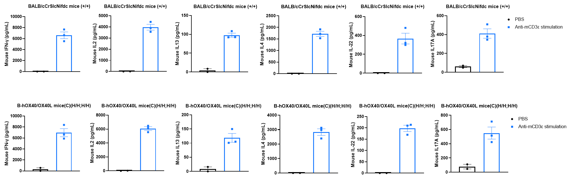
Strain specific cytokines expression analysis in wild-type BALB/cCrSlcNifdc mice and homozygous humanized B-hOX40/0X40L mice(C) by ELISA. Serum was collected from wild-type BALB/cCrSlcNifdc mice (+/+) (female, n=2, 7-week-old) and homozygous B-hOX40/0X40L mice(C) (H/H;H/H) (female, n=3, 7-week-old) stimulated with anti-mouse CD3ε antibody (37.5 μg/mL, 200 μL/mouse, i.p.) for 2 hrs in vivo. Expression level of IL4, IL13, IL2, IL22, IL17A, and IFN-γ were analyzed by ELISA. After mCD3ε stimulation, a significant increase of mouse IL4, IL13, IL2, IL22, IL17A, and IFN-γ were detected in BALB/cCrSlcNifdc mice (n=3) and homozygous B-hOX40/0X40L mice(C) (n=3). Values are expressed as mean ± SEM.
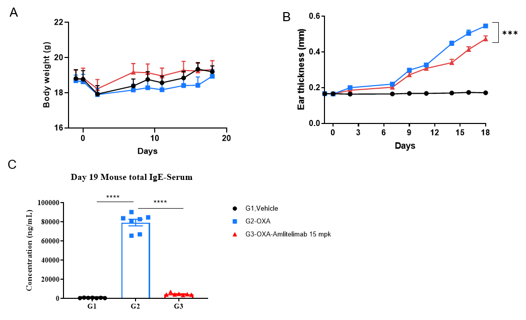
Efficacy of anti-human OX40L antibody in B-hOX40/hOX40L mice(C). Mice in each group were intraperitoneally injected with anti-hOX40L antibody (in house, n=7). (A) Body weight changes during the treatment. (B) Statistical analysis of ear thickness in each group. (C) Total IgE levels in serum. The results showed that compared to the untreated group (G2), the group of mice treated with anti-OX40L antibody showed a significant reduction in ear thickness. Serum was collected at the study endpoint. IgE level was analyzed by ELISA. The results showed that the levels of total IgE in mice treated with anti-OX40L antibody was lower than that in untreated mice. Values are expressed as mean ± SEM. Significance was determined by two-way ANOVA test. *P < 0.05, **P < 0.01, ***P < 0.001.