Targeting strategy
Gene targeting strategy for B-hPSGL-1, Rag2 KO mice.
The exons 2 of mouse Psgl-1 gene that encode extracellular domain were replaced by human counterparts in B-hPSGL-1, Rag2 KO mice. The genomic region of mouse Psgl-1 gene that encodes transmembrane domain and cytoplasmic portion was retained. The promoter, 5’UTR and 3’UTR region of the mouse gene were also retained. The chimeric PSGL-1 expression was driven by endogenous mouse Psgl-1 promoter, while mouse Psgl-1 gene transcription and translation will be disrupted.
The exon 3 and 3’UTR region of mouse Rag2 were knocked out in B-hPSGL-1, Rag2 KO mice, resulting in a disruption of the Rag2 gene.
Protein expression analysis-spleen
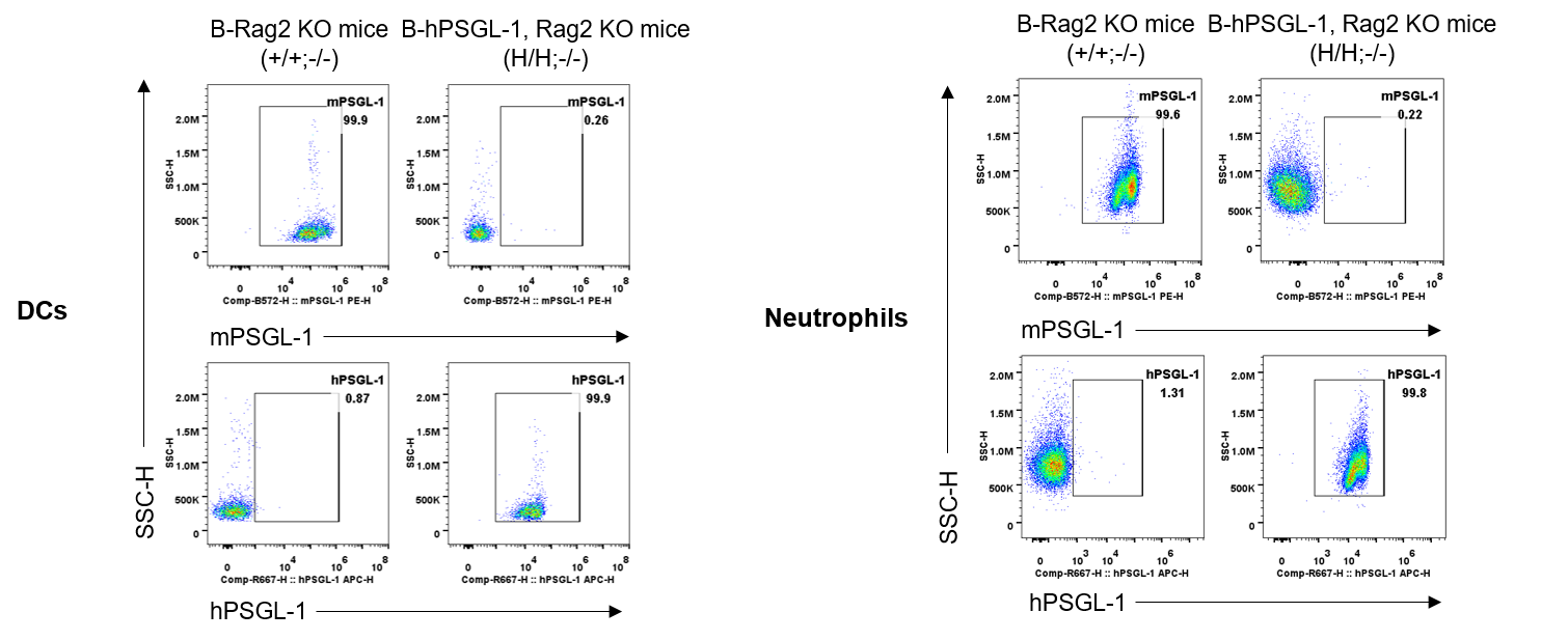
Strain specific PSGL-1 expression analysis in homozygous B-hPSGL-1, Rag2 KO mice by flow cytometry. Splenocytes were collected from B-Rag2 KO mice(+/+;-/-) and homozygous B-hPSGL-1, Rag2 KO mice(H/H;-/-), protein expression was analyzed by flow cytometry with species-specific anti-mouse PSGL-1 antibody (eBioscience, 12-1621-80) and anti-human PSGL-1 antibody (Biolegend, 328811). Mouse PSGL-1 was detectable on DCs and neutrophils of B-Rag2 KO mice. Human PSGL-1 was detectable on DCs and neutrophils of homozygous B-hPSGL-1, Rag2 KO mice.
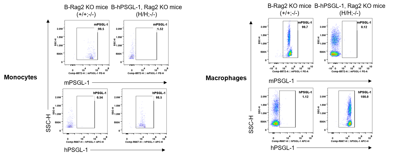
Strain specific PSGL-1 expression analysis in homozygous B-hPSGL-1, Rag2 KO mice by flow cytometry. Splenocytes were collected from B-Rag2 KO mice(+/+;-/-) and homozygous B-hPSGL-1, Rag2 KO mice(H/H;-/-), protein expression was analyzed by flow cytometry with species-specific anti-mouse PSGL-1 antibody (eBioscience, 12-1621-80) and anti-human PSGL-1 antibody (Biolegend, 328811). Mouse PSGL-1 was detectable on monocytes and macrophage of B-Rag2 KO mice. Human PSGL-1 was detectable on monocytes and macrophage of homozygous B-hPSGL-1, Rag2 KO mice.
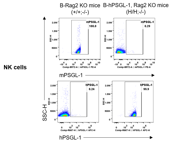
Strain specific PSGL-1 expression analysis in homozygous B-hPSGL-1, Rag2 KO mice by flow cytometry. Splenocytes were collected from B-Rag2 KO mice(+/+;-/-) and homozygous B-hPSGL-1, Rag2 KO mice(H/H;-/-), protein expression was analyzed by flow cytometry with species-specific anti-mouse PSGL-1 antibody (eBioscience, 12-1621-80) and anti-human PSGL-1 antibody (Biolegend, 328811). Mouse PSGL-1 was detectable on NK cells of B-Rag2 KO mice. Human PSGL-1 was detectable on NK cells of homozygous B-hPSGL-1, Rag2 KO mice.
Frequency of leukocyte subpopulations in spleen
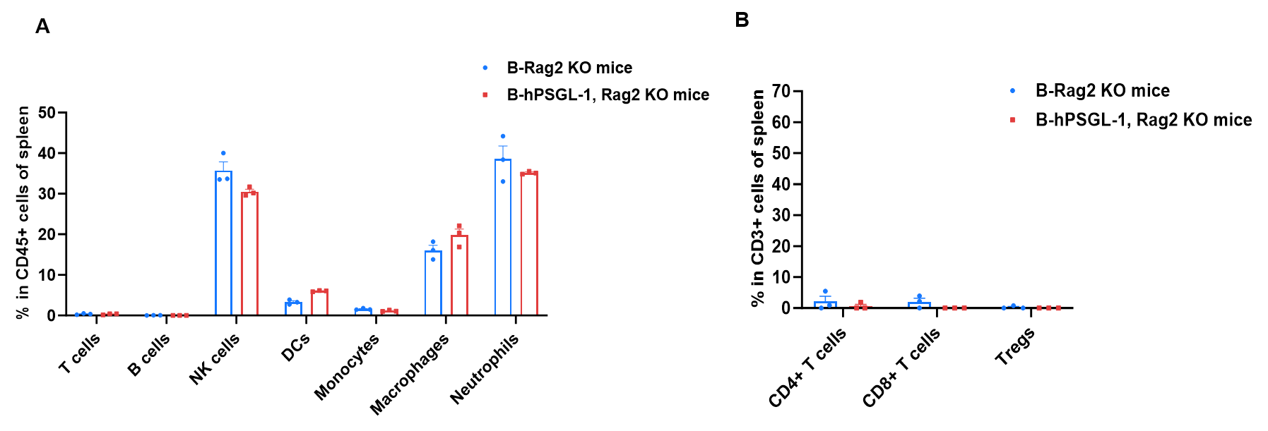
Frequency of leukocyte subpopulations in spleen by flow cytometry. Splenocytes were isolated from B-Rag2 KO mice and homozygous B-hPSGL-1, Rag2 KO mice (female, 7-week-old, n=3). A. Flow cytometry analysis of the splenocytes was performed to assess the frequency of leukocyte subpopulations. B. Frequency of T cell subpopulations. Frequencies of T cells, B cells, NK cells, DCs, neutrophils, monocytes, macrophages, CD4+ T cells, CD8+ T cells and Tregs in B-hPSGL-1, Rag2 KO mice were similar to those in B-Rag2 KO mice, demonstrating that humanization of PSGL-1 does not change the frequency or distribution of these cell types in spleen. Values are expressed as mean ± SEM.
Frequency of leukocyte subpopulations in blood
Frequency of leukocyte subpopulations in blood by flow cytometry. Blood cells were isolated from B-Rag2 KO mice and homozygous B-hPSGL-1, Rag2 KO mice (female, 7-week-old, n=3). A. Flow cytometry analysis of the blood cells was performed to assess the frequency of leukocyte subpopulations. B. Frequency of T cell subpopulations. Percentages of T cells, B cells, NK cells, dendritic cells, neutrophils, monocytes, macrophages, CD4+ T cells, CD8+ T cells and Tregs in B-hPSGL-1, Rag2 KO mice were similar to those in B-Rag2 KO mice, demonstrating that humanization of PSGL-1 does not change the frequency or distribution of these cell types in blood. Values are expressed as mean ± SEM.
Frequency of leukocyte subpopulations in lymph nodes
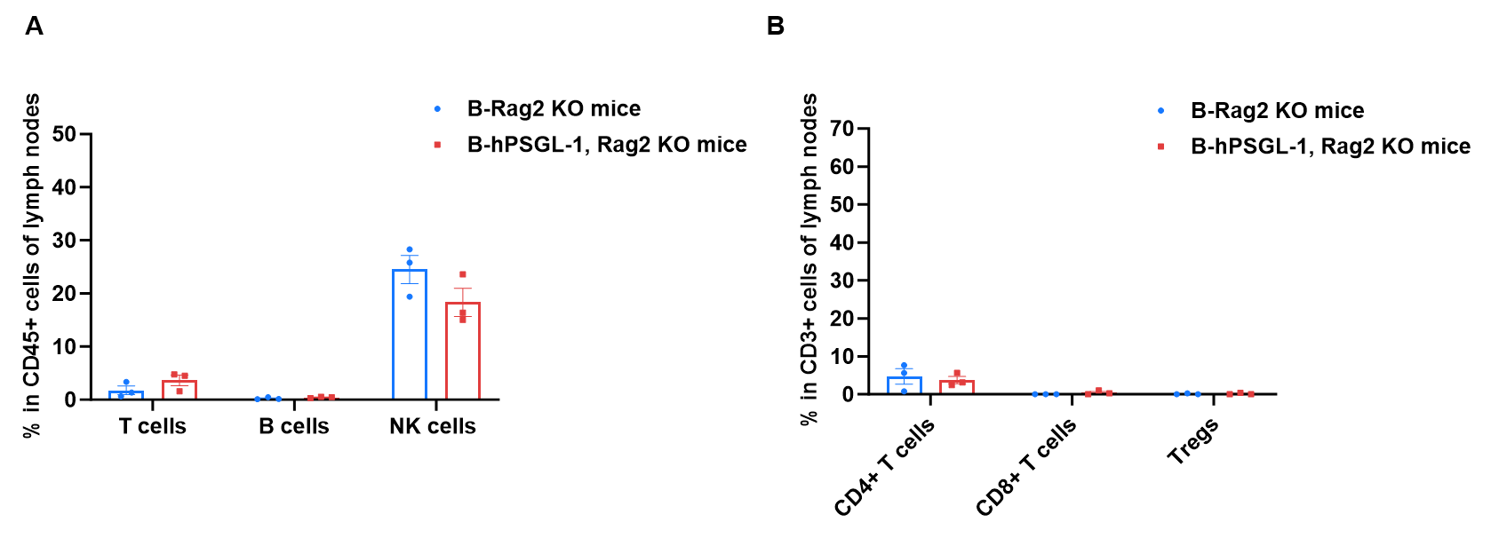
Frequency of leukocyte subpopulations in blood by flow cytometry. Lymph nodes cells were isolated from B-Rag2 KO mice and homozygous B-hPSGL-1, Rag2 KO mice (female, 7-week-old, n=3). A. Flow cytometry analysis of the lymph nodes cells was performed to assess the frequency of leukocyte subpopulations. B. Frequency of T cell subpopulations. Percentages of T cells, B cells, NK cells, CD4+ T cells, CD8+ T cells and Tregs in B-hPSGL-1, Rag2 KO mice were similar to those in B-Rag2 KO mice. Values are expressed as mean ± SEM.
* When publishing results obtained using this animal model, please acknowledge the source as follows: The animal model [B-hPSGL-1, Rag2 KO mice] (Cat# 114033) was purchased from Biocytogen.