Description
- Background: Retinoschisin (RS1) is a secreted glycoprotein that is primarily expressed in the retina and is secreted by photoreceptor cells and bipolar cells. RS1 protein plays a crucial role in intercellular connections in the retina, regulation of signal transduction, as well as in the development and maintenance of the retina.
- Targeting strategy: The exons 1-3 of mouse Rs1 gene were knocked out in B-Rs1 KO mice.
- Validation: Mouse Rs1 mRNA and RS1 protein were detected in the eyes of wild-type C57BL/6JNifdc mice but not in homozygous B-Rs1 KO mice.
- Application: Loss-of-function mutations in the Rs1 gene lead to X-linked retinoschisis (XLRS), a hereditary retinal disorder that primarily affects young males. It is characterized by splitting of the retinal layers and a decline in vision.
Targeting strategy
Gene targeting strategy for B-Rs1 KO mice.The exons 1-3 of mouse Rs1 gene were knocked out in B-Rs1 KO mice.
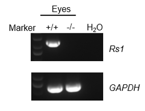
mRNA expression analysis
Strain specific analysis of Rs1 mRNA expression in wild-type C57BL/6JNifdc mice and homozygous B-Rs1 KO mice by RT-PCR. Eyes RNA were isolated from wild-type C57BL/6JNifdc (+/+) and homozygous B-Rs1 KO mice (-/-), then cDNA libraries were synthesized by reverse transcription, followed by PCR with mice Rs1 primers. Mouse Rs1 mRNA was detectable in wild-type but not detected in homozygous B-Rs1 KO mice.
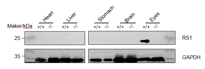
Protein expression analysis
Western blot analysis of RS1 protein expression in homozygous B-Rs1 KO mice. Various tissue lysates were collected from wild-type C57BL/6JNifdc mice (+/+) and B-Rs1 KO mice (-/-), and then analyzed by western blot with cross reactive anti-RS1 antibody (Proteintech. 24430-1-AP). 40μg total proteins were loaded for western blotting analysis. Mouse RS1 was detected in eyes in wild-type but not detected in homozygous B-Rs1 KO mice.
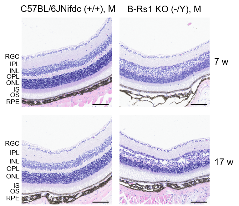
H&E staining of B-Rs1 KO mice
Representative images of HE staining of wild-type C57BL/6JNifdc mice and B-Rs1 KO mice. Retina tissues of wild-type C57BL/6JNifdc mice (+/+) and B-Rs1 KO mice (-/Y) (7 and 17 weeks old, male) were collected and analyzed with H&E staining. The results showed that the retinal layers were disordered, and multiple layers such as the outer nuclear layer and the inner nuclear layer disappeared in B-Rs1 KO mice compared to the C57BL/6JNifdc mice. Also, B-Rs1 KO mice exhibited retinal splitting. Scale bar, 100 μm.
RPE: Retinal pigment epithelium, OS: Outer segment, IS: Inner segment, ONL: Outer nuclear layer, OPL: Outer plexiform layer, INL: Inner nuclear layer, IPL: Inner plexiform layer,
GCL: Ganglion cell layer.
* When publishing results obtained using this animal model, please acknowledge the source as follows: The animal model [B-Rs1 KO mice] (Cat# 113761) was purchased from Biocytogen.