mRNA Expression Analysis
- (A) Human TFR1 mRNA is exclusively detectable in splenocytes of homozygous B-hTFR1 mice, but not in wild-type C57BL/6 mice.
- (B) Relative TFR1 mRNA expression levels in the cortex and hippocampus were comparable between B-hTFR1 mice and wild-type C57BL/6JNifdc mice across 2, 6, 10, and 16 months old, regardless of gender.
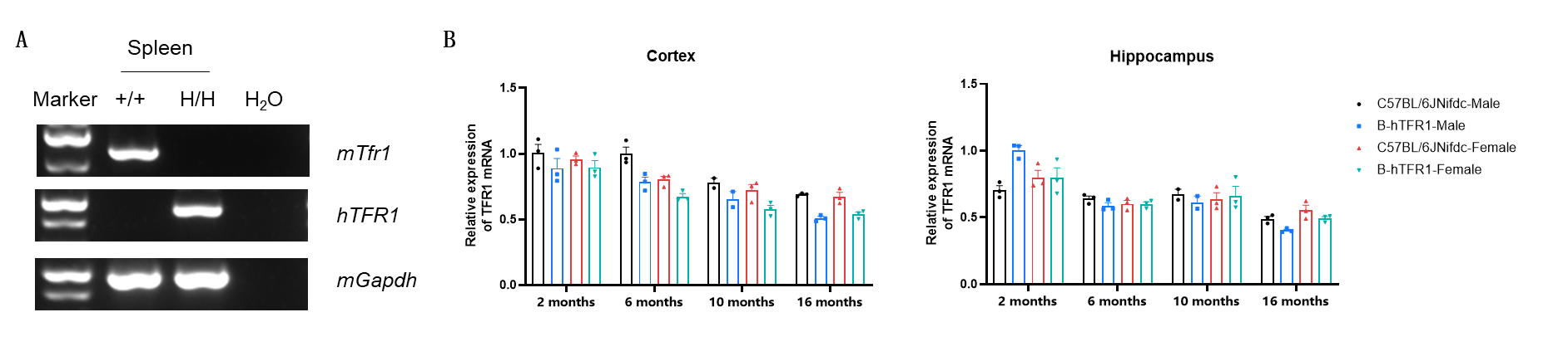
Strain-specific TFR1 expression analysis in wild-type C57BL/6 mice and homozygous B-hTFR1 mice. (A) Spleen RNA were isolated from wild-type C57BL/6 mice (+/+) and homozygous B-hTFR1 mice (H/H), then cDNA libraries were synthesized by reverse transcription, followed by PCR with mouse or human TFR1 primers. (B) Quantitative real-time PCR (qRT-PCR) analysis of relative TFR1 mRNA expression in the cortex (left) and hippocampus (right) of male and female wild-type C57BL/6JNifdc mice and homozygous B-hTFR1 mice at 2, 6, 10, and 16 months of age. Expression levels were normalized to TFR1 expression in cortex of 2-month-old C57BL/6JNifdc male mice. Values are expressed as mean ± SEM.
Protein Expression Analysis
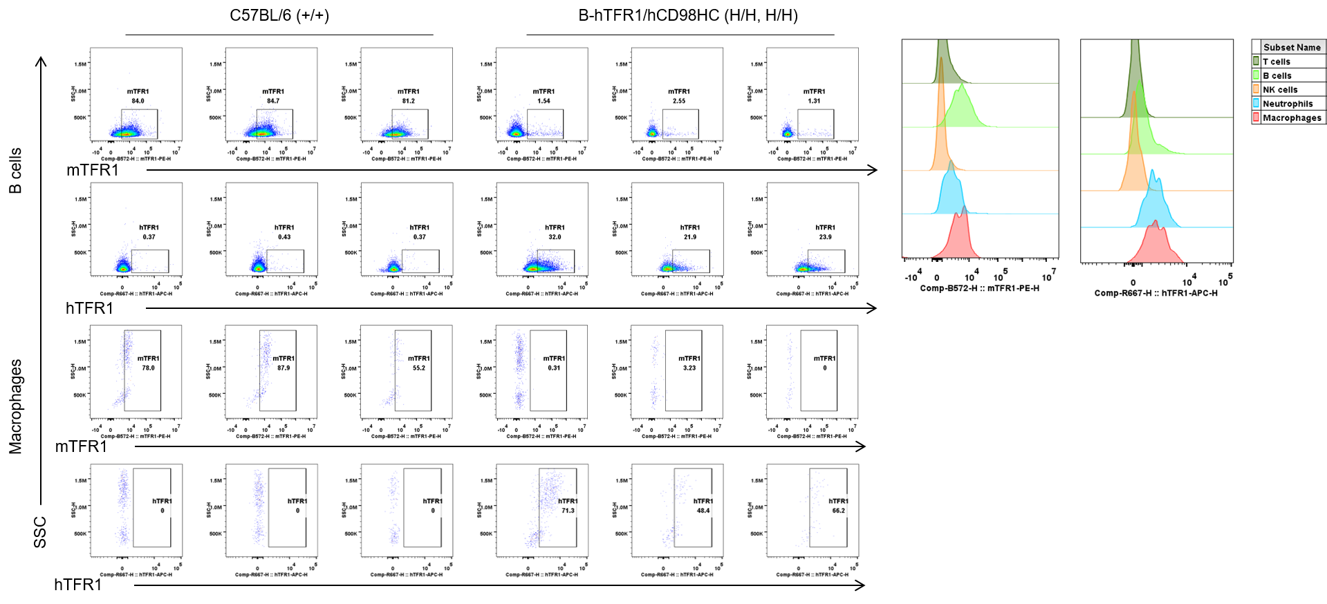
Strain-specific TFR1 expression analysis in wild-type C57BL/6 mice and homozygous B-hTFR1 mice. Bone marrow erythrocytes were collected from wild-type C57BL/6 mice (+/+) and homozygous B-hTFR1 mice (H/H). Protein expression was analyzed with anti-mouse TFR1 antibody (Biolegend, 113807) and anti-human TFR1 antibody (Biolegend, 334107) by flow cytometry.
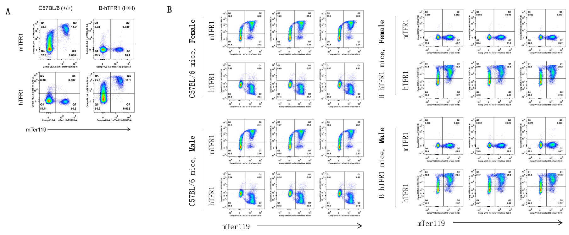
TFR1 Protein Expression Analysis in Leukocytes from Blood
Strain specific TFR1 expression analysis in wild-type C57BL/6 and homozygous B-hTFR1/hCD98HC mice by flow cytometry. Blood cells were collected from wild-type C57BL/6 (+/+) and homozygous B-hTFR1/hCD98HC mice (H/H, H/H) and analyzed by flow cytometry with anti-mouse TFR1 antibody (Biolegend, 113808) and anti-human TFR1 antibody (Biolegend, 334108). mTFR1 was only detectable in wild-type mice, and hTFR1 was exclusively detectable in B cells and macrophages from homozygous mice but not from wild-type mice.
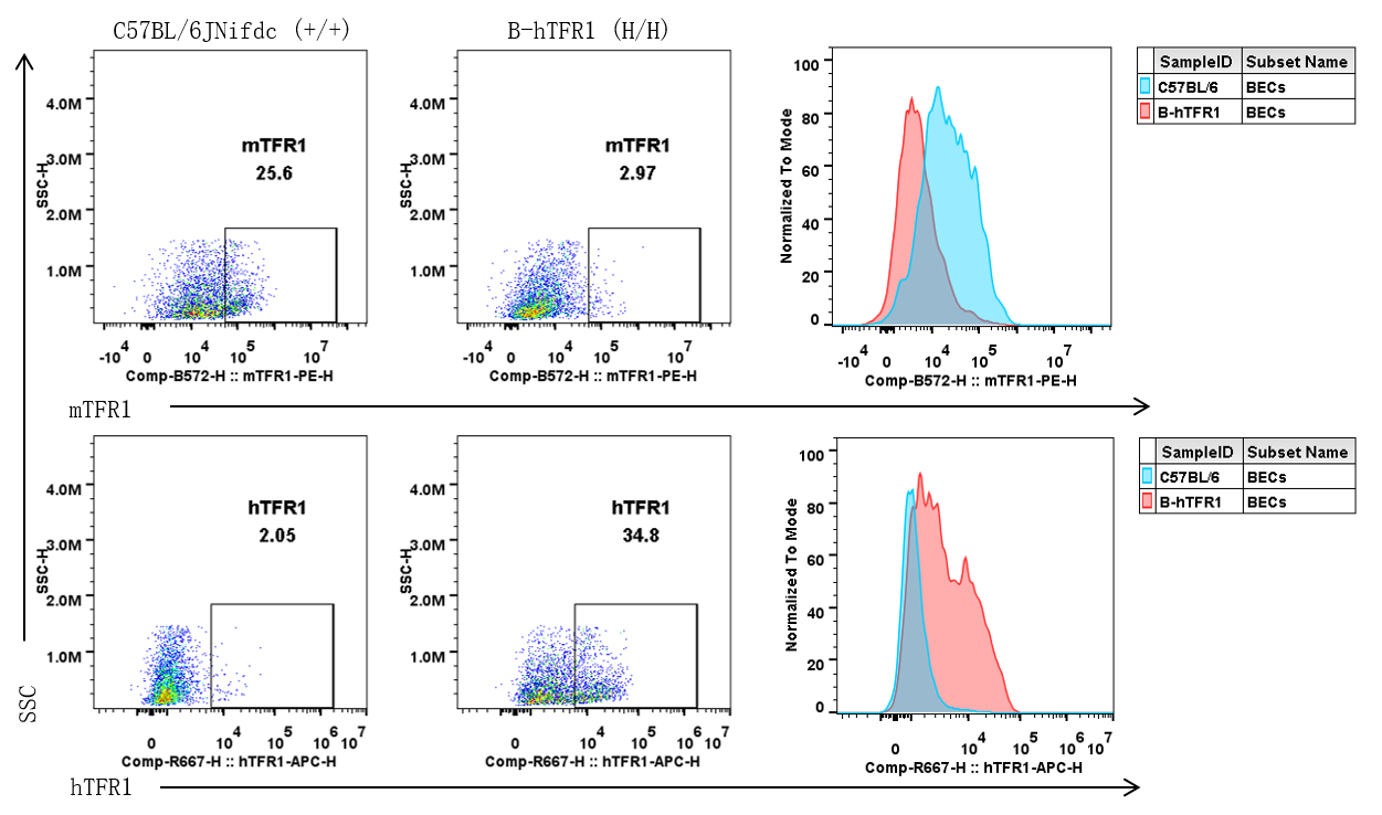
TFR1 Protein Expression in Brain Endothelial Cells
- Mouse TFR1 was detectable only in brain endothelial cells of wild-type mice.
- Human TFR1 was exclusively detectable in brain endothelial cells from homozygous B-hTFR1 mice but absent in wild-type mice.
Strain specific TFR1 expression analysis in wild-type C57BL/6JNifdc and homozygous B-hTFR1 mice by flow cytometry. Brain cells were collected from wild-type C57BL/6JNifdc (+/+) and homozygous B-hTFR1 mice (H/H), and analyzed by flow cytometry with anti-mouse TFR1 antibody (Biolegend, 113808) and anti-human TFR1 antibody (Biolegend, 334108).
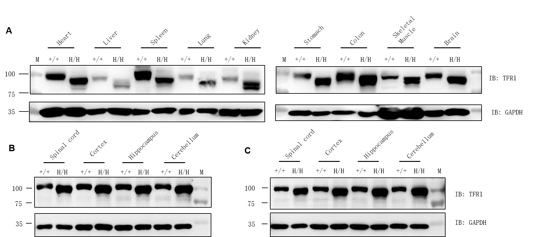
Protein Expression Profiles of TFR1
- TFR1 exhibits ubiquitous expression across diverse tissues. In humanized mice, hTFR1 displays an expression pattern consistent with mouse Tfr1 in wild-type mice.
- TFR1 exhibits ubiquitous expression across diverse tissues. In humanized mice, hTFR1 displays an expression pattern consistent with mouse Tfr1 in wild-type mice.
TFR1 protein expression analysis by western blotting using a cross-reactive anti-TFR1 antibody (abcam, ab214039). M, marker. (B) Male. (C) Female.
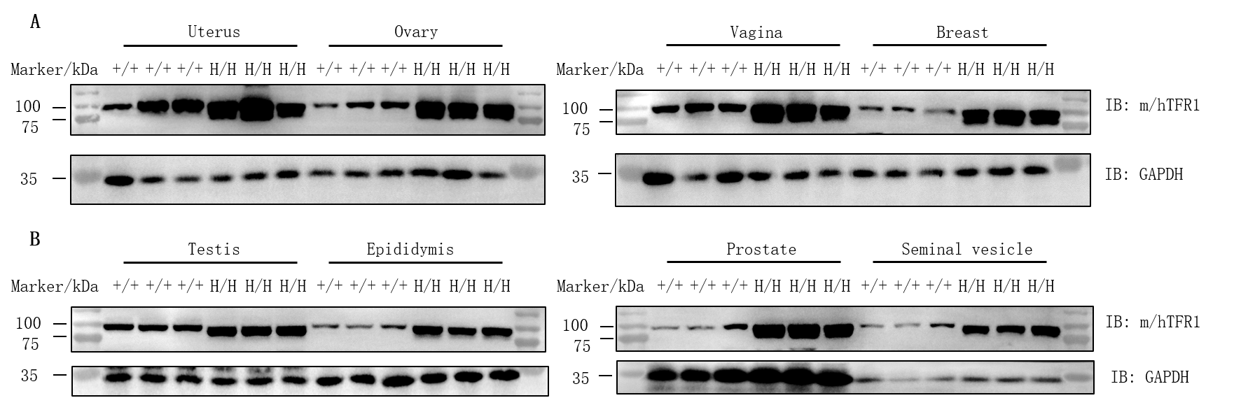
TFR1 Protein Expression in Reproductive Organs
- TFR1 was detectable in the uterus, ovary, vagina, breast, testis, epididymis, prostate and seminal vesicle of both C57BL/6JNifdc and B-hTFR1 mice.
Protein expression analysis of TFR1 in homozygous B-hTFR1/hCD98HC mice. Various tissue lysates were collected from wild-type C57BL/6JNifdc mice (+/+) and homozygous B-hTFR1/hCD98HC mice (H/H), and then analyzed by western blot with anti-transferrin receptor antibody (abcam, ab214039) and anti-CD98 antibody (abcam, ab307587). 30 μg total proteins were loaded for western blotting analysis. TFR1 was detected in uterus, ovary, vagina, breast, testis, epididymis, prostate and seminal vesicle from both wild-type C57BL/6JNifdc mice and homozygous B-hTFR1/hCD98HC mice, as the antibody was cross-reactive between human and mouse.
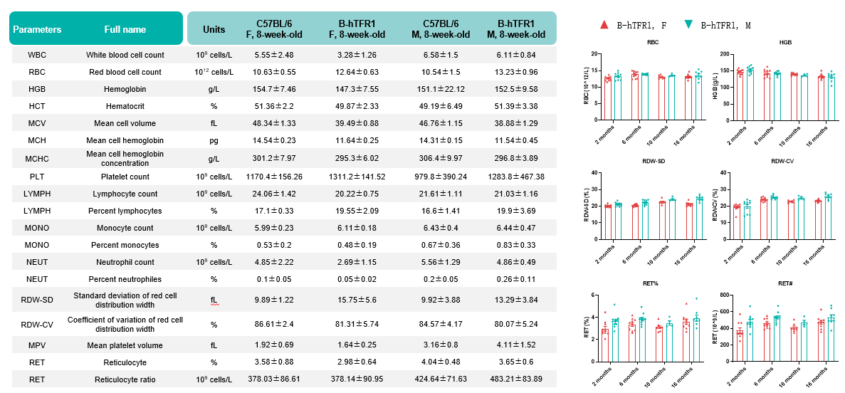
Functional Assay of Hematology Analysis
- Humanization of TFR1 does not alter hematological parameters, with no anemia phenotypes throughout 2-16 months old.
Values are expressed as mean ± SD.
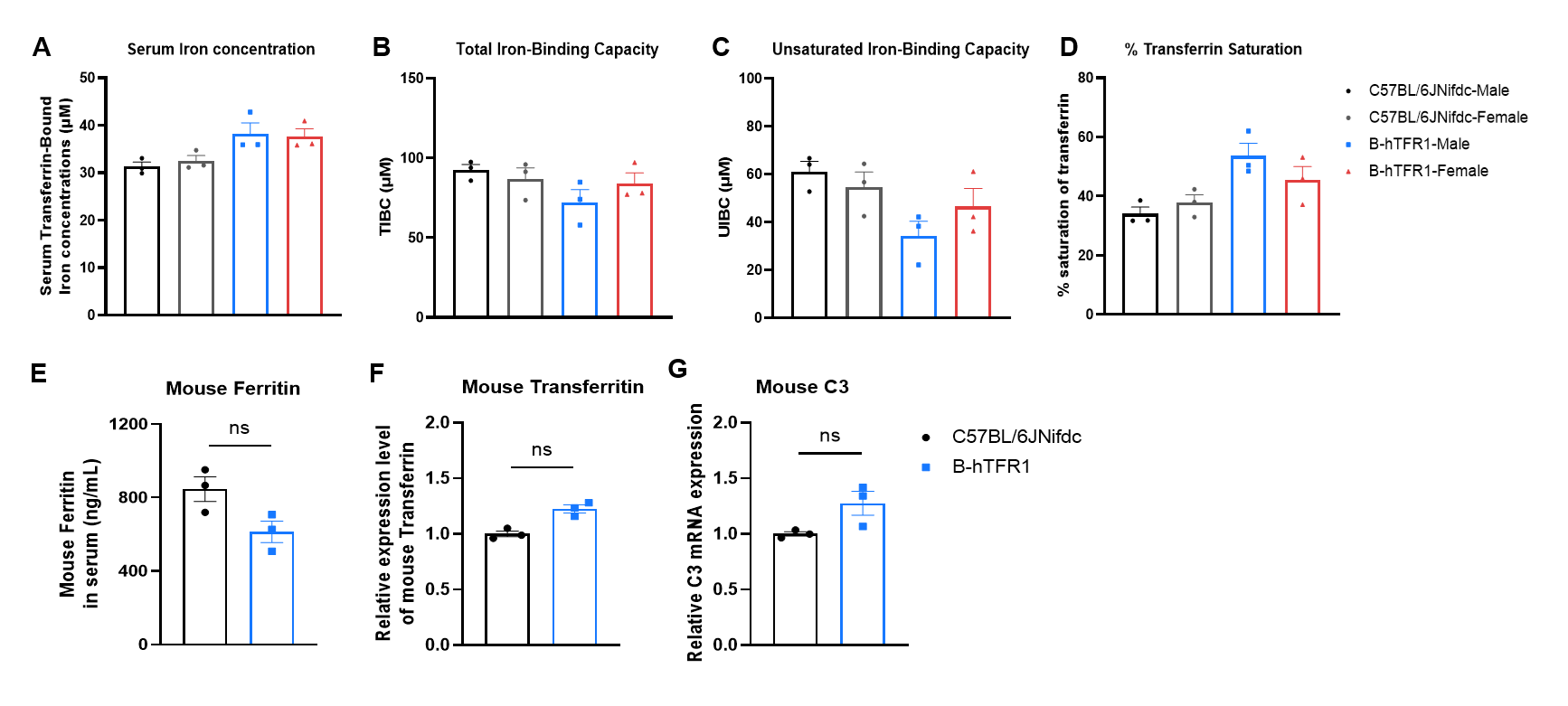
Functional Assay of Serum Iron Homeostasis
- All the detected parameters in homozygous B-hTFR1 mice were similar to those in the wild-type C57BL/6JNifdc mice.
- B-hTFR1 mice show no significant differences in iron homeostasis compared to wild-type C57BL/6JNifdc mice
Serum iron homeostasis assay in wild-type C57BL/6JNifdc and homozygous humanized B-hTFR1 mice. (A-D) Serum were collected from C57BL/6JNifdc (female, n=3; male, n=3; 14-week-old) and B-hTFR1 mice (female, n=3; male, n=3; 9-week-old), and then analyzed by Total Iron-Binding Capacity (TIBC) and Serum Iron Assay kit (abcam, ab239715). (E-G) Mouse ferritin, transferritin and complement 3 detection in wild-type C57BL/6JNifdc and B-hTFR1 mice (male, n=3, 8-week-old). Values are expressed as mean ± SEM. Significance was determined by two-way ANOVA test. *P < 0.05, **P < 0.01, ***p < 0.0001.
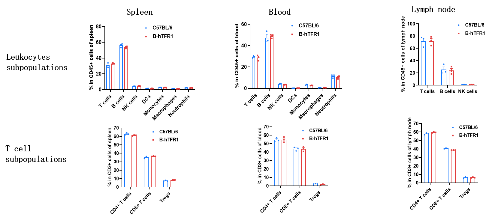
Animal State Evaluation-Leukocyte Profiling
- Humanization of TFR1 does not alter the frequency or distribution of immune cell types in spleen, blood and lymph nodes.
Values are expressed as mean ± SD.
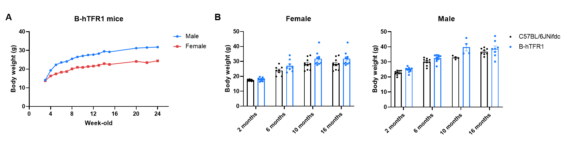
Animal State Evaluation-Growth Curve
- B-hTFR1 mice exhibit normal body weight growth patterns comparable to C57BL/6JNifdc control mice across both sexes and all monitored ages.
Body weight growth of B-hTFR1 mice compared with C57BL/6JNifdc mice. (A) Longitudinal body weight of male and female B-hTFR1 mice from 3 to 24 weeks of age. (B) Body weight of female and male B-hTFR1 mice at 2, 6, 10, and 16 months of age, compared with age- and sex-matched wild-type C57BL/6JNifdc mice (black bars). Values are expressed as mean ± SEM.
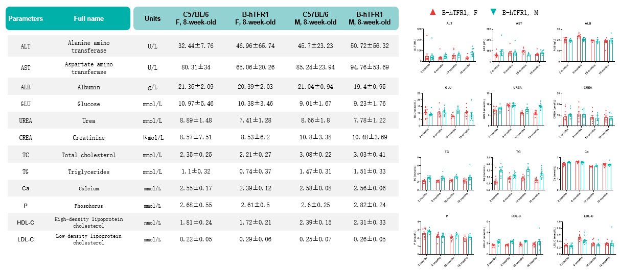
Animal State Evaluation-Blood Chemistry
- Humanization of TFR1 does not alter blood chemistry parameters throughout 2-16 months old.
Values are expressed as mean ± SD.
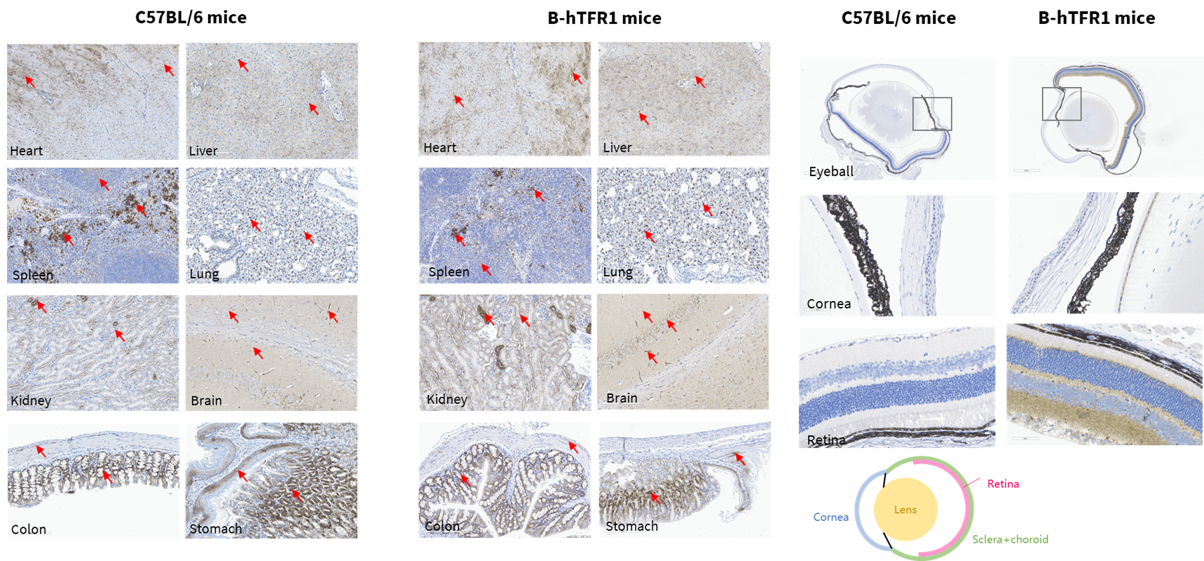
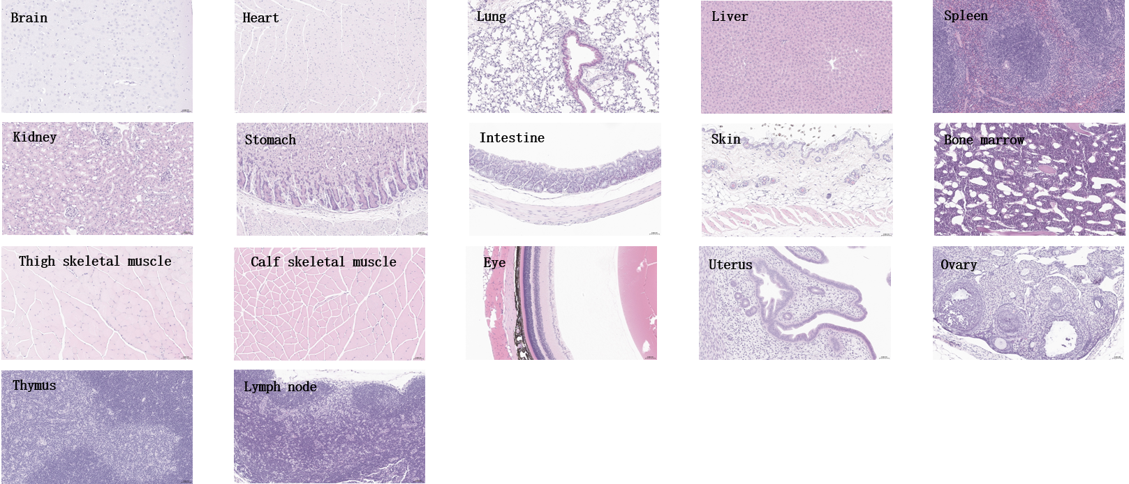
Animal State Evaluation-Organ Anatomy
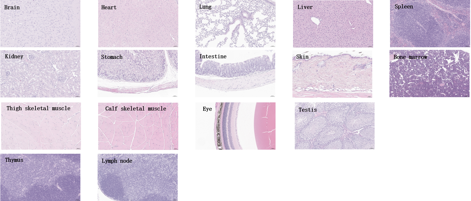
- No abnormal pathological findings were observed in all tissues examined in B-hTFR1 mice (female).
Histopathological analysis of major organs from homozygous B-hTFR1 mice. Tissues were collected from B-hTFR1 mice (female, 8-week-old, n=3), fixed in 4% paraformaldehyde and stained with hematoxylin and eosin (H&E). Representative photomicrographs are shown. Scale bar: 40x.
- No abnormal pathological findings were observed in all tissues examined in B-hTFR1 mice (male).
Histopathological analysis of major organs from homozygous B-hTFR1 mice. Tissues were collected from B-hTFR1 mice (male, 8-week-old, n=3), fixed in 4% paraformaldehyde and stained with hematoxylin and eosin (H&E). Representative photomicrographs are shown. Scale bar: 40x.
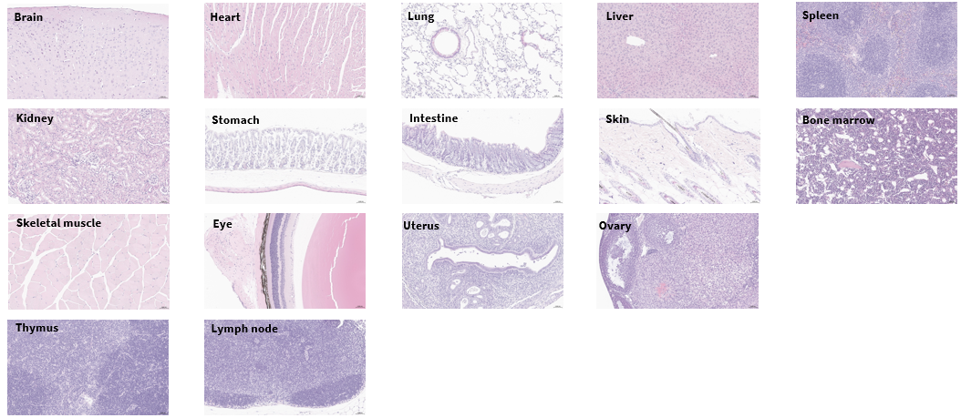
- No abnormal pathological findings were observed in all tissues examined in 6-month-old B-hTFR1 mice (female).
Histopathological analysis of major organs from homozygous B-hTFR1 mice. Tissues were collected from B-hTFR1 mice (female, 6-month-old, n=3), fixed in 4% paraformaldehyde and stained with hematoxylin and eosin (H&E). Representative photomicrographs are shown. Scale bar: 40x.
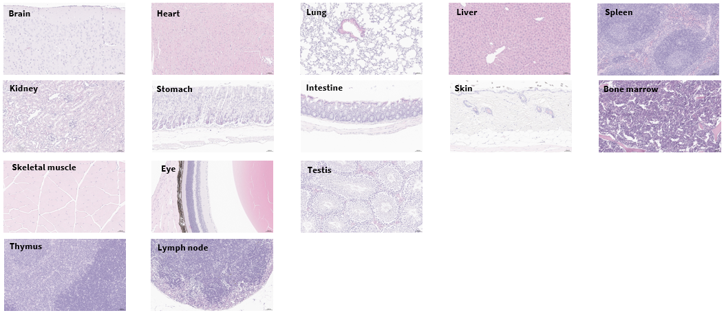
- No abnormal pathological findings were observed in all tissues examined in B-hTFR1 mice (male).
Histopathological analysis of major organs from homozygous B-hTFR1 mice. Tissues were collected from B-hTFR1 mice (male, 6-month-old, n=3), fixed in 4% paraformaldehyde and stained with hematoxylin and eosin (H&E). Representative photomicrographs are shown. Scale bar: 40x.
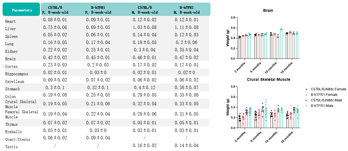
- The weights of various organs in B-hTFR1 mice were comparable to those of wild-type C57BL/6JNifdc mice.
Values are expressed as mean ± SD.
Animal State Evaluation-Body composition analysis
Body composition analysis of wild-type C57BL/6JNifdc mice and homozygous B-hTFR1 mice (6-month-old, n=10) measured by Body Composition Analyzer. (A) Absolute body weight, lean mass, fat mass, and free water mass (expressed in grams). (B) Relative lean content, fat content, free water content, total content, and total water content (expressed as percentages). B-hTFR1 mice exhibited normal body composition profiles with no notable differences from wild-type C57BL/6JNifdc mice.
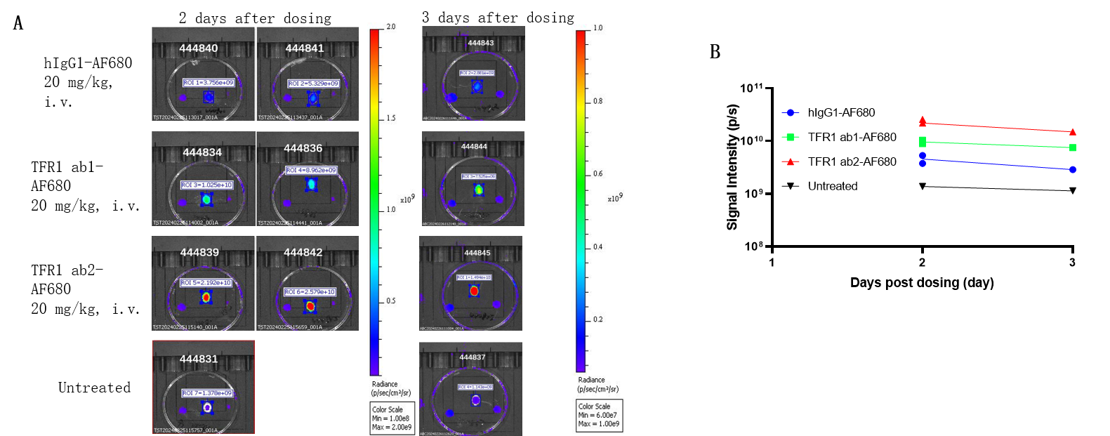
Brain Uptake of Anti-Human TFR1 Antibody in B-hTFR1 Mice
- Brain uptake of anti-human TFR1 antibody Ab2 was higher than Ab1 in B-hTFR1 mice.
B-hTFR1 mice were intravenously injected with AF680-conjugated control hIgG1 or anti human TFR1 antibodies ab1 and ab2 (provided by a client). After 2 days or 3 days post-injection, the mice were perfused and their brains were collected for analysis. (A) Mouse brain images under imaging system. (B) Fluorescence intensity of mouse brain under imaging system. The results indicate that the uptake of anti-human TFR1 antibody ab2 in the brain of B-hTFR1 mice was higher than that of anti-human TFR1 antibody ab1.
Note: These data were obtained from collaborative validation with the client, who provided the antibody.
In vivo PK evaluation of anti-human TFR1 BsAbs
- B-hTFR1 mouse brain enables uptake of intravenously administered anti-human TFR1 BsAbs.
- B-hTFR1 mice serve as a powerful preclinical model for in vivo evaluation of protein therapeutic
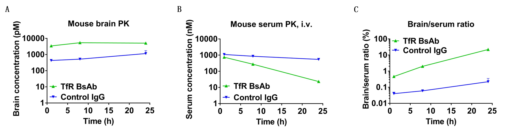
In vivo pharmacokinetic (PK) evaluation of anti-human TFR1 bispecific antibodies (BsAbs). B-hTFR1 mice were injected with control IgG (10 mpk) and anti-human TFR1 BsAbs (10.9 mpk) provided by a client via tail vein. Brain and serum were taken for in vivo PK evaluation. Brain concentrations(A), serum concentrations (B), and brain-to-serum ratio (C) of anti-human TFR1 BsAbs were quantified. As shown in panel, anti-human TFR1 BsAbs exhibited higher serum clearance and enhanced brain exposure after dose. The results confirmed that brain of B-hTFR1 mice enables uptake of an intravenously administered anti-human TFR1 BsAbs and B-hTFR1 mice provide a powerful preclinical model for in vivo evaluation of effective delivery of protein therapeutics to the central nervous system (CNS). Graphs represent mean ± SEM.
Note: This experiment was performed by the client using B-hTFR1 mice. All the other materials were provided by the client.
B-hTFR1/hCD98HC Mice Enable Comparative Analysis of TFR1 and CD98HC
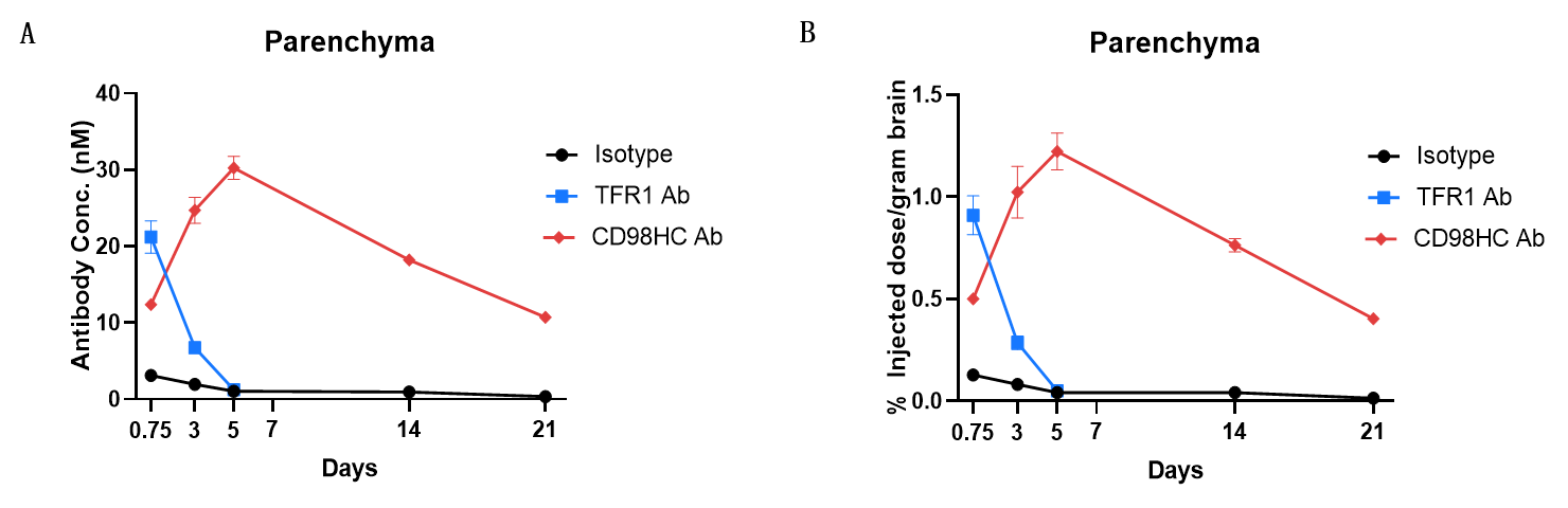
In vivo PK evaluation and comparison of anti-human TFR1 and anti-CD98HC antibody. B-hTFR1/hCD98HC mice (n=2, female, 8-week-old) were injected with control IgG (10 mpk) anti-human TFR1 antibody (TFR1 Ab, JR-141 analog, monovalent, produced in house, 12.56 mpk) and anti-human CD98HC antibody (CD98HC Ab, CD98BBBB-h1.L analog, monovalent, produced in house, 13.3 mpk) via tail vein. Brain were taken for in vivo PK evaluation after dosing 18 h and 3, 5, 14, 21 days. Brain concentrations (A) and % of injection/gram brain (B) were quantified. As shown in panel, anti-human TFR1 antibody exhibited higher brain exposure in 24 h after dose, while anti-CD98HC antibody exhibited higher brain exposure in 72 h after dose. The results confirmed that B-hTFR1/hCD98HC mice enables uptake of an intravenously administered anti-human TFR1 antibody or anti-human CD98HC antibody, and this mice can be used for the comparison of penetration efficacy of shuttle molecules targeting TFR1 or CD98HC. Graphs represent mean ± SEM.
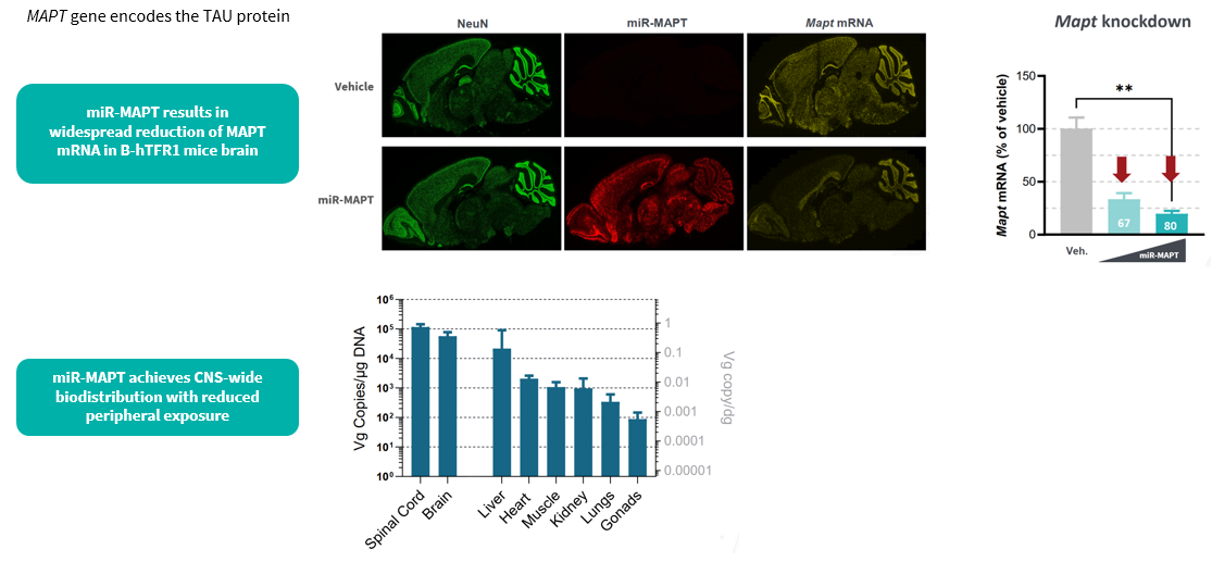
B-hTFR1 Mice Enable BBB Penetration of Human TFR1-Targeted AAV
- Brain and spinal cord— highest of all tissues assessed
- Peripheral tissues, including the Liver, show lower Vg, 10 – 100×below CNS
- No miR-MAPT guide expression detected outside the CNS — on-target silencing restricted to target tissue
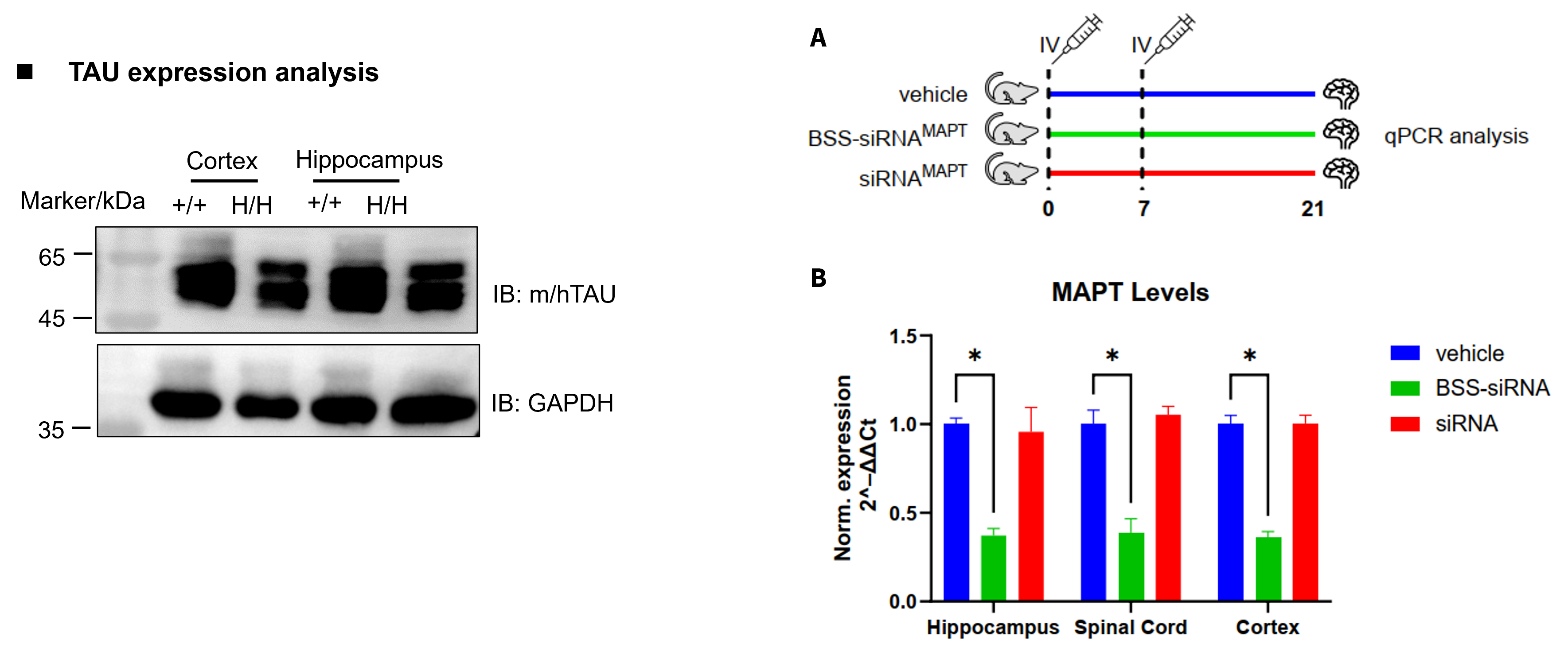
B-hTFR1/hTAU Mice Enable BBB Penetration of Antibody Oligonucleotide Conjugates
Gene targeting strategy for B-hTFR1/hTAU mice. The exons 2~10 of mouse Mapt gene that encode the full-length protein were replaced by human MAPT exons 2~15 in B-hTFR1/hTAU mice. The 3’UTR region of the mouse gene are replaced by human counterparts. The chimeric MAPT expression is driven by endogenous mouse Mapt promoter, while mouse Mapt gene transcription and translation will be disrupted. The exons 4-19 of mouse Tfr1 gene that encode the extracellular region were replaced by human TFR1 exons 4-19 in B-hTFR1/hTAU mice.
B-hTFR1/hDMPK Mice Enable Muscle Penetration of Antibody Oligonucleotide Conjugates
- DMPK-targeting AOC markedly reduced human DMPK mRNA in tibialis anterior muscle.
- B-hTFR1/hDMPK mice provide a powerful preclinical model to evaluate human DMPK-targeted AOCs
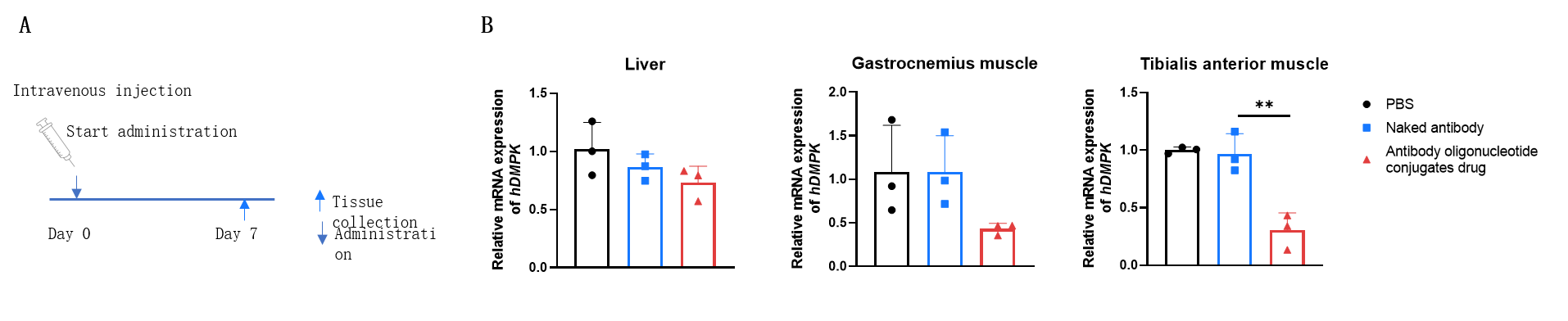
The inhibitory efficiency of the antibody oligonucleotide conjugates drug against human DMPK in heterozygous B-hTFR1/hDMPK mice. The antibody oligonucleotide conjugates drug (3 mpk, produced in-house), naked antibody (3 mpk, produced in-house) and PBS were administered to the heterozygous B-hTFR1/hDMPK mice individually on day 0. The mice were sacrificed on day 7, and the liver, gastrocnemius muscle and tibialis anterior muscle were collected to detect the expression level of human DMPK mRNA by qPCR. The human DMPK mRNA in the treatment groups (antibody oligonucleotide conjugates drug) were significantly reduced compared to the control groups (naked antibody and PBS) in tibialis anterior muscle, demonstrating that B-hTFR1/hDMPK mice provide a powerful preclinical model for in vivo evaluation of human DMPK targeted antibody oligonucleotide conjugates drug. Values are expressed as mean ± SEM.
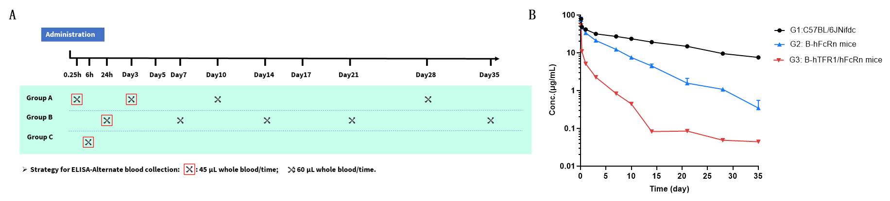
B-hTFR1/hFcRn Mice Enable PK Prediction of TFR1-Antibodies
Pharmacokinetic (PK) analysis of JR-141 analog in wild-type and humanized TFR1/FcRn mouse strains. (A) Scheme design. Male mice (n = 9 per group) from wild-type C57BL/6 mice (G1), B-hFcRn mice (G2) and B-hTFR1/hFcRn mice (G3) received a single intravenous (i.v.) dose of 3 mg/kg JR-141 analog. Blood samples were collected at 0.25 h, 6 h, 24 h, and on days 3, 7, 10, 14, 21, 28, and 35 post-administration. (B) Serum concentrations of JR-141 analog were measured by ELISA. Compared to wild-type mice (G1), B-hFcRn mice (G2) and B-hTFR1/hFcRn mice (G3) exhibited accelerated clearance. B-hTFR1/hFcRn mice better reflect in vivo antibody concentration changes compared to wild-type C57BL/6JNifdc and B-hFcRn mice, especially valuable for evaluating the PK of antibodies exhibiting target-mediated drug disposition (TMDD) in a more physiologically relevant context.
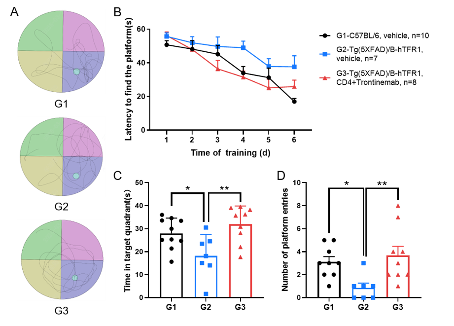
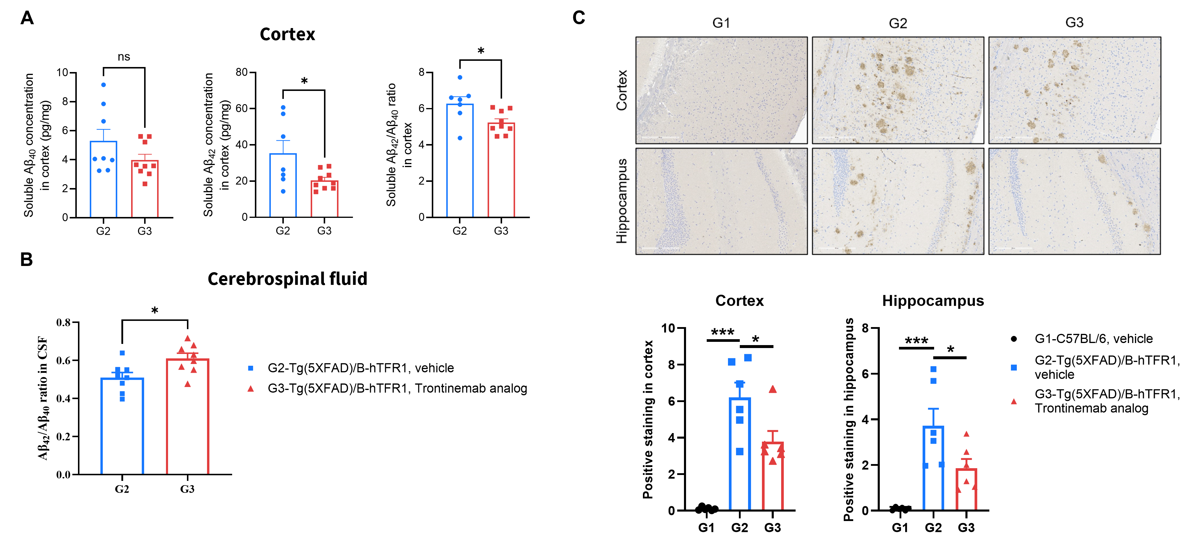
Tg(5XFAD)/B-hTFR1 Mice Enable Drug Efficacy Test of Brainshuttle antibody targeting Aβ
This experiment was conducted in collaboration with the client using Tg(5XFAD)/B-hTFR1 mice.
Drug efficacy of Trontinemab analog in Tg(5XFAD)/B-hTFR1 mice
This experiment was conducted in collaboration with the client using Tg(5XFAD)/B-hTFR1 mice.
* When publishing results obtained using this animal model, please acknowledge the source as follows: The animal model [B-hTFR1 mice] (Cat# 110861) was purchased from Biocytogen.