Description
TFR1/TAU: The combination of targeted antibody (TFR1) and therapeutic oligonucleotide (TAU)
- Gene Information: The transferrin receptor protein 1 (TFR1) gene encodes human TFR1, a transmembrane glycoprotein. The microtubule-associated protein tau (MAPT) gene encodes human TAU, which is associated with several neurodegenerative disorders.
- Protein Expression: TFR1 is highly expressed in the brain endothelial cells of the blood-brain barrier, and can be used as a target for enhancing drug delivery. TAU is mainly distributed in the central nervous system. Under pathological conditions, tau undergoes hyperphosphorylation, which causes the protein to detach from microtubules and form insoluble intracellular aggregates.
- Signaling Pathway: TFR1 primarily mediates the binding and endocytosis of transferrin with its ligand, driving the uptake of iron into cells. Overactivation of kinases or inhibition of phosphatases triggers aberrant phosphorylation of TAU, ultimately leading to synaptic dysfunction and neuronal cell death.
- Therapeutic Inhibition: When conjugated to an oligonucleotide drug targeting human MAPT, the TFR1-targeting antibody facilitates blood–brain barrier penetration and brain delivery. It sustains inhibition of MAPT mRNA expression, diminishes tau protein production, and demonstrates potent efficacy for the treatment of pathological tau aggregation and diverse neurodegenerative diseases.
Targeting strategy
TFR1
- The exons 4-19 of mouse Tfr1 gene that encode extracellular domain are replaced by human counterparts in B-hTFR1 mice. The genomic region of mouse Tfr1 gene that encodes cytoplasmic portion is retained.
- The promoter, 5’UTR and 3’UTR region of the mouse gene are also retained. The chimeric TFR1 expression is driven by endogenous mouse Tfr1 promoter, while mouse Tfr1 gene transcription and translation will be disrupted.
TAU
- The exons 2~10 of mouse Mapt gene that encode the full-length protein were replaced by human MAPT exons 2~15 in B-hTFR1/hTAU mice. The 3’UTR region of the mouse gene is replaced by human counterparts.
- The chimeric MAPT expression is driven by endogenous mouse Mapt promoter, while mouse Mapt gene transcription and translation will be disrupted.
mRNA Expression Analysis
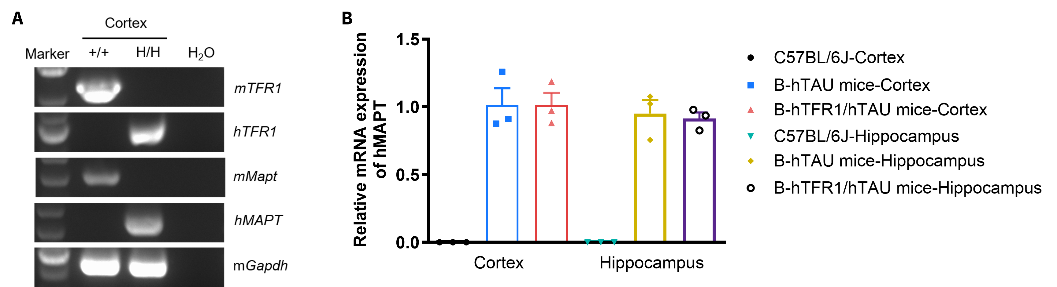
- (A) Human TFR1 and MAPT mRNA are exclusively detectable in cortex of homozygous B-hTFR1/hTAU mice, but not in wild-type C57BL/6 mice.
- (B) Relative MAPT mRNA expression level in the cortex and hippocampus were comparable between B-hTAU mice and B-hTFR1/hTAU mice.
Strain-specific TFR1 and MAPT expression analysis in wild-type C57BL/6 mice and homozygous B-hTFR1/hTAU mice. (A) Cortex RNA was isolated from wild-type C57BL/6 mice (+/+) and homozygous B-hTFR1/hTAU mice (H/H), then cDNA libraries were synthesized by reverse transcription, followed by PCR with mouse or human TFR1 and MAPT primers. (B) Quantitative real-time PCR (qRT-PCR) analysis of relative MAPT mRNA expression in the cortex and hippocampus of male wild-type C57BL/6 mice, homozygous B-hTAU mice and homozygous B-hTFR1/hTAU mice.
TFR1 Protein Expression
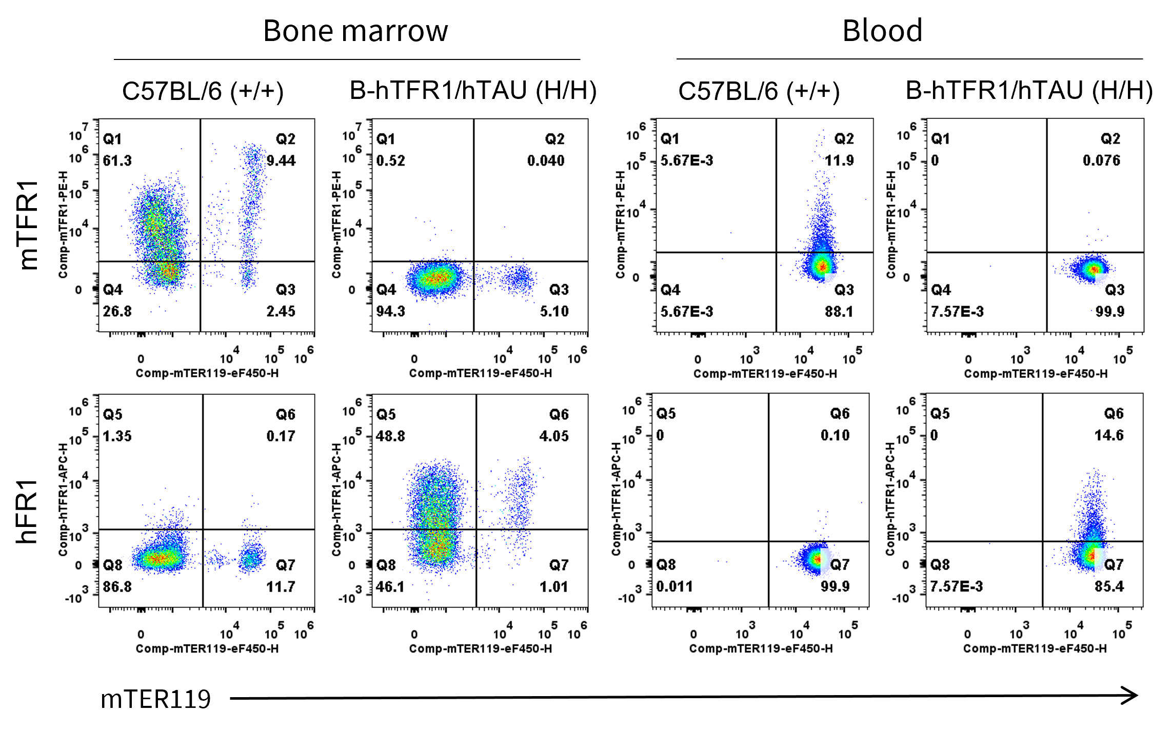
- Mouse TFR1 was detected on bone marrow and blood in wild-type C57BL/6 mice, but not in B-hTFR1/hTAU mice.
- Human TFR1 was detected on bone marrow and blood in B-hTFR1/hTAU mice, but not in wild-type C57BL/6 mice.
Mouse and human TFR1 expression analysis in bone marrow and blood. Bone marrow and blood cells were collected from wild-type C57BL/6 mice and homozygous B-hTFR1/hTAU mice. TFR1 expression was analyzed by flow cytometry using anti-mouse TFR1 antibody (Biolegend, 113808) and anti-human TFR1 antibody (Biolegend, 334108).
TFR1 and TAU Protein Expression
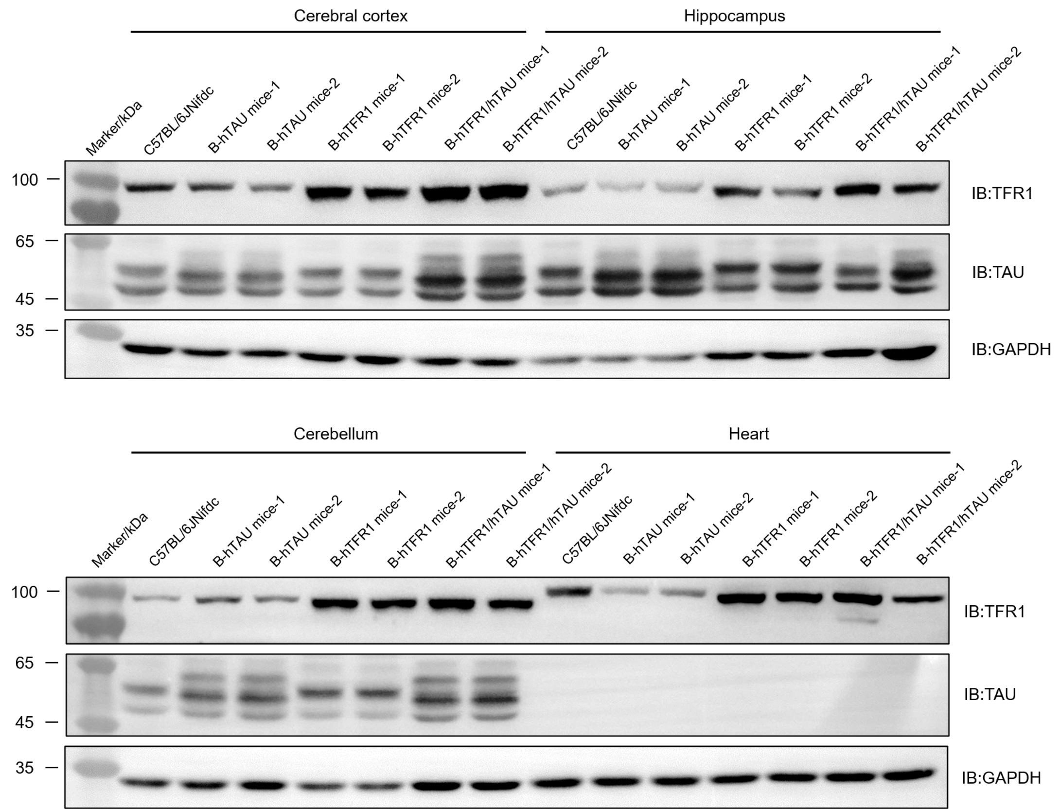
- TFR1 was detectable in the cortex, hippocampus, cerebellum and heart of both wild-type C57BL/6 mice and B-hTFR1/hTAU mice, as the antibody was cross-reactive between human and mouse.
- TAU was detectable in the cortex, hippocampus and cerebellum of both wild-type C57BL/6 mice and B-hTFR1/hTAU mice, as the antibody was cross-reactive between human and mouse.
Protein expression analysis of TFR1 and TAU in wild-type C57BL/6 mice, B-hTFR1 mice, B-hTAU mice and B-hTFR1/hTAU mice. Brain and heart were collected from wild-type C57BL/6 mice, B-hTFR1 mice, B-hTAU mice and B-hTFR1/hTAU mice, and then analyzed by western blot with anti-transferrin receptor antibody (abcam, ab214039) and anti-TAU antibody (CST, 46687). 40 μg total protein was loaded for western blotting analysis.
Inhibitory efficiency of antibody oligonucleotide conjugate drug against the human TFR1/TAU
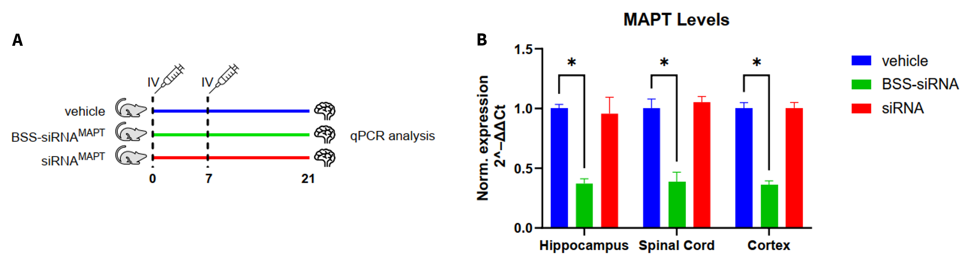
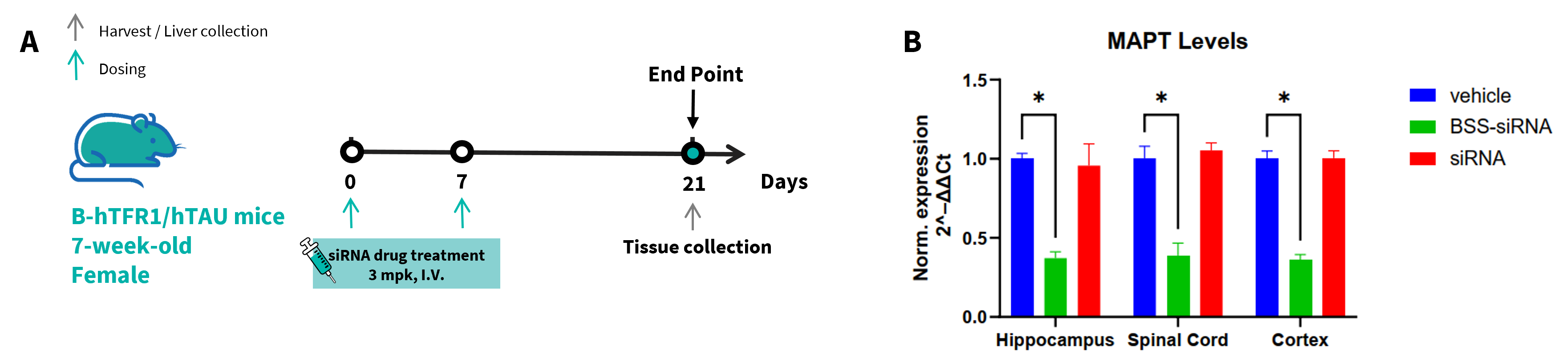
- The human MAPT mRNA in the treatment group was significantly reduced compared to the control group, demonstrating that B-hTFR1/hTAU mice provide a powerful preclinical model for in vivo evaluation of human MAPT targeted antibody oligonucleotide conjugate drugs.
The inhibitory efficiency of the antibody oligonucleotide conjugate (AOC) drug against human MAPT in B-hTFR1/hTAU mice. B-hTFR1/hTAU mice were randomly divided into three groups (n=3/group, 7-week-old, female). The vehicle, antibody oligonucleotide conjugates drug and oligonucleotide drug were administered to B-hTFR1/hTAU mice individually on day 0 and day 7. The mice were sacrificed on day 21, and the hippocampus, cortex and spinal cord were collected to detect the expression level of human MAPT mRNA by qPCR. (A) The schematic diagram of experimental processing. (B) The expression of human MAPT mRNA in hippocampus, cortex and spinal cord. Values are expressed as mean ± SEM. Significance was determined by unpaired t test. *P < 0.05.
This experiment was conducted in collaboration with the client using B-hTFR1/hTAU mice.
- The human MAPT mRNA in the treatment group were significantly reduced compared to the control group, demonstrating that B-hTFR1/hTAU mice provide a powerful preclinical model for in vivo evaluation of human MAPT targeted antibody oligonucleotide conjugates drug.
The inhibitory efficiency of the antibody oligonucleotide conjugate (AOC) drug against human MAPT in B-hTFR1/hTAU mice. B-hTFR1/hTAU mice were randomly divided into three groups (n=3/group, 7-week-old, female). The vehicle, antibody oligonucleotide conjugates drug and oligonucleotide drug were administered to B-hTFR1/hTAU mice individually on day 0 and day 7. The mice were sacrificed on day 21, and the hippocampus, cortex and spinal cord were collected to detect the expression level of human MAPT mRNA by qPCR. (A) The schematic diagram of experimental processing. (B) The expression of human MAPT mRNA in hippocampus, cortex and spinal cord. Values are expressed as mean ± SEM. Significance was determined by unpaired t test. *P < 0.05.
This experiment was conducted in collaboration with the client using B-hTFR1/hTAU mice.
* When publishing results obtained using this animal model, please acknowledge the source as follows: The animal model [B-hTFR1/hTAU mice] (Cat# 113290) was purchased from Biocytogen.