
C.B6-Cd3etm1(CD3E)Bcgen Cd3dtm1(CD3D)Bcgen Cd3gtm1(CD3G)Bcgen/Bcgen • 111719
| Product name | B-hCD3EDG mice(C) |
|---|---|
| Catalog number | 111719 |
| Strain name | C.B6-Cd3etm1(CD3E)Bcgen Cd3dtm1(CD3D)Bcgen Cd3gtm1(CD3G)Bcgen/Bcgen |
| Strain background | C57BL/6; BALB/cCrSlcNifdc |
| NCBI gene ID | 916,915,917 (Human) |
| Aliases | T3E; TCRE; IMD18; CD3epsilon; T3D; IMD19; CD3DELTA; CD3-DELTA; T3G; IMD17; CD3GAMMA; CD3-GAMMA |
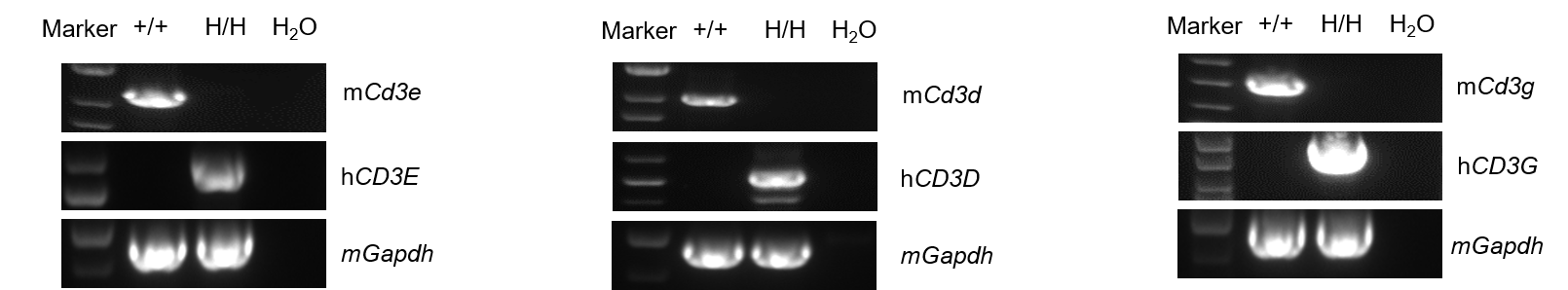
Strain specific analysis of CD3E, D, G mRNA expression in wild-type BALB/cCrSlcNifdc mice and B-hCD3EDG mice(C) by RT-PCR. Thymus RNA were isolated from wild-type BALB/cCrSlcNifdc mice (+/+) and homozygous B-hCD3EDG mice(C) (H/H), then cDNA libraries were synthesized by reverse transcription, followed by PCR with mouse or human CD3E, D, G primers. Mouse Cd3e, d, g mRNA was only detectable in wild-type mice. Human CD3E, D, G mRNA was exclusively detectable in homozygous B-hCD3EDG mice(C), but not in wild-type mice.
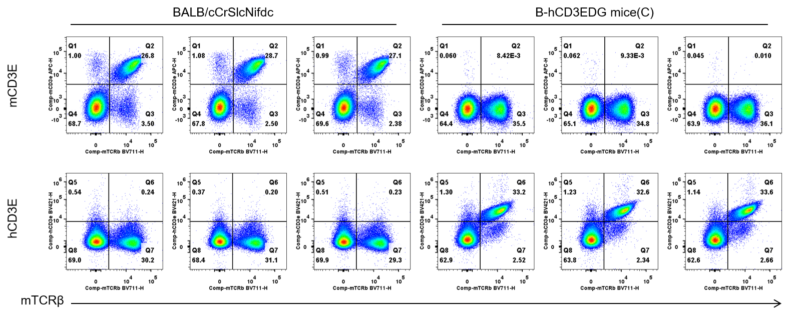
Strain specific CD3E expression analysis in wild-type BALB/cCrSlcNifdc mice and homozygous humanized B-hCD3EDG mice(C) by flow cytometry. Splenocytes were collected from wild-type BALB/cCrSlcNifdc mice and homozygous B-hCD3EDG mice(C) (female, n=3, 6-week-old). Protein expression was analyzed with anti-mouse CD3e antibody (Biolegend, 100312) and anti-human CD3e antibody (BD Horizon™ , 562426) by flow cytometry. Mouse CD3E was only detectable in wild-type BALB/cCrSlcNifdc mice. Human CD3E was exclusively detectable in homozygous B-hCD3EDG mice(C), but not in wild-type BALB/cCrSlcNifdc mice.
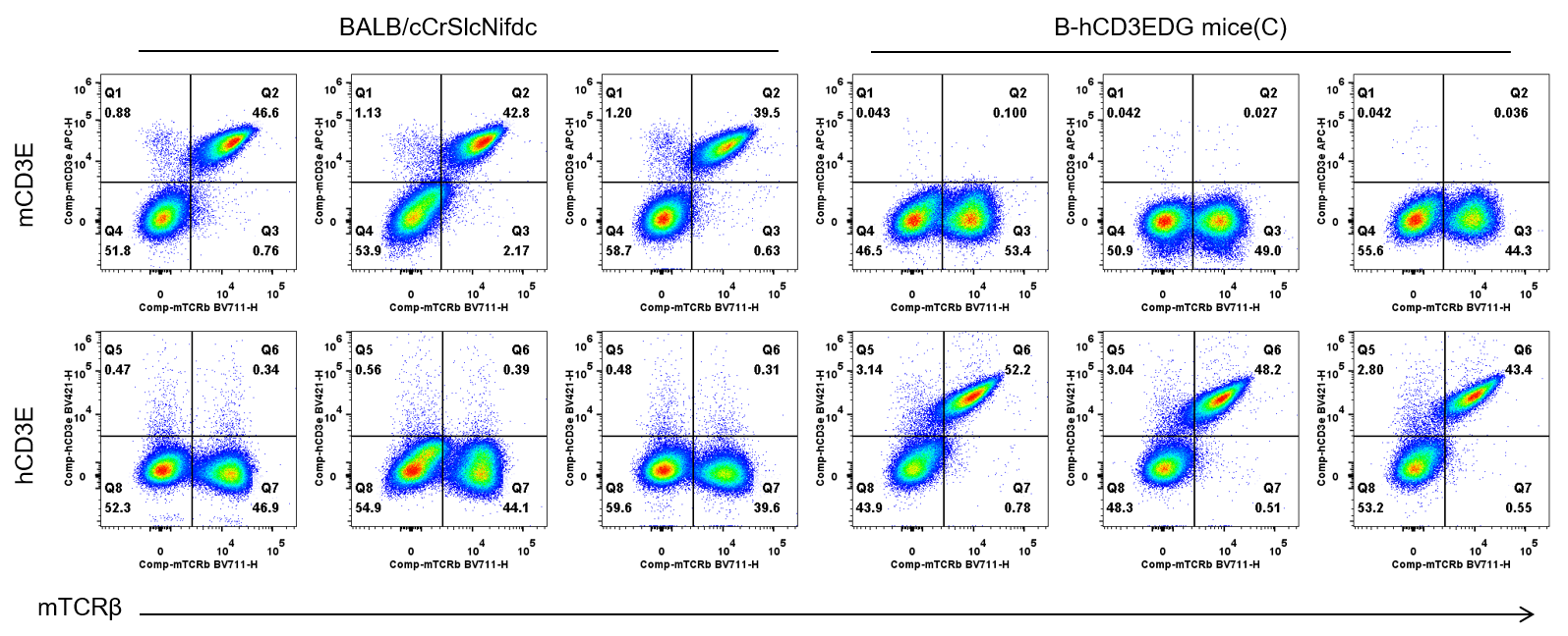
Strain specific CD3E expression analysis in wild-type BALB/cCrSlcNifdc mice and homozygous humanized B-hCD3EDG mice(C) by flow cytometry. Blood cells were collected from wild-type BALB/cCrSlcNifdc mice and homozygous B-hCD3EDG mice(C) (female, n=3, 6-week-old). Protein expression was analyzed with anti-mouse CD3e antibody (Biolegend, 100312) and anti-human CD3e antibody (BD Horizon™ , 562426) by flow cytometry. Mouse CD3E was only detectable in wild-type BALB/cCrSlcNifdc mice. Human CD3E was exclusively detectable in homozygous B-hCD3EDG mice(C), but not in wild-type BALB/cCrSlcNifdc mice.
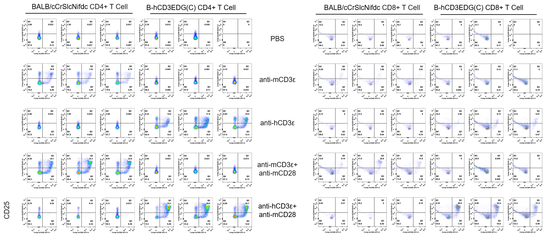
CD25 and CD69 expression analysis in wild-type BALB/cCrSlcNifdc mice and homozygous humanized B-hCD3EDG mice(C) by flow cytometry. T cells were isolated from splenocytes of BALB/cCrSlcNifdc and B-hCD3EDG mice(C) (n=3, 6-week-old), and were incubated in the presence of anti-mCD3ε antibody (2ug/ml,BioXcell,BE0001-2) , anti-hCD3ε antibody (2ug/ml,BioXcell,BE0001-1) and anti-mCD28 antibody (5ug/ml,BioXcell,BE0015-1) for 48h. T cell proliferation was tested by flow cytometry. T cell activation in B-hCD3EDG mice(C) was significantly up-regulated by anti-hCD3ε antibody or anti-hCD3ε antibody and anti-mCD28 antibody.
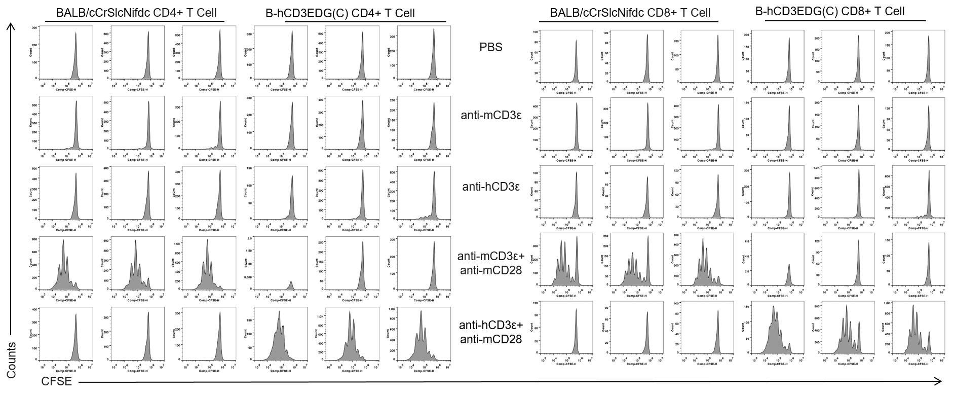
The proliferation of CD4+T cells and CD8+T cells was analyzed by flow cytometry. T cells were isolated from splenocytes of BALB/cCrSlcNifdc and B-hCD3EDG mice(C) (n=3, 6-week-old), and were incubated in the presence of anti-mCD3ε antibody(2ug/ml,BioXcell,BE0001-2) , anti-hCD3ε antibody (2ug/ml,BioXcell,BE0001-1) and anti-mCD28 antibody (5ug/ml,BioXcell,BE0015-1) for 72h. T cell proliferation was tested by flow cytometry. T cell activation in B-hCD3EDG mice(C) was significantly up-regulated by anti-hCD3ε antibody and anti-mCD28 antibody.

Frequency of leukocyte subpopulations in spleen by flow cytometry. Splenocytes were isolated from wild-type BALB/cCrSIcNifdc mice and homozygous B-hCD3EDG mice(C) (female, 6-week-old, n=3). A. Flow cytometry analysis of the splenocytes was performed to assess the frequency of leukocyte subpopulations. B. Frequency of T cell subpopulations. Frequencies of T cells, B cells, NK cells, DCs, neutrophils, monocytes, macrophages, CD4+ T cells, CD8+ T cells and Tregs in B-hCD3EDG mice(C) were similar to those in BALB/cCrSIcNifdc. The frequency of leukocyte subpopulations in thymus, lymph node and blood of B-hCD3EDG mice(C) were also comparable to wild-type BALB/cCrSIcNifdc mice (Data not shown). Values are expressed as mean ± SEM.

Antitumor activity of anti-human CD3×CD19 bispecific antibody (TNB-486 analog) in B-hCD3EDG mice(C). B-hCD19-luc-GFP A20 cells were injected intravenously into B-hCD3EDG mice(C) (female, 9-week-old, n=6). Mice were randomized for treatment when the average bioluminescence signal reached ~10⁶ p/s. The BsAb or PBS was administered intravenously per the dosing scheme. Tumor burden (via bioluminescence imaging) and body weight were monitored twice weekly. (A) Anti-human CD3×CD19 BsAb significantly inhibited the growth of B-hCD19-luc-GFP A20 cells compared to the PBS group. (B) Body weight changes during treatment. Values are expressed as mean ± SEM.
The overage of this tumor model is 40%.
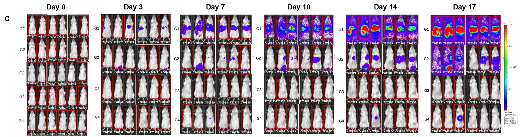
Antitumor activity of anti-human CD3×CD19 bispecific antibody (TNB-486 analog) in B-hCD3EDG mice(C). B-hCD19-luc-GFP A20 cells were injected intravenously into B-hCD3EDG mice(C) (female, 9-week-old, n=6). Mice were randomized for treatment when the average bioluminescence signal reached ~10⁶ p/s. The BsAb or PBS was administered intravenously per the dosing scheme. Tumor burden (via bioluminescence imaging) and body weight were monitored twice weekly. (C) Raw bioluminescence images. These results indicate that the B-hCD3EDG mice(C) provide a powerful preclinical model for in vivo evaluation of CD3-based antibodies. Values are expressed as mean ± SEM.
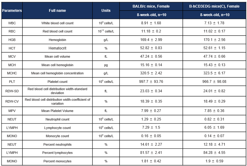
Complete blood count (CBC) of B-hCD3EDG mice(C). Values are expressed as mean ± SD.
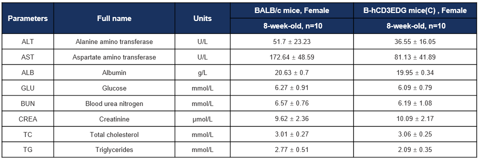
Biochemical test of B-hCD3EDG mice(C). Values are expressed as mean ± SD.