


B-HLA-A2.1/hNY-ESO-1 B16-F10

NA • 322593

B-HLA-A2.1/hNY-ESO-1 B16-F10

Catalog Number: 322593
Strain Name: NA
Strain Background: C57BL/6
NCBI gene ID: 567,3105,1485 (Human)
Aliases: IMD43; AMYLD6; MHC1D4; HLAA; CTAG; ESO1; CT6.1; CTAG1; LAGE-2; LAGE2B; NY-ESO-1
---
可提供授权方案
B-HLA-A2.1/hNY-ESO-1 B16-F10

在此页面上

  • Description
  • Phenotypic analysis
  • Tumorigenicity

海报

查看全部

    发表文章

      Description
      • Origin: The B16-F10 cell line is derived from C57BL/6J murine skin cells. The cell line is a commonly used murine model for melanoma. 
      • Background Information: HLA-A belongs to the HLA class I heavy chain paralogues. This class I molecule is a heterodimer consisting of a heavy chain and a light chain (beta-2 microglobulin). The heavy chain is anchored in the membrane. Class I molecules play a central role in the immune system by presenting peptides derived from the endoplasmic reticulum lumen so that they can be recognized by cytotoxic T cells. The protein product of NY-ESO-1 functions as an antigen that is upregulated across a broad range of malignancies, yet it is also constitutively expressed in healthy testicular tissue. The gene resides within a duplicated segment of the X-chromosome, resulting in the presence of an adjacent paralog with an identical nucleotide sequence.
      • Gene targeting strategy: The B2m gene (Exon2 to Exon3) of mouse were replaced by the sequence encompassing the human B2M CDS, HLA-A*0201 gene that included leader sequence, α1 and α2 domains ligated to a fragment of the murine H-2Db gene containing the α3, transmembrane and cytoplasmic domains and human NY-ESO-1 CDS. Human HLA-A2.1 is highly expressed on the surface of B-HLA-A2.1/hNY-ESO-1 B16-F10. Human NY-ESO-1 detectable by western blot.
      • Tumorigenicity: Confirmed in B-HLA-A2.1 mice. 
      • Application: The B-HLA-A2.1/hNY-ESO-1 B16-F10 tumor model can be used for preclinical evaluation of cancer vaccines and TCR mimics.
      • Notes

      Inoculated cell lines can be suspended with RPMI-1640 stock solution. 

      Before implementing the project, it is recommended to perform tumor growth experiments. The recommended cell inoculation amount is between 5E5-1E6. 

      In the experiment, it is necessary to ensure that the number of animals inoculated subcutaneously is at least 1.6 times the actual grouping number.

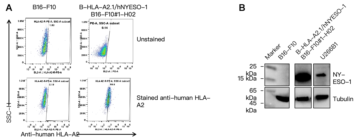
      HLA-A2 and NY-ESO-1 Protein Expression Analysis
      • Human HLA-A2.1 and NY-ESO-1 were detected on the surface of B-HLA-A2.1/hNY-ESO-1 B16-F10.

      HLA-A2.1 and NY-ESO-1 expression analysis in B-HLA-A2.1/hNY-ESO-1 B16-F10 by flow cytometry and western blot, respectively. Single cell suspensions from wild-type B16-F10 and B-HLA-A2.1/hNY-ESO-1 B16-F10 #1-H02 cultures were stained with anti-human HLA-A2 antibody (Biolegend, 343306) and anti-NY-ESO-1 (Proteintech, 19521-1-AP), respectively. (A) Human HLA-A2 was detected on the surface of B-HLA-A2.1/hNY-ESO-1 B16-F10 cells but not wild-type B16-F10 cells. (B) Human NY-ESO-1 was detected in the B-HLA-A2.1/hNY-ESO-1 B16-F10 cells but not wild-type B16-F10 cells.


      Tumor Growth Curve & Body Weight Changes

      Subcutaneous tumor growth of B-HLA-A2.1/hNY-ESO-1 B16-F10 cells. B-HLA-A2.1/hNY-ESO-1 B16-F10 cells (2×10⁵) and wild-type B16-F10 cells (2×10⁵) were subcutaneously implanted into B-HLA-A2.1 mice (male, 7-week-old, n=6). Tumor volume and body weight were measured twice a week. (A) Average tumor volume. (B) Body weight. Volume was expressed in mm3 using the formula: V=0.5 × long diameter × short diameter2. Results indicate that B-HLA-A2.1/hNY-ESO-1 B16-F10 cells were able to establish tumors in vivo and can be used for efficacy studies. Values are expressed as mean ± SEM.
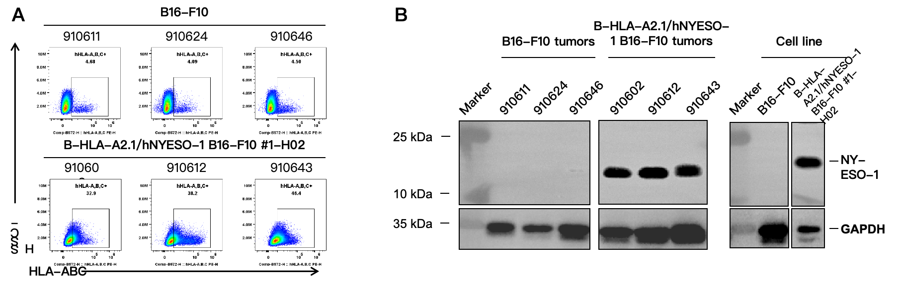
      HLA-A2.1 and NY-ESO-1 Protein Expression Analysis of Tumor Tissue
      • Human HLA-A2.1 and NY-ESO-1 were detected on the surface of B-HLA-A2.1/hNY-ESO-1 B16-F10 tumors cells.

      HLA-A2.1 and NY-ESO-1 expression were evaluated​​ on B-HLA-A2.1/hNY-ESO-1 B16-F10 by flow cytometry and western blot, respectively. ​​These cells​​ were subcutaneously transplanted into B-HLA-A2.1 mice (n=6). At the end of the experiment, tumor cells were harvested and ​​analyzed with species-specific anti-HLA-ABC antibody (Biolegend, 311406) and anti-NY-ESO-1 (Proteintech, 19521-1-AP) expression​​ by flow cytometry and western blot, respectively. Note: ​​In the tumor tissues from the B16‑F10 wild‑type cell group, we also detected a small subpopulation of cells, which, upon further analysis, were identified as non‑specific signals.
