

NA • 322418

Inoculated cell lines can be suspended with DMEM stock solution.
Before implementing the project, it is recommended to perform tumor growth experiments. The recommended cell inoculation amount is between 1E5-5E5.
In the experiment, it is necessary to ensure that the number of animals inoculated subcutaneously is at least 1.6 times the actual grouping number.

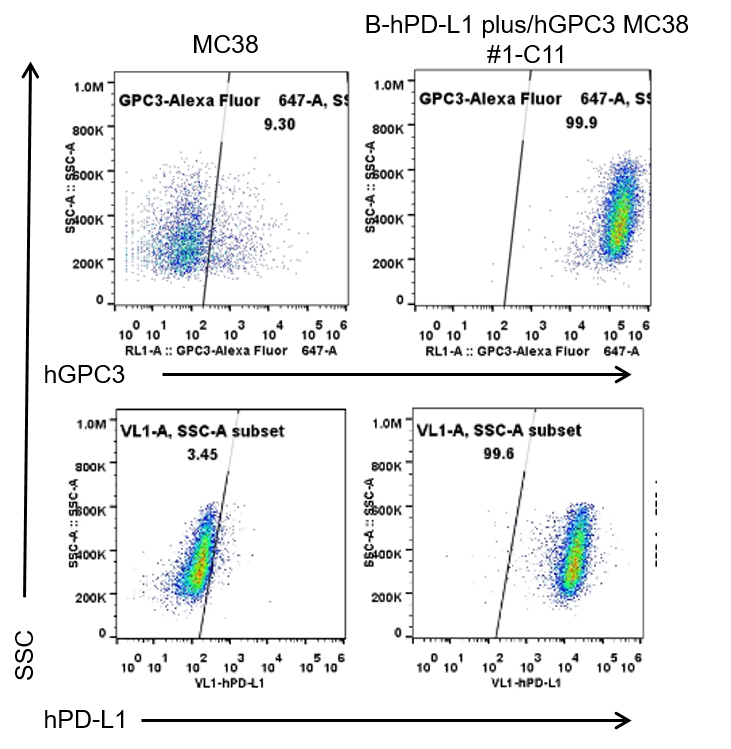
GPC3 and PD-L1 expression analysis in B-hPD-L1 plus/hGPC3 MC38 cells by flow cytometry. Single cell suspensions from wild-type MC38 and B-hPD-L1 plus/hGPC3 MC38 #1-C11 cultures were stained with anti-GPC3 antibody (in house, GC33-HuIgG1) and anti-PD-L1 antibody (Biolegend, 329706). Human GPC3 and human PD-L1 were detected on the surface of B-hPD-L1 plus/hGPC3 MC38 cells but not wild-type MC38 cells.

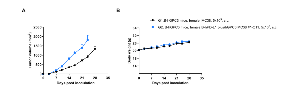
Subcutaneous tumor growth of B-hPD-L1 plus/hGPC3 MC38 cells. B-hPD-L1 plus/hGPC3 MC38 (5x10⁵) and wild-type MC38 cells (5x10⁵) were subcutaneously implanted into homozygous B-hGPC3 mice (7-week-old, n=6). Tumor volume and body weight were measured twice a week. (A) Average tumor volume. (B) Body weight. Volume was expressed in mm3 using the formula: V=0.5 X long diameter X short diameter2. Results indicate that B-hPD-L1 plus/hGPC3 MC38 cells were able to establish tumors in vivo and can be used for efficacy studies. Values are expressed as mean ± SEM.
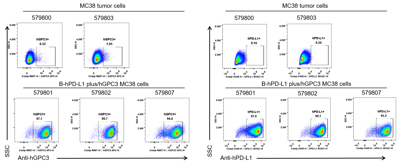
GPC3 and PD-L1 expression evaluated on B-hPD-L1 plus/hGPC3 MC38 cells by flow cytometry. B-hPD-L1 plus/hGPC3 MC38 cells were subcutaneously transplanted into homozygous B-hGPC3 mice (n=6). Upon conclusion of the experiment, tumor cells were harvested and analyzed with anti-GPC3 antibody (in house, GC33-HuIgG1) and anti-PD-L1 antibody (Biolegend, 329706) by flow cytometry. Therefore, B-hPD-L1 plus/hGPC3 MC38 cells can be used for in vivo efficacy studies evaluating novel GPC3 and PD-L1 therapeutics.
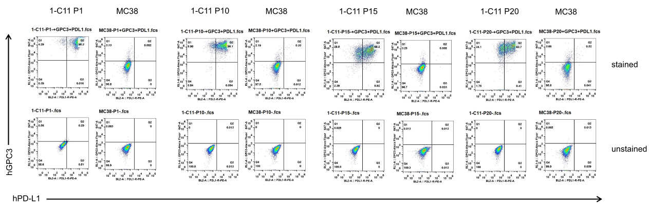
Passage stability analysis of B-hPD-L1 plus/hGPC3 MC38 cells by flow cytometry. Single-cell suspensions from wild-type MC38 and B-hPD-L1 plus/hGPC3 MC38 cultures were stained with species-specific anti-PD-L1 and anti-GPC3 antibodies. Human GPC3 and human PD-L1 were specifically detectable on the surface of B-hPD-L1 plus/hGPC3 MC38 cells but not on wild-type MC38 cells. Human GPC3 expression remained stable across multiple passages with no significant changes observed. Human PD-L1 expression also remained stable across early passages, although a partial decrease in expression was observed after 10 passages. The passage number for this cell line is calculated starting from the first subculture after revival from the master cell bank, which is designated as P1.
It is recommended to use cells within the first 10 passages for optimal results.
