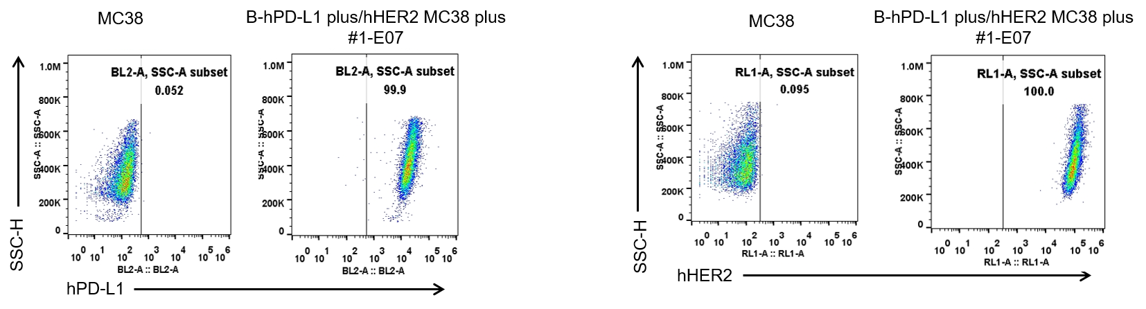
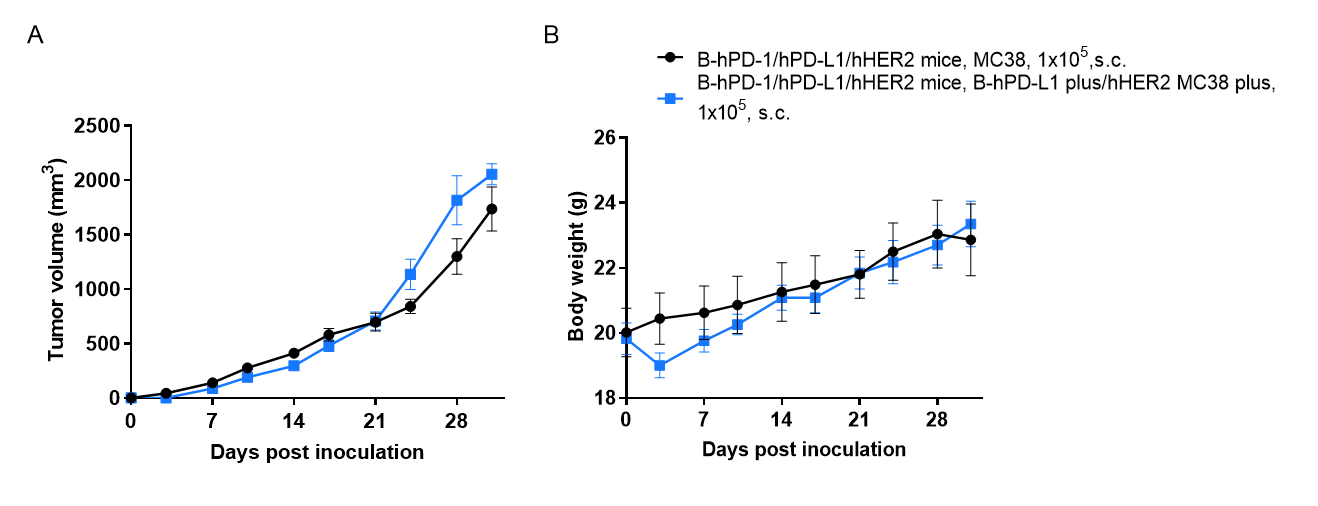
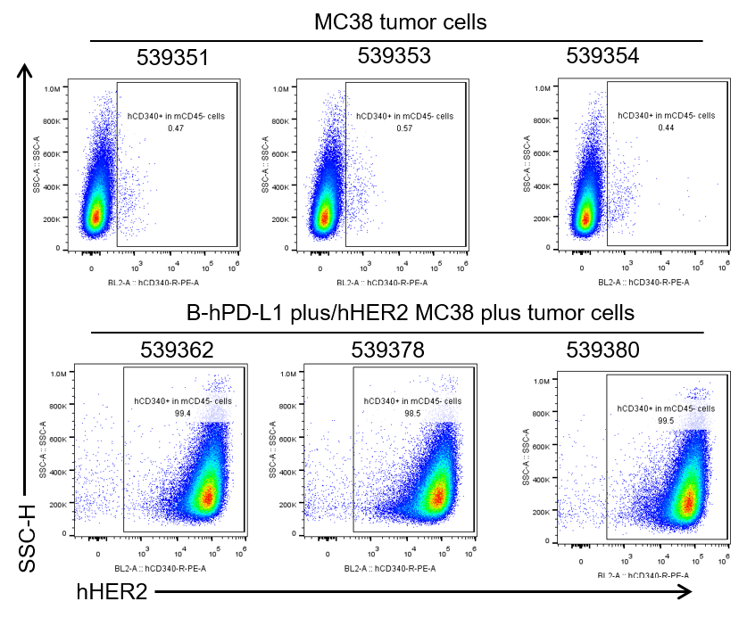
B-hPD-L1 plus/hHER2 MC38 plus
Catalog Number: 322375
Strain Name: NA
Strain Background: C57BL/6
NCBI gene ID: 60533,13866 (Human)
Aliases: B7h1; Pdl1; Pdcd1l1; Pdcd1lg1; A530045L16Rik; Neu; HER2; HER-2; c-neu; Erbb-2; c-erbB2; l11Jus8; mKIAA3023
---
可提供授权方案