

C57BL/6-Il11tm1(IL11)Bcgen Il11ratm1(IL11RA)Bcgen/Bcgen • 111865


IL11
IL11RA
B-hIL11/hIL11RA mice were obtained by crossing B-hIL11 mice and B-hIL11RA mice

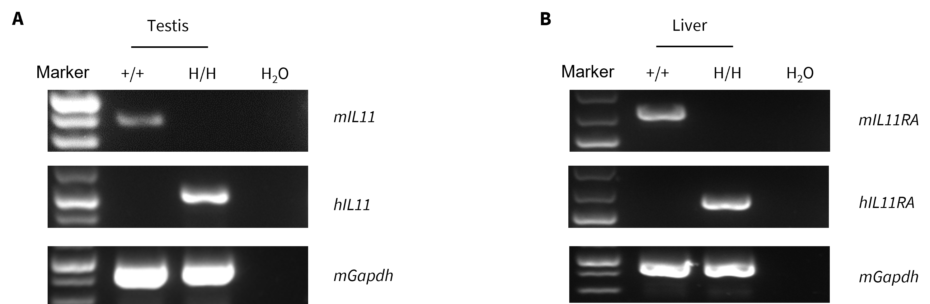
Strain specific analysis of IL11 and IL11RA mRNA expression in wild-type C57BL/6 mice and homozygous humanized B-hIL11 mice and B-hIL11RA mice by RT-PCR. (A) Testis RNA was isolated from wild-type C57BL/6 mice (+/+) and homozygous B-hIL11 mice (H/H), and then cDNA libraries were synthesized by reverse transcription, followed by PCR with human IL11 primers. (B) Liver RNA was isolated from wild-type C57BL/6 mice (+/+) and homozygous B-hIL11RA mice (H/H), and then cDNA libraries were synthesized by reverse transcription, followed by PCR with human IL11RA primers.
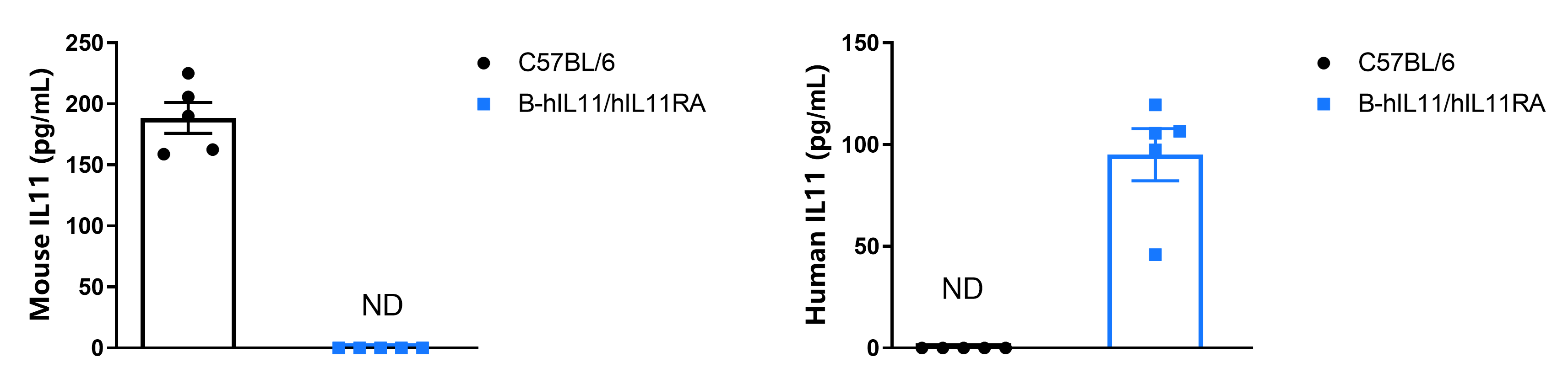
Strain specific IL11 expression analysis in homozygous B-hIL11/hIL11RA mice by ELISA. Wild-type C57BL/6 mice (+/+) and homozygous B-hIL11/hIL11RA mice (H/H) (female, 7 week-old, n=5) were fasted overnight and induced with APAP (400 mg/kg) for 24 h. Then Serum was collected and analyzed by ELISA with species-specific IL11 ELISA kit (mouse IL11: abcam, ab215084; human IL11:R&D, D1100).
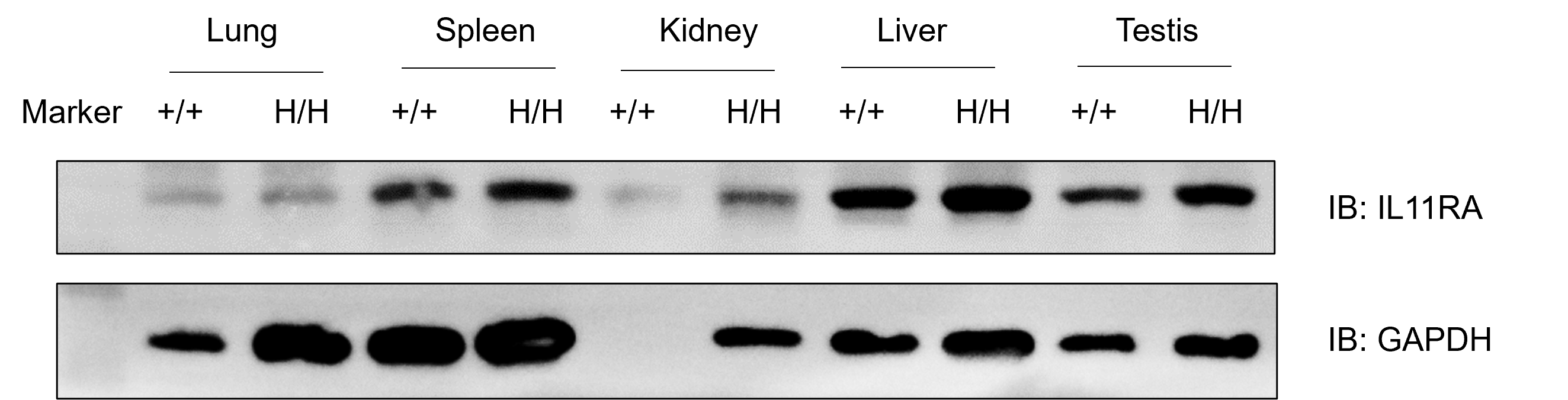
Protein expression analysis of IL11RA in homozygous B-hIL11/hIL11RA mice. Various tissue lysates were collected from wild-type C57BL/6 mice (+/+) and homozygous B-hIL11/hIL11RA mice (H/H), and then analyzed by western blot with anti-IL11RA antibody (abcam, ab125015).
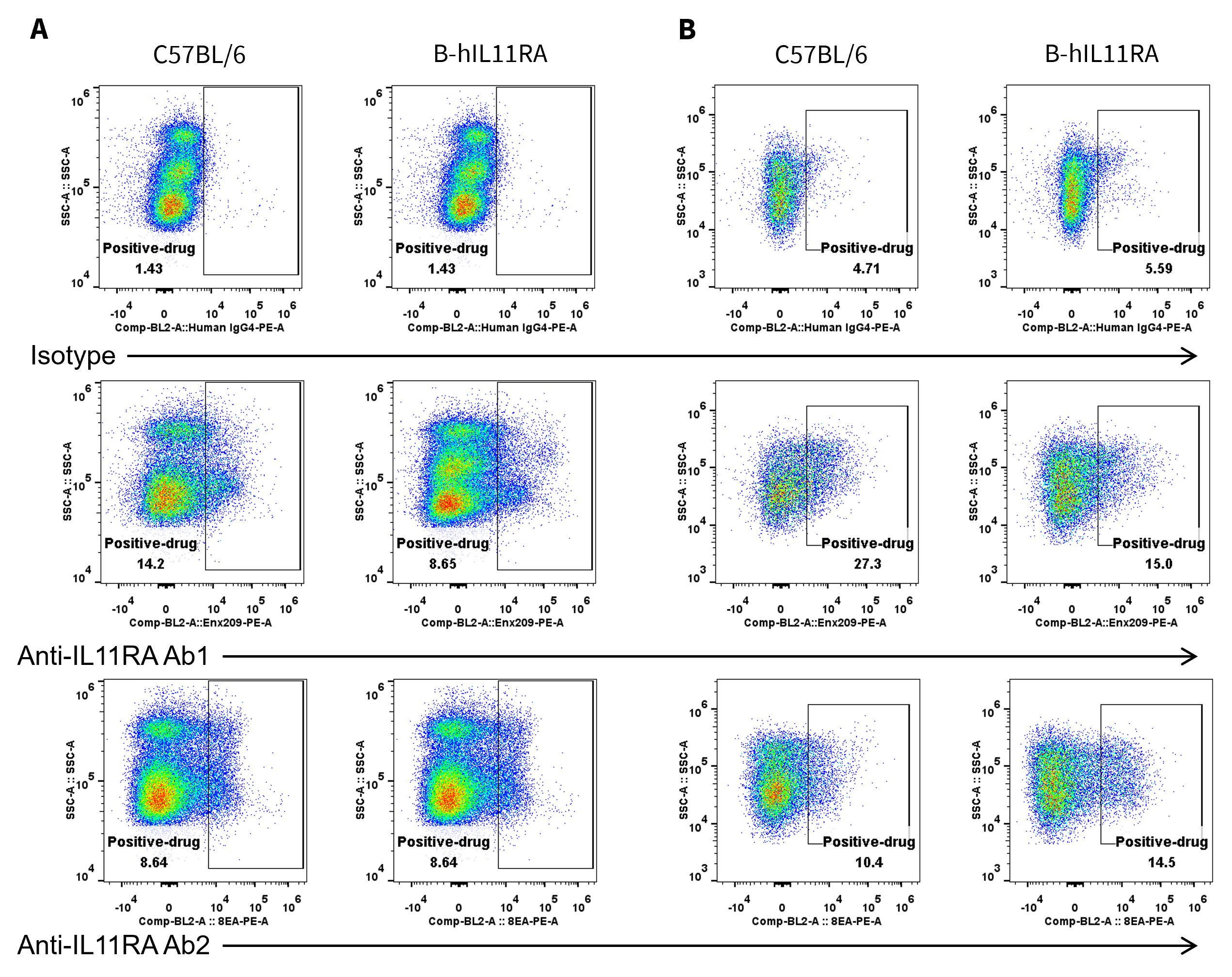
Antibody binding detection in homozygous B-hIL11RA mice by flow cytometry. Liver was collected from wild-type C57BL/6 mice (+/+) and homozygous B-hIL11RA mice (H/H), and analyzed by flow cytometry with anti-IL11RA antibodies (Ab1 and Ab2, provided by client). (A) Nonparenchymal cells. (B) Parenchymal cells.
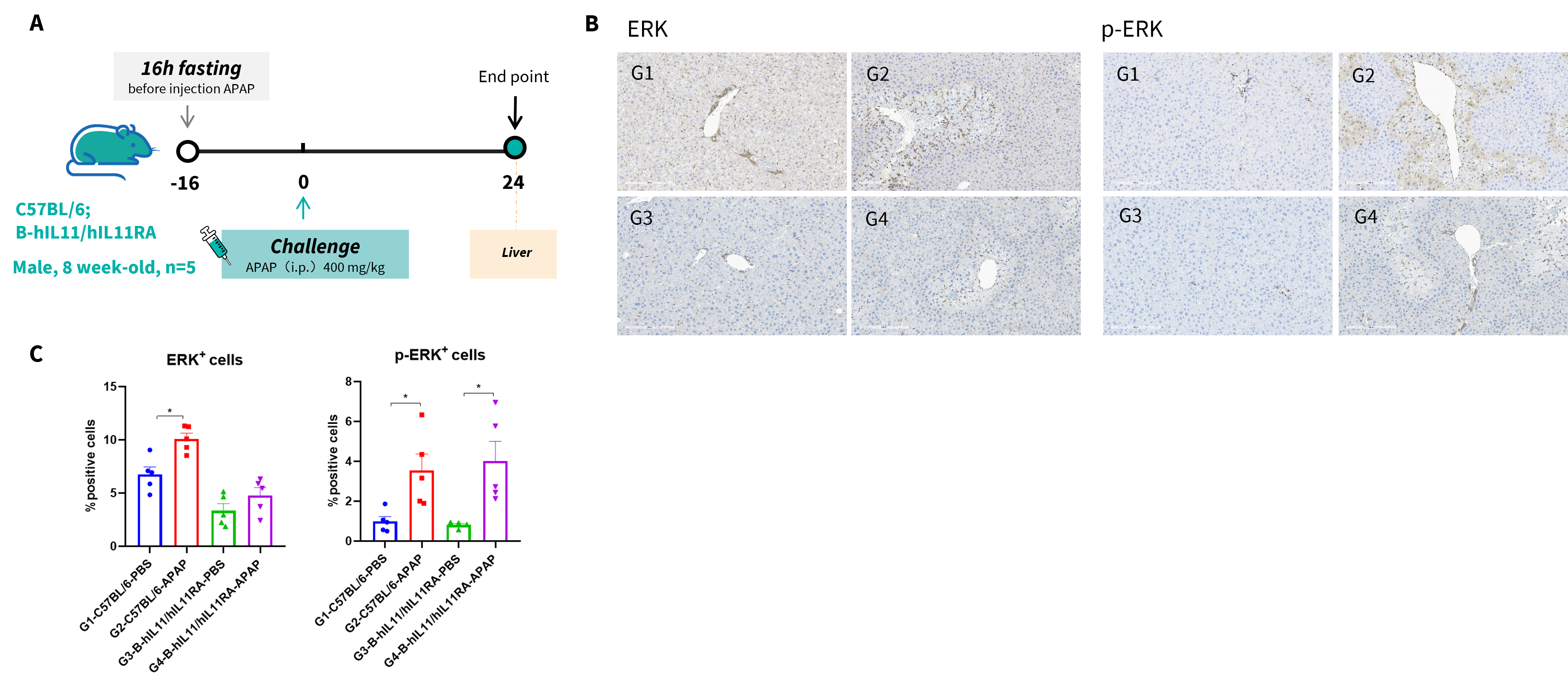
Functional analysis in APAP-induced liver damage in male B-hIL11/hIL11RA mice. (A) Schematic showing the process of APAP-induced liver damage in B-hIL11/hIL11RA mice. B-hIL11/hIL11RA mice (male, 8 weeks, n=5) were fasted overnight and inducted with APAP (400 mg/kg) for 24 h. Liver tissues were collected for functional analysis. (B) Representative IHC images of liver (200 X). (C) Quantification of ERK and p-ERK positive cells from hepatocytes. Values are expressed as mean ± SD. *p<0.05.
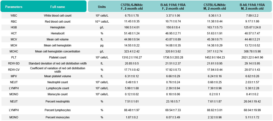
Values are expressed as mean ± SD.
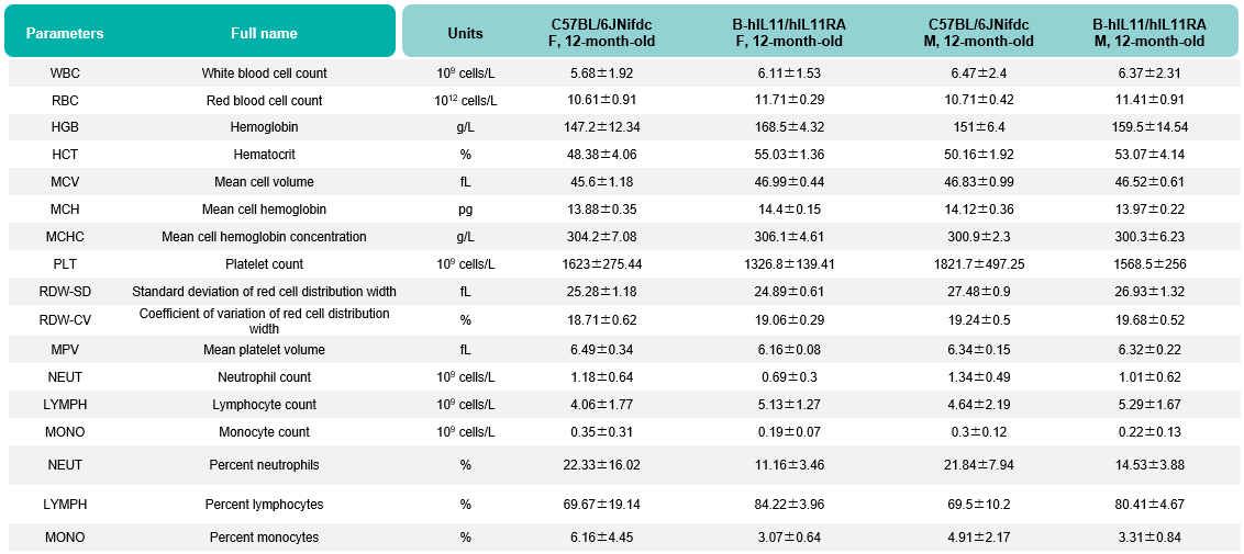
Values are expressed as mean ± SD.
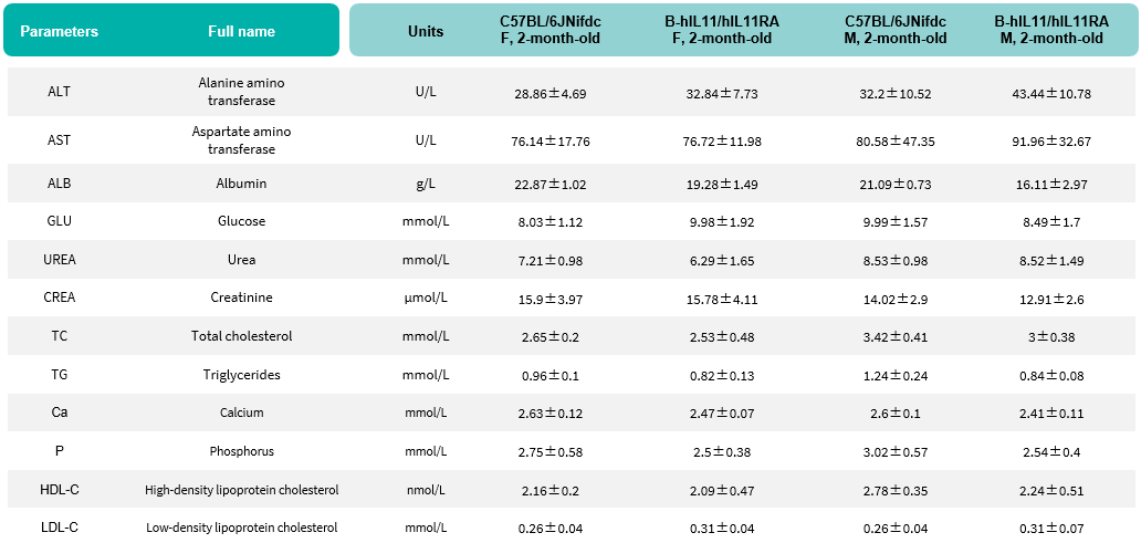
Values are expressed as mean ± SD.
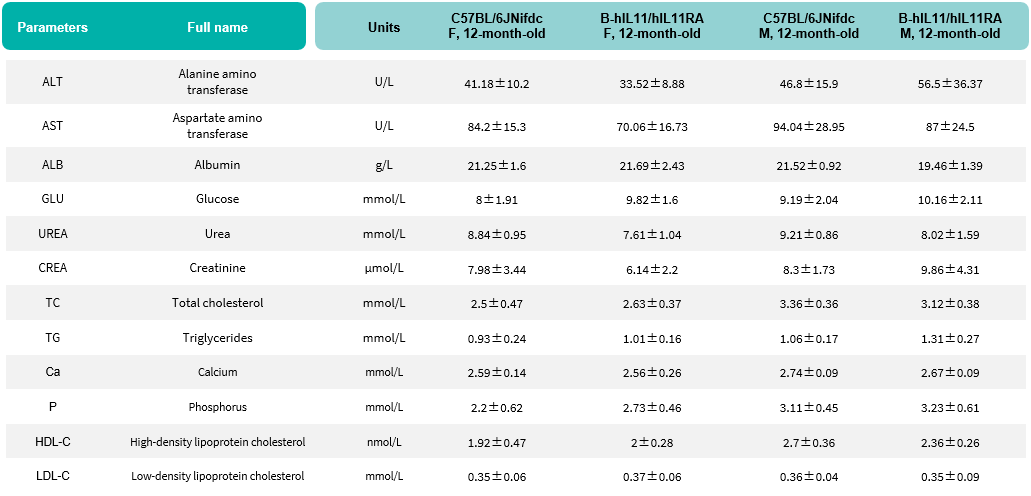
Values are expressed as mean ± SD.
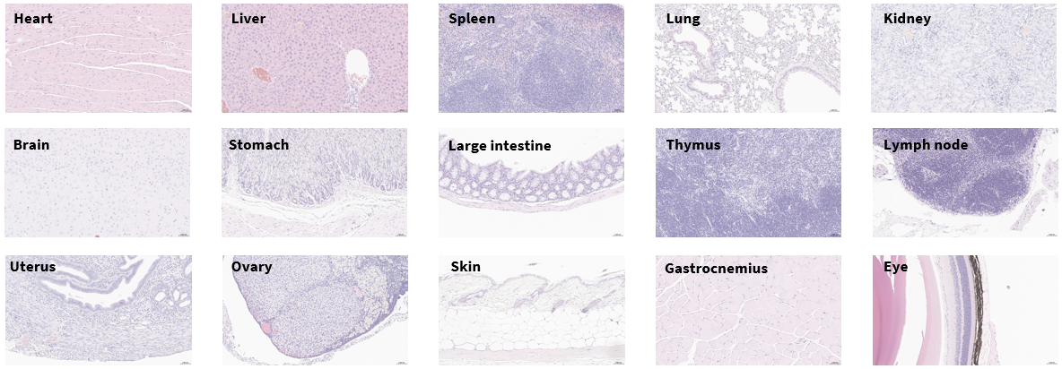
Histopathological analysis of major organs from homozygous B-hIL11/hIL11RA mice. Tissues were collected from B-hIL11/hIL11RA mice (female, 2-month-old, n=3), fixed in 4% paraformaldehyde and stained with hematoxylin and eosin (H&E). Representative photomicrographs are shown. Scale bar: 200x.
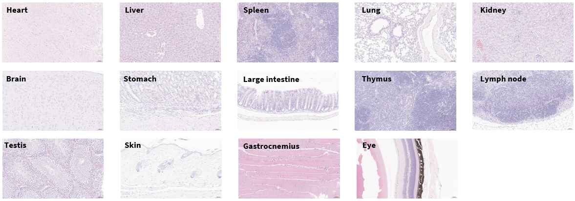
Histopathological analysis of major organs from homozygous B-hIL11/hIL11RA mice. Tissues were collected from B-hIL11/hIL11RA mice (male, 2-month-old, n=3), fixed in 4% paraformaldehyde and stained with hematoxylin and eosin (H&E). Representative photomicrographs are shown. Scale bar: 200x.
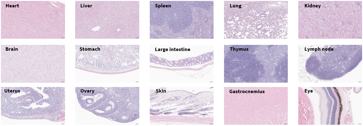
Histopathological analysis of major organs from homozygous B-hIL11/hIL11RA mice. Tissues were collected from B-hIL11/hIL11RA mice (female, 12-month-old, n=3), fixed in 4% paraformaldehyde and stained with hematoxylin and eosin (H&E). Representative photomicrographs are shown. Scale bar: 200x.
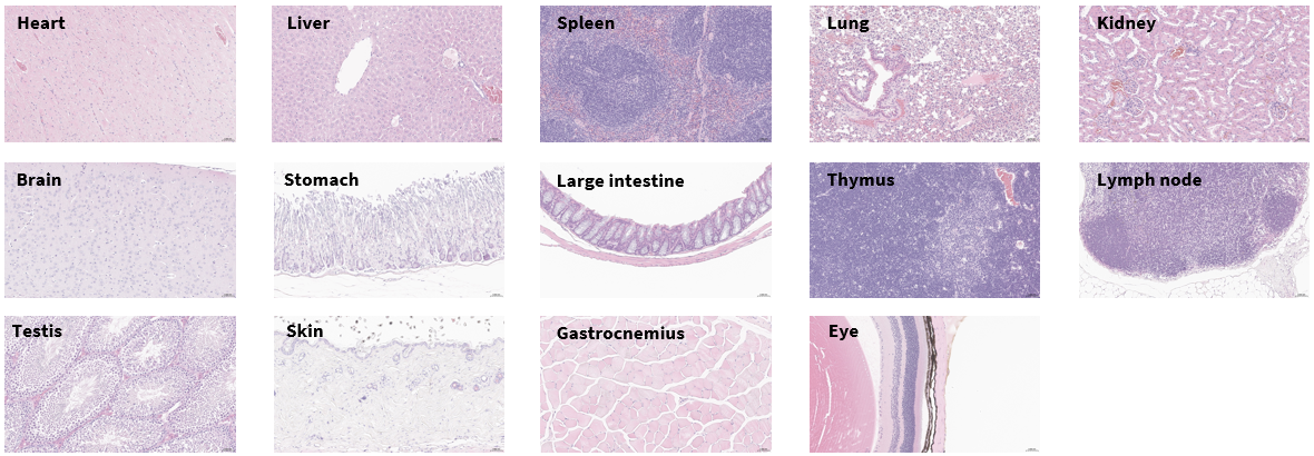
Histopathological analysis of major organs from homozygous B-hIL11/hIL11RA mice. Tissues were collected from B-hIL11/hIL11RA mice (male, 12-month-old, n=3), fixed in 4% paraformaldehyde and stained with hematoxylin and eosin (H&E). Representative photomicrographs are shown. Scale bar: 200x.

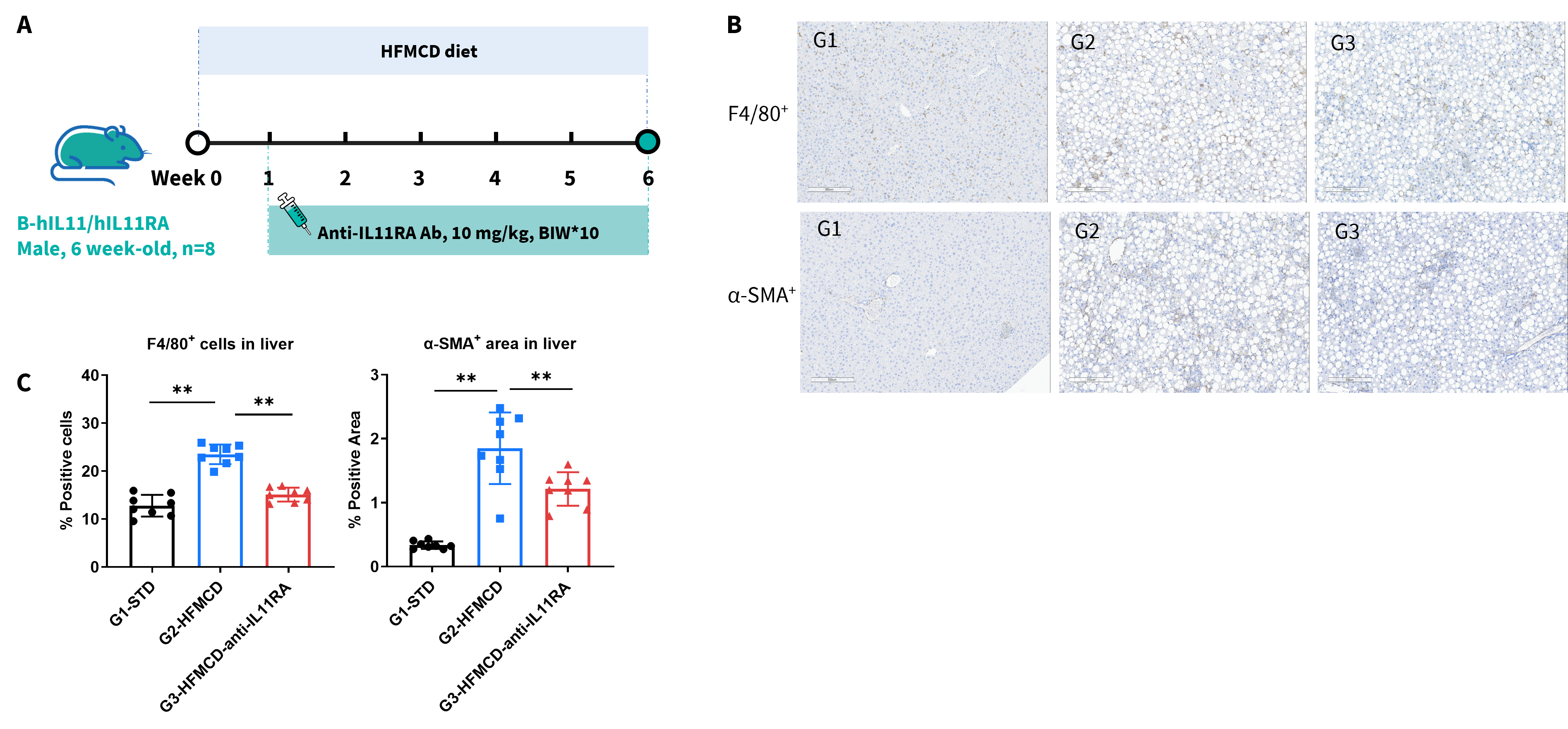
Anti-human IL11RA antibody inhibited the liver damage in B-hIL11/hIL11RA mice. (A) Schematic of treatment workflow for B-hIL11/hIL11RA mice. Mice (n=8/group) were randomized to standard diet (STD) or HFMCD for 6 weeks; anti-hIL11RA antibody administration began 1 week post-diet initiation, followed by liver tissue harvest for detection.(B, C) Compared with STD group (G1), HFMCD model group (G2) exhibited elevated liver injury markers, increased F4/80⁺ macrophages and α-SMA⁺ areas, confirming successful NASH modeling in B-hIL11/hIL11RA mice (200 X). Data presented as mean ± SEM. HFMCD: high-fat methionine-choline-deficient diet.
