

C57BL/6-Pdcd1tm3(PDCD1)Bcgen Cd274tm1(CD274)Bcgen Tacstd2tm1(TACSTD2)Bcgen/Bcgen • 113032

PD-1/PD-L1 and TROP2: Key targets linking cancer immunotherapy and tumor-targeted therapy

PD-1
• Exon 2 of the mouse PD-1 gene, which encodes the IgV domain, was replaced with human PD-1 exon 2 in B-hPD-1 plus/hPD-L1/hTROP2 mice.
• The endogenous mouse promoter, 5' UTR, 3' UTR, and genomic regions encoding the signal peptide, non-IgV portion of the extracellular domain, and transmembrane and cytoplasmic domains were retained. Chimeric PD-1 expression is driven by the endogenous mouse PD-1 promoter, while endogenous mouse PD-1 transcription and translation are disrupted.
PD-L1
• Exon 3 of the mouse PD-L1 gene, which encodes the IgV domain, was replaced with human PD-L1 exon 3 in B-hPD-1 plus/hPD-L1/hTROP2 mice.
• The endogenous mouse promoter, 5' UTR, 3' UTR, and genomic regions encoding the signal peptide, non-IgV portion of the extracellular domain, and transmembrane and cytoplasmic domains were retained. Chimeric PD-L1 expression is driven by the endogenous mouse PD-L1 promoter, while endogenous mouse PD-L1 transcription and translation are disrupted.
TROP2
• Exon 1 of the mouse Trop2 gene that encodes the full-length protein was replaced by human TROP2 exon 1 in B-hPD-1 plus/hPD-L1/hTROP2 mice.

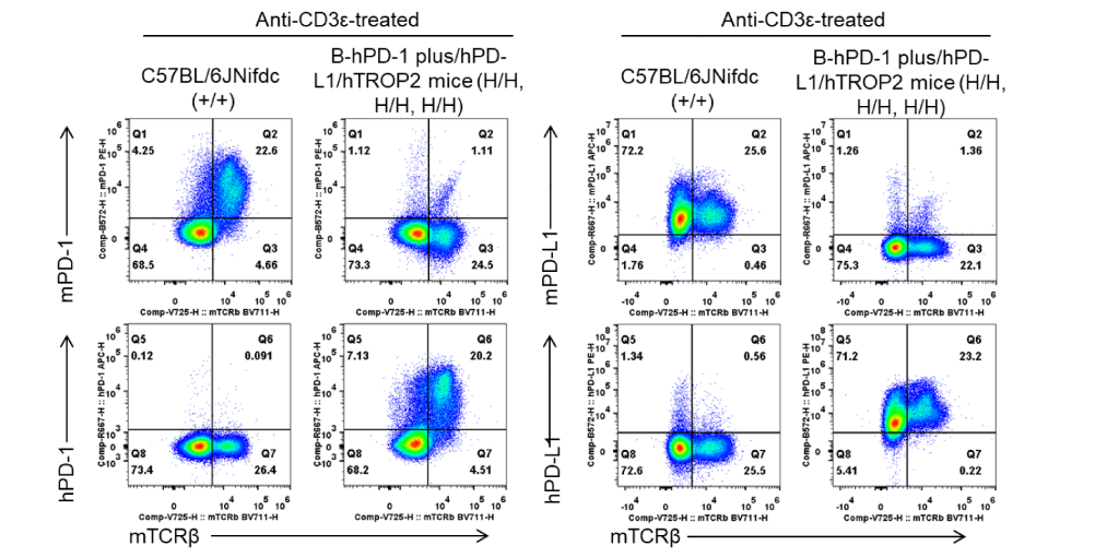
PD-1 and PD-L1 expression analysis in wild-type C57BL/6JNifdc mice and homozygous B-hPD-1 plus/hPD-L1/hTROP2 mice by flow cytometry. Splenocytes were collected from wild-type C57BL/6JNifdc mice (+/+) and homozygous B-hPD-1 plus/hPD-L1/hTROP2 mice (H/H;H/H;H/H) (female, 6-8-week-old, n = 1) stimulated with anti-CD3ε in vivo (7.5 μg/mouse, i.p., for 24 hours). Protein expression was analyzed with anti-human PD-1 antibody (BioLegend, 329908), anti-mouse PD-1 antibody (BioLegend, 109104), anti-human PD-L1 antibody (BioLegend, 329706), and anti-mouse PD-L1 antibody (BioLegend, 124312) by flow cytometry.
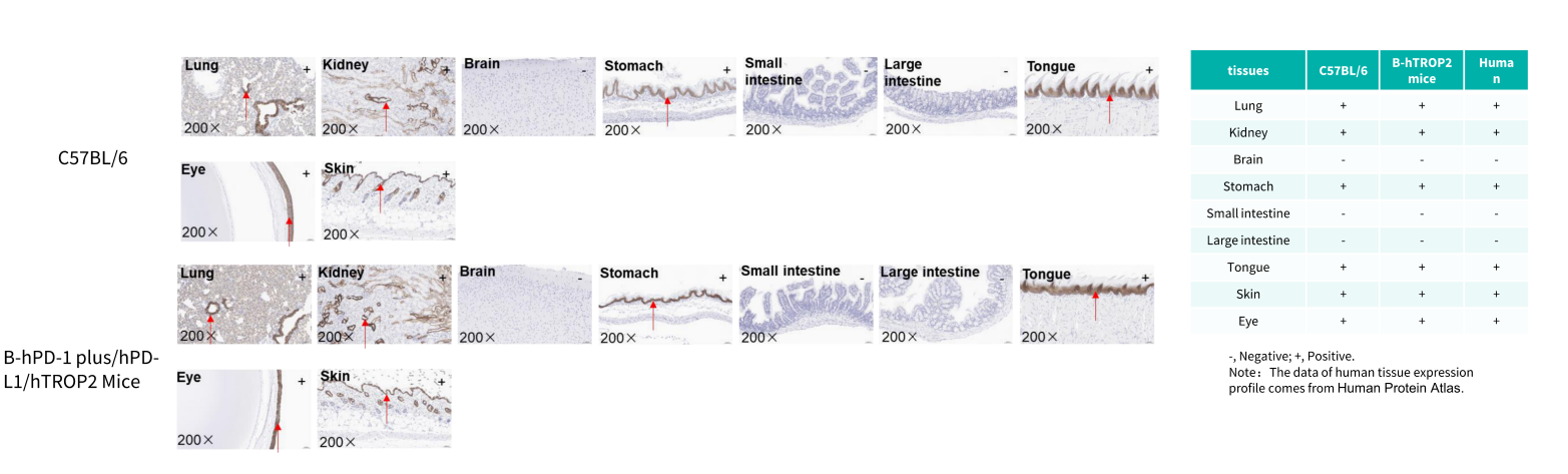
Immunohistochemical (IHC) analysis of TROP2 protein expression in wild-type mice and B-hPD-1 plus/hPD-L1/hTROP2 Mice. Major tissues were collected from wild-type mice and homozygous B-hPD-1 plus/hPD-L1/hTROP2 mice and analyzed by IHC with anti-TROP2 antibody (Abcam, ab214488).

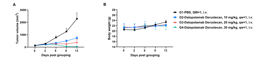
Establishment of an MC38 model in B-hPD-1 plus/hPD-L1/hTROP2 mice and in vivo efficacy study of an anti-human TROP2 antibody-drug conjugate (Datopotamab Deruxtecan, in-house). Murine colon cancer B-hPD-L1 plus/hTROP2 MC38 cells (Cat# 322415) were subcutaneously implanted into homozygous B-hPD-1 plus/hPD-L1/hTROP2 mice (female, 9-week-old, n = 5). Mice were grouped when tumor volume reached approximately 100 mm³, at which time they were intravenously injected with anti-human TROP2 ADC as indicated in the panel. Values are expressed as mean ± SEM.
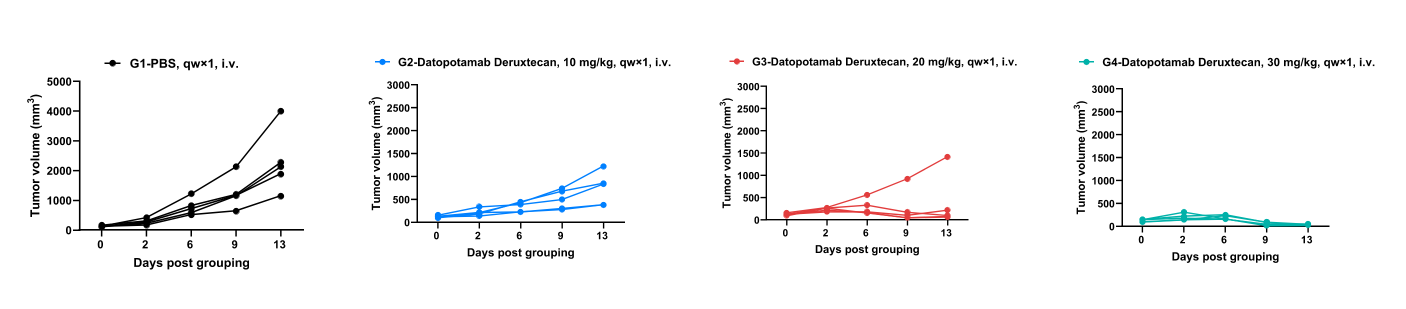
In vivo efficacy of anti-human TROP2 ADC-individual tumor growth curves.
