

C57BL/6-Igs2tm1(C3*R102G)Bcgen/Bcgen • 113286


Gene targeting strategy for B-hC3*R102G mice.

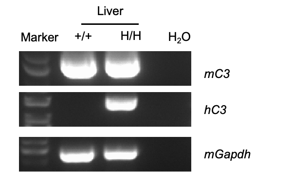
Strain specific analysis of C3 mRNA expression in wild-type C57BL/6JNifdc and B-hC3*R102G mice by RT-PCR. Liver RNA was isolated from wild-type C57BL/6JNifdc(+/+) and homozygous B-hC3*R102G mice(H/H), then cDNA libraries were synthesized by reverse transcription, followed by PCR with human C3 primers. Human C3 mRNA was exclusively detectable in homozygous B-hC3*R102G mice. Mouse C3 was detectable in wild-type mice and B-hC3*R102G mice.
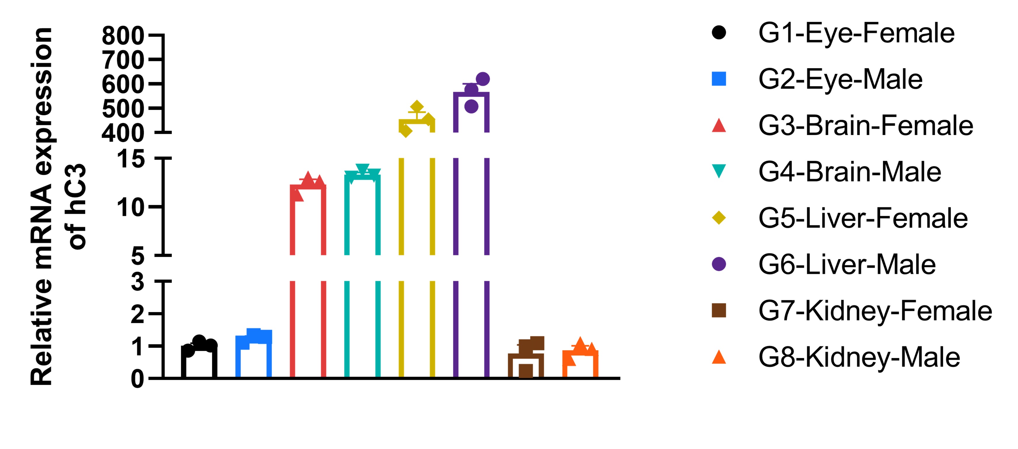
Strain specific analysis of C3 mRNA expression by RT-qPCR. The brain, liver, kidney, and eye tissues RNA were isolated from heterozygous B-hC3*R102G mice (H/+) (male and female n=3), then cDNA libraries were synthesized by reverse transcription, followed by RT-qPCR with human C3 primers. Human C3 is predominantly expressed in the liver. All results were normalized to the female eye C3 mRNA. Values are expressed as mean ± SEM.

Strain specific C3 expression analysis in wild-type C57BL/6JNifdc mice and homozygous B-hC3*R102G mice by ELISA. Serum was collected from wild-type C57BL/6JNifdc mice (+/+) and homozygous B-hC3*R102G mice (H/H), and analyzed by ELISA. Human C3 was exclusively detectable in B-hC3*R102G mice. Values are expressed as mean ± SEM.
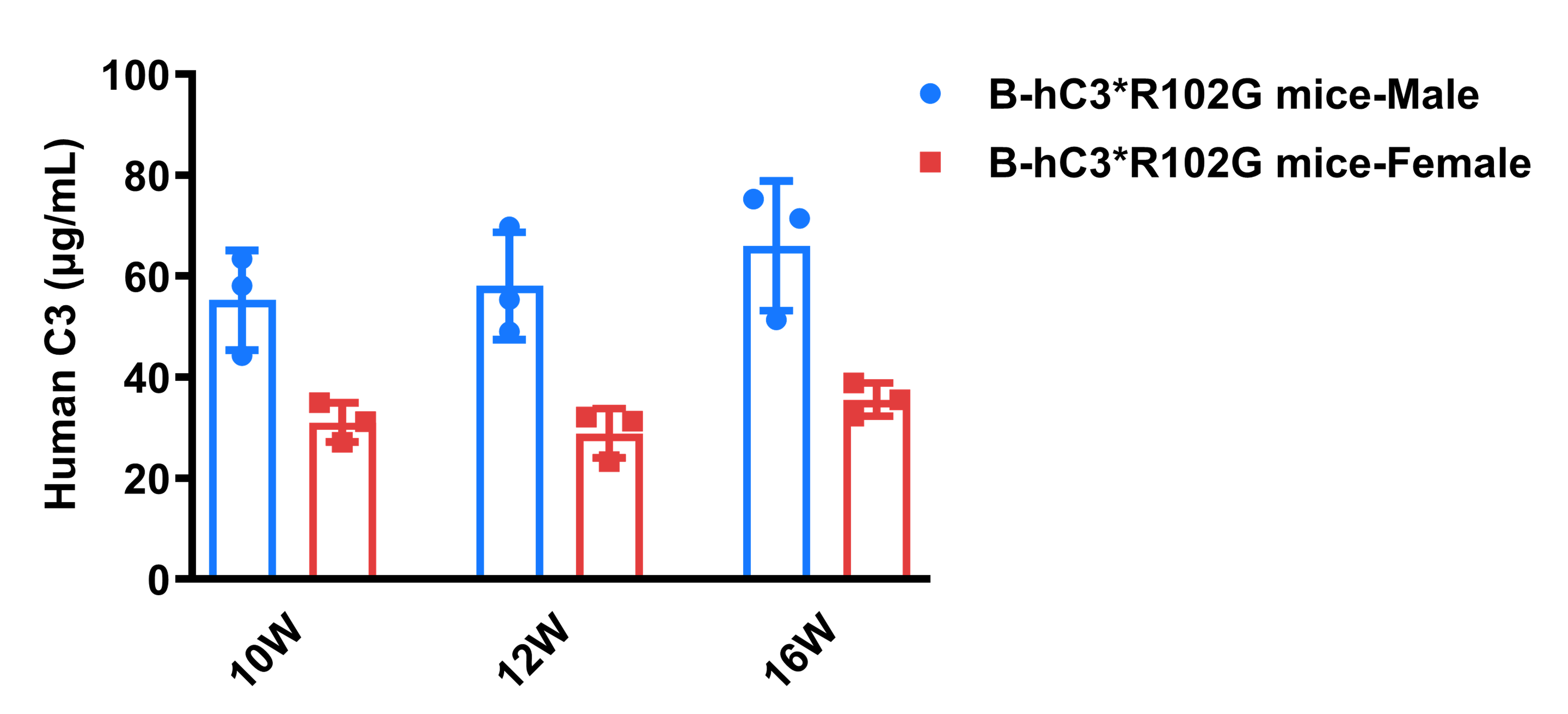
Strain specific C3 expression analysis in B-hC3*R102G mice by ELISA. Serum was collected from homozygous B-hC3*R102G mice (H/H) (male and female, n=3). The expression level of human C3 were analyzed by ELISA. Human C3 was exclusively detectable in humanized B-hC3*R102G mice (n=3). Values are expressed as mean ± SEM.
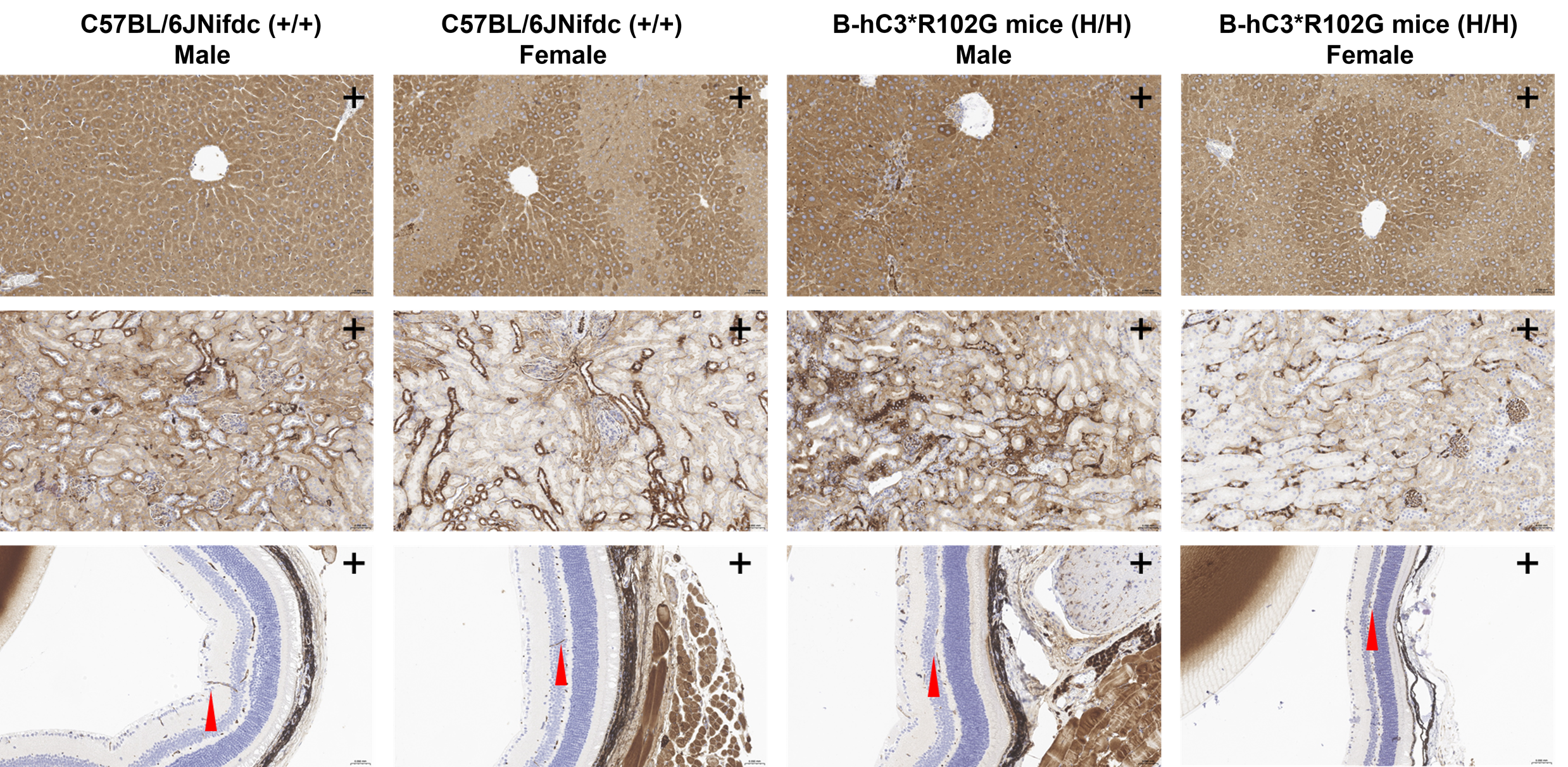
IHC Staining in B-hC3*R102G mice. The liver, kidney, and eye tissues of wild-type C57BL/6JNifdc mice (+/+) and homozygous B-hC3*R102G mice (H/H) were isolated at 15 weeks old and analyzed with IHC staining. Results showed that C3 was detected in the liver, eye, and kidney of B-hC3*R102G mice and wild-type C57BL/6JNifdc mice, as the antibody cross-recognizes both human and mouse C3. Red arrows indicate a positive signal.
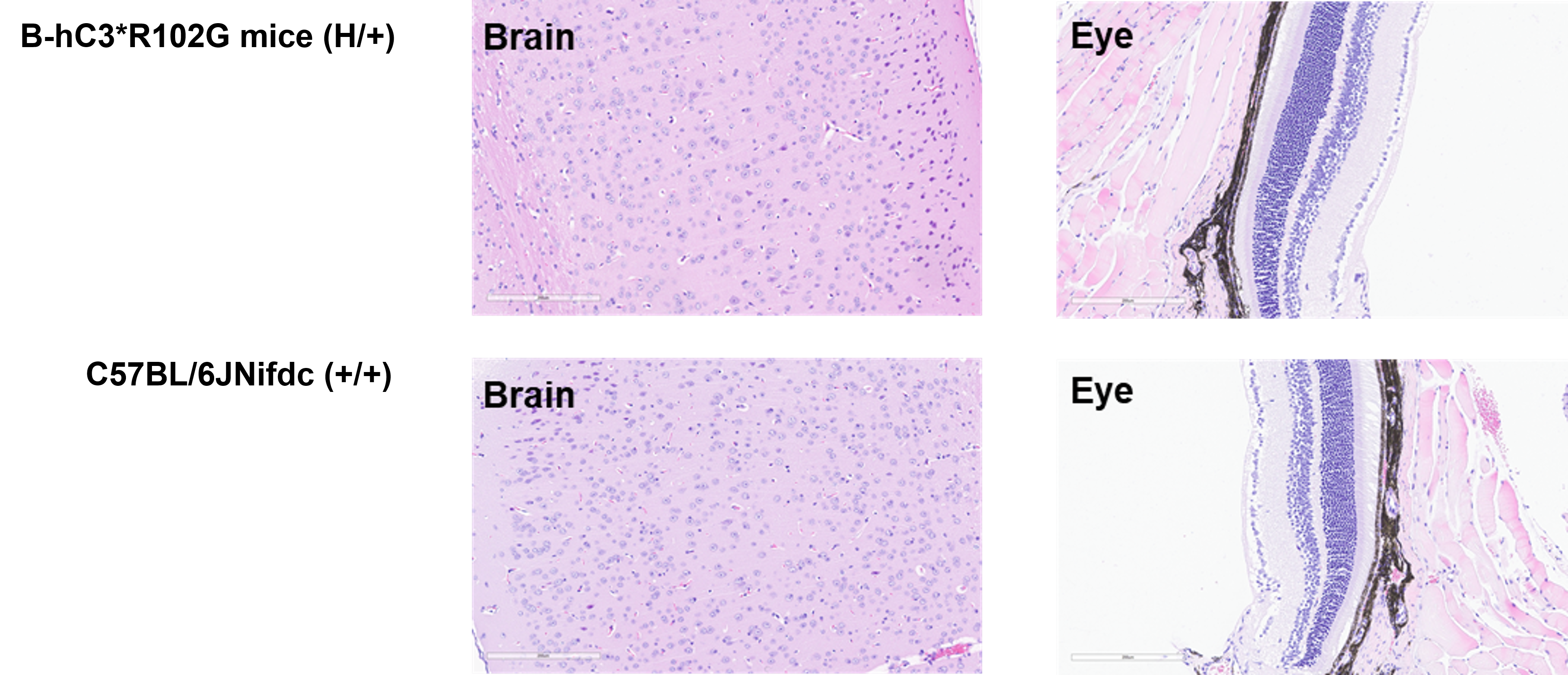
Histopathological analysis of organs in B-hC3*R102G mice. The brain and eye tissues of wild-type C57BL/6JNifdc mice (+/+) and heterozygous B-hC3*R102G mice (H/+) were isolated at 15 weeks old and analyzed with H&E staining (male, n=1). There were no obvious abnormalities in the brain and eye in heterozygous B-hC3*R102G mice and wild-type C57BL/6JNifdc mice.
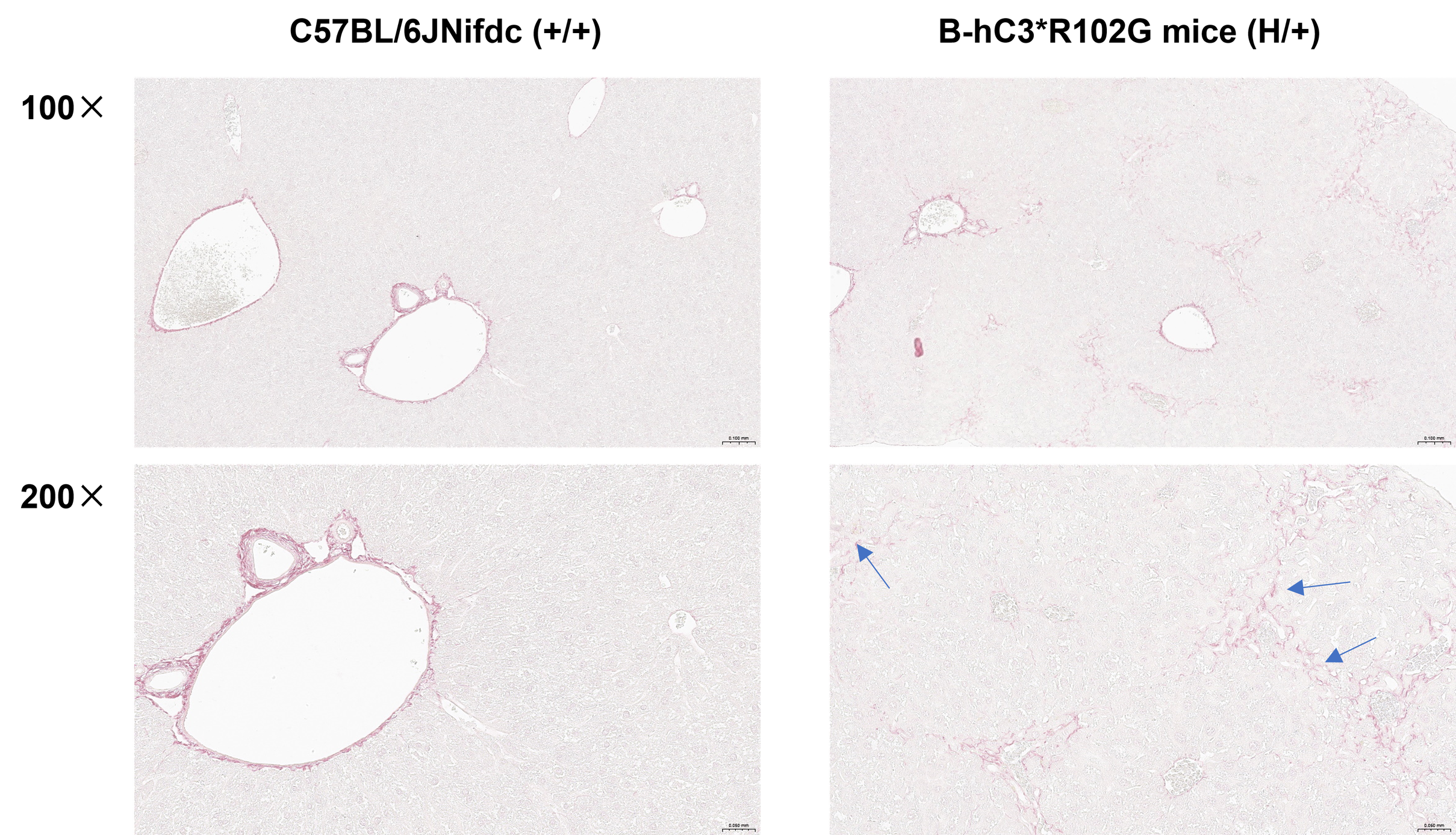
Sirius Red Staining of liver in B-hC3*R102G mice. The liver of wild-type C57BL/6JNifdc mice (+/+) and heterozygous B-hC3*R102G mice (H/+) were isolated at 16 weeks old and analyzed with Sirius Red Staining (male, n=6). Results showed that in the liver tissue of heterozygous B-hC3*R102G mice, fibrous hyperplasia was observed in all portal areas of the liver. Blue arrows indicate the Sirius red-positive signals of proliferated collagen fibers. There were no obvious abnormalities in wild-type C57BL/6JNifdc mice.
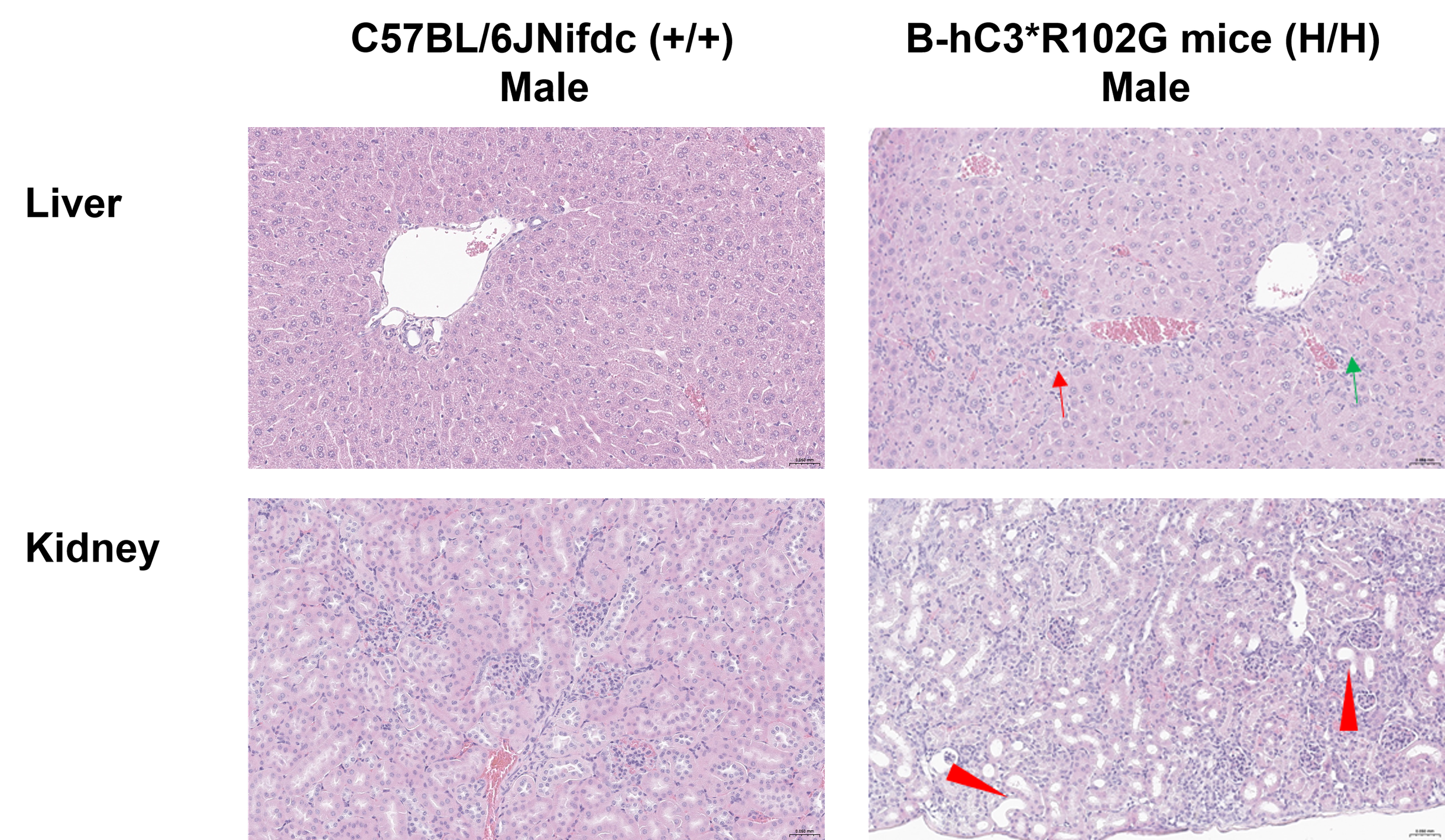
Histopathological analysis of organs in B-hC3*R102G mice. The liver and kidney of wild-type C57BL/6JNifdc mice (+/+) and homozygous B-hC3*R102G mice (H/H) were isolated at 15 weeks old and analyzed with H&E staining (male, n=8). There were no obvious abnormalities found in wild-type C57BL/6JNifdc mice. The liver of male B-hC3*R102G mice (7/8) showed fibroblast proliferation and inflammatory cell infiltration. The kidney of male B-hC3*R102G mice (3/8) showed basophilic change of renal tubules, increased interstitial cells, and dilatation of renal tubules. Red arrows: increased fibroblasts; Green arrows: biliary duct proliferation; Red triangles: renal tubular dilatation and basophilic change.
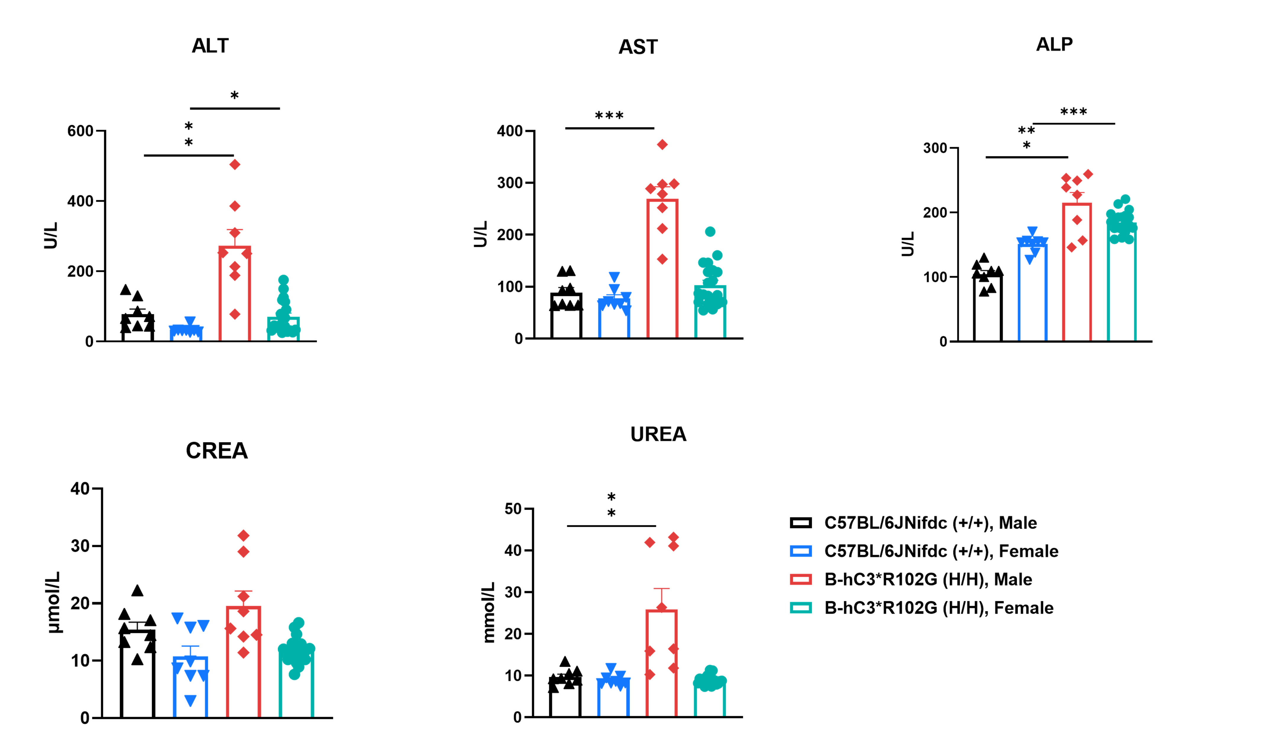
Analysis of blood biochemicals in homozygous B-hC3*R102G mice and wild-type C57BL/6JNifdc mice. Serum was collected from wild-type C57BL/6JNifdc mice (+/+) and homozygous B-hC3*R102G mice (H/H) (male, n=8; female, n=8, 15 weeks old) and analyzed for biochemistry. The ALT, AST, ALP, and UREA were increased in male B-hC3*R102G mice. Values are expressed as mean ± SEM. Significance was determined by t-test, *p<0.05,**p<0.01, ***p<0.001.

Survival Curve of B-hC3*R102G mice. Graph showing the survival curve of heterozygous B-hC3*R102G mice (H/+) (male, n=12; female, n=12), and homozygous B-hC3*R102G mice (H/H) (male, n=20; female, n=20). The survival rate of male B-hC3*R102G mice was around 50% at 14 weeks old. And we didn’t observe that mice died in the female B-hC3*R102G mice until 16 weeks old.

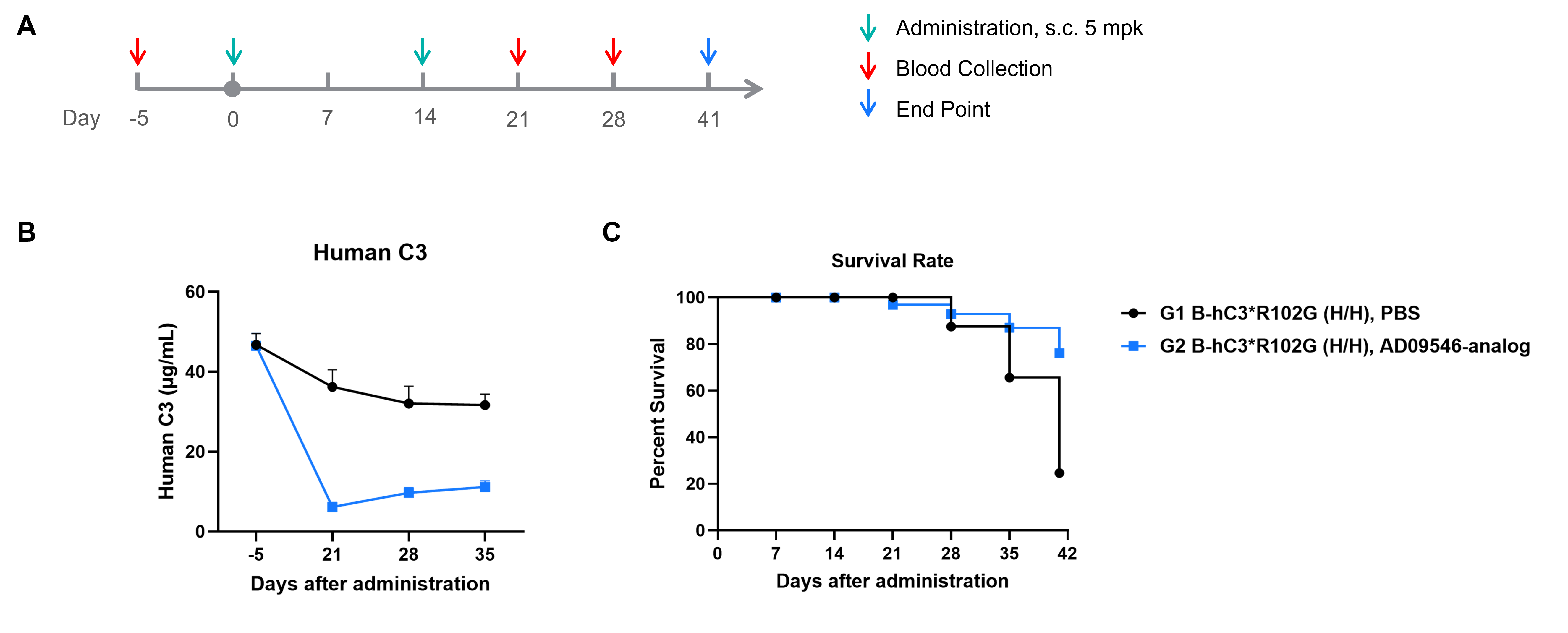
The inhibitory efficiency of the nucleic acid drugs against human C3 in homozygous B-hC3*R102G mice. B-hC3*R102G mice (H/H) were divided into two groups based on human C3 expression level (male, 8 weeks old, n=8). The human C3 targeted nucleic acid drugs (synthesized according to patents), and PBS were administered to the mice individually. The nucleic acid drug was administered in the form of a PBS aqueous solution. (A) The schematic diagram of experimental processing. (B) The expression of human C3 protein in serum after treatment. (C) The survival curve after treatment.
