Description
IL23: A key inflammation cytokine in the inflammatory response driven by Th17 cells
- Gene Information: IL-23 is a heterodimeric cytokine composed of p19 and p40 subunits, and is encoded by interleukin-23 subunit alpha (IL23A) and interleukin-12 subunit beta (IL12B) respectively. IL23A is located on chromosome 12q13.3, IL12B is located on chromosome 5q33.3.
- Protein Expression: Mature, biologically active IL-23 protein is secreted only when a cell expresses both the p19 and p40 subunits and assembles them correctly. IL-23 is mainly produced by activated antigen-presenting cells, such as macrophages and dendritic cells, as well as damaged keratinocytes or intestinal epithelial cells.
- Signaling Pathway: IL23 binds to the IL23R/IL12RB1 receptor complex to trigger JAK-STAT, p38 MAPK, and NF-κB signaling. This initiates the transcription of downstream pro-inflammatory factors, such as IL-17A, IL-17F, IL-22 and RORγt, thereby promoting the differentiation and maintenance of pathogenic Th17 cells and exacerbating chronic tissue inflammation.
- Therapeutic Inhibition: Blocking the interaction between IL23 and IL23 receptor can reduce the severity of autoimmune diseases, such as the IBD model and psoriasis.
RAG2: An essential chromatin-sensing cofactor in V(D)J recombination
- Gene Information: Recombination Activating 2 (RAG2) is a protein-coding gene located on chromosome 11p12, which is involved in the initiation of V(D)J recombination during B and T cell development.
- Protein Expression: RAG2 exists exclusively in T and B cells at specific early developmental stages within the bone marrow and thymus. It is expressed and survives only during the G0/G1 phase, and the Rag2 protein is rapidly degraded as soon as the cell undergoes DNA replication.
- Signaling Pathway: RAG2 forms a DNA-cleaving complex with RAG1. In this complex, RAG1 provides the catalytic activity, RAG2 acts as a structural scaffold: its N-terminus binds tightly to DNA, and its C-terminal PHD finger anchors the complex to chromatin via trimethylated histone H3 (H3K4me3), which is the core component in the immune system that controls the assembly of diverse immune receptors.
- Therapeutic Inhibition: Complete deficiency of RAG2 leads to severe immunodeficiency characterized by a near-total absence of mature T and B cells, thereby precluding the development of autoimmunity. RAG2 mutations can cause Omenn syndrome, a severe combined immunodeficiency associated with autoimmune-like symptoms.
Targeting strategy
IL23A
- The exons 1-4 of mouse Il23a gene that encode the whole molecule (ATG to STOP codon) were replaced by human counterparts in B-hIL23A/hIL12B, Rag2 KO mice.
- The promoter, 5’UTR and 3’UTR region of the mouse gene were retained. The human IL23A expression was driven by endogenous mouse Il23a promoter, while mouse Il23a gene transcription and translation will be disrupted.
IL12B
- The exons 2-8 of mouse Il12b gene that encode the whole molecule (ATG to STOP codon), including 3’UTR were replaced by human counterparts in B-hIL23A/hIL12B, Rag2 KO mice. The promoter and 5’UTR region of the mouse gene were retained.
- The human IL12B expression was driven by endogenous mouse Il12b promoter, while mouse Il12b gene transcription and translation will be disrupted.
Rag2
- The exon 3 and 3’UTR region of mouse Rag2 were knocked out in B-hIL23A/hIL12B, Rag2 KO mice, resulting in a disruption of the Rag2 gene.
IL23 Protein Expression Analysis in BMDC Supernatant
- Mouse IL23 was only detectable in wild-type B-Rag2 KO mice.
- Human IL23 was exclusively detectable in homozygous B-hIL23A/hIL12B, Rag2 KO mice and B-hIL23A/hIL12B mice.
Mouse IL-23 and human IL-23 expression analysis in B-hIL23A/hIL12B, Rag2 KO mice by ELISA. Bone marrow derived dendritic cells were produced by culturing the bone marrow from wild-type B-Rag2 KO mice (-/-), homozygous B-hIL23A/hIL12B mice (H/H;H/H), and homozygous B-hIL23A/hIL12B, Rag2 KO mice (H/H;H/H;-/-), which were stimulated with LPS in vitro. After stimulation, the supernatants were collected and the levels of mouse and human IL23 were analyzed by ELISA (R&D, M2300; R&D, D2300B). Values are expressed as mean ± SEM. ND: not detectable.
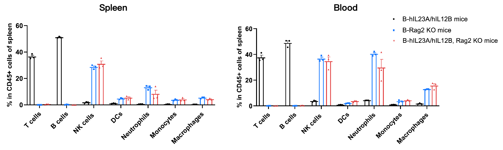
Analysis of Leukocyte Subpopulations
- The percentages of T cells, B cells, NK cells, DCs, monocytes, macrophages, and neutrophils in homozygous B-hIL23A/hIL12B, Rag2 KO mice were similar to those in B-Rag2 KO mice, and B-hIL23A/hIL12B, Rag2 KO mice presented a near-total absence of T and B cells.
- Humanization of IL23A and IL12B does not affect normal immune cell development or splenic distribution.
Analysis of leukocyte subpopulations by flow cytometry in immune organs and blood. Splenocytes and peripheral blood were isolated from wild-type B-Rag2 KO mice, homozygous B-hIL23A/hIL12B mice, and homozygous B-hIL23A/hIL12B, Rag2 KO mice (female, n=3, 8-week-old). Single live cells were gated on the CD45⁺ population and analyzed by flow cytometry as indicated. Values are expressed as mean ± SEM.
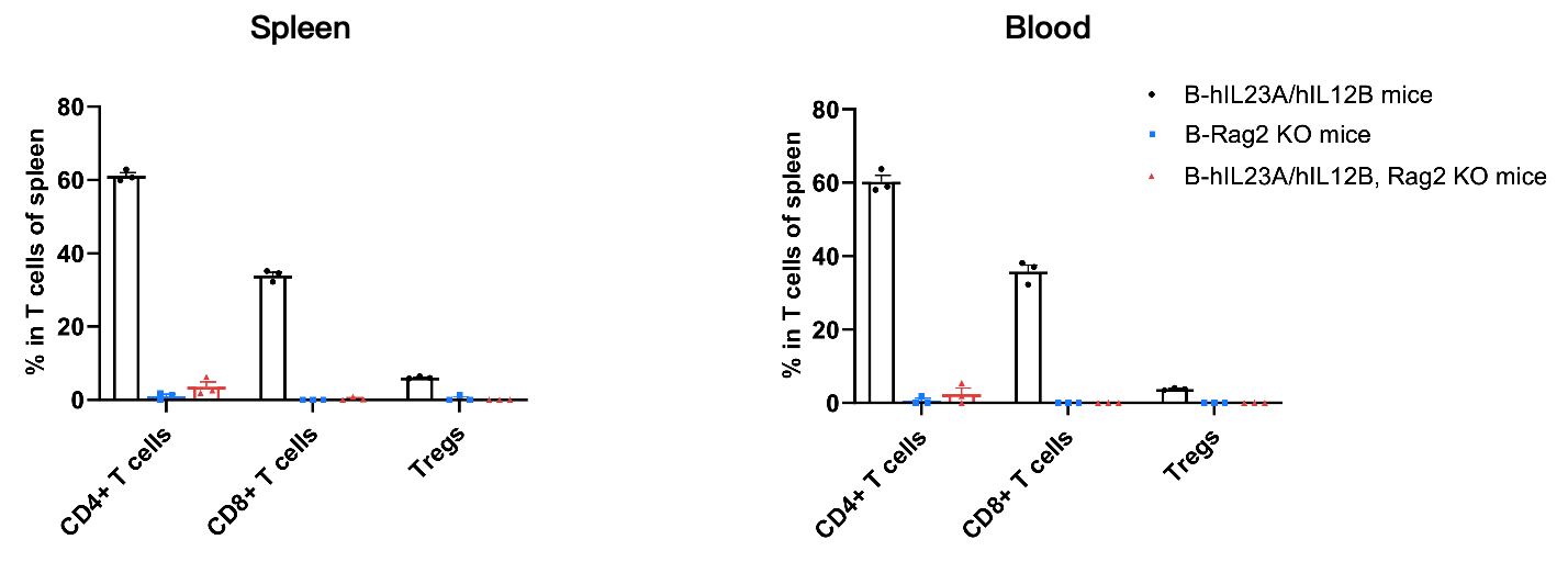
Analysis of T Cell Subpopulations
- The proportions of CD4⁺ T cells, CD8⁺ T cells, and Tregs in homozygous B-hIL23A/hIL12B, Rag2 KO mice were comparable to those in B-Rag2 KO mice, and B-hIL23A/hIL12B, Rag2 KO mice presented a near-total absence of CD4+ T cells, CD8+ T cells, and Tregs.
- Humanization of IL23A and IL12B does not affect normal T cell development, differentiation, or splenic distribution.
Analysis of T-cell subpopulations by flow cytometry in immune organs and blood. Splenocytes and peripheral blood were isolated from wild-type B-Rag2 KO mice, homozygous B-hIL23A/hIL12B mice, and homozygous B-hIL23A/hIL12B, Rag2 KO mice (female, n=3, 8-week-old). Single live cells were gated on the TCRβ⁺ T-cell population and analyzed by flow cytometry as indicated. Values are expressed as mean ± SEM.
In Vivo Efficacy of Anti-human IL23 Antibody and Anti-human IL23R Oral Peptide in T Cells Transfer Induced Colitis
- Administration of anti-human IL23 antibodies and anti-human IL23R oral peptide efficiently improved T cells transfer induced colitis.
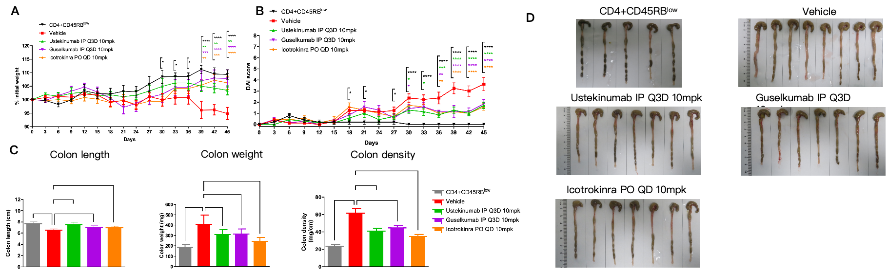
The therapeutic efficacy of Ustekinumab, Guselkumab, and Icotrokinra on T cells transfer induced colitis model in B-hIL23A/hIL12B, Rag2 KO mice. CD4+CD45RBlow T and CD4+CD45RBhigh T cells were isolated from the spleen of B-hIL23R/hIL12RB1 plus mice. B-hIL23A/hIL12B, Rag2 KO mice in group G1 were injected with CD4+CD45RBlow T cells, while B-hIL23A/hIL12B, Rag2 KO mice in groups G2-G5 were injected with CD4+CD45RBhigh T cells. The treatment groups received anti-human IL12/IL23p40 antibody Ustekinumab (10 mpk, provided by WuXi AppTec), anti-human IL23p19 antibody Guselkumab (10 mpk, provided by WuXi AppTec) and anti-human IL23R oral peptide Icotrokinra (10 mpk, provided by WuXi AppTec). (A) Body weight change. (B) DAI score. (C) Colon index. (D) Colon photo. Two-way ANOVA or one-way ANOVA was used for multiple comparisons, with each group compared to Vehicle. Values are expressed as mean ± SEM. *p<0.05, **p<0.01, ***p<0.001, ****p<0.0001. Note: This experiment was conducted by WuXi AppTec, T cell transfer induced chronic colitis in B-hIL23A/hIL12B, Rag2 KO mice (Donor: B-hIL23R/hIL12RB1 plus mice).
- Administration of anti-human IL23 antibodies and anti-human IL23R oral peptide efficiently reduced inflammatory infiltration and epithelial damage in colon tissue compared with untreated controls.
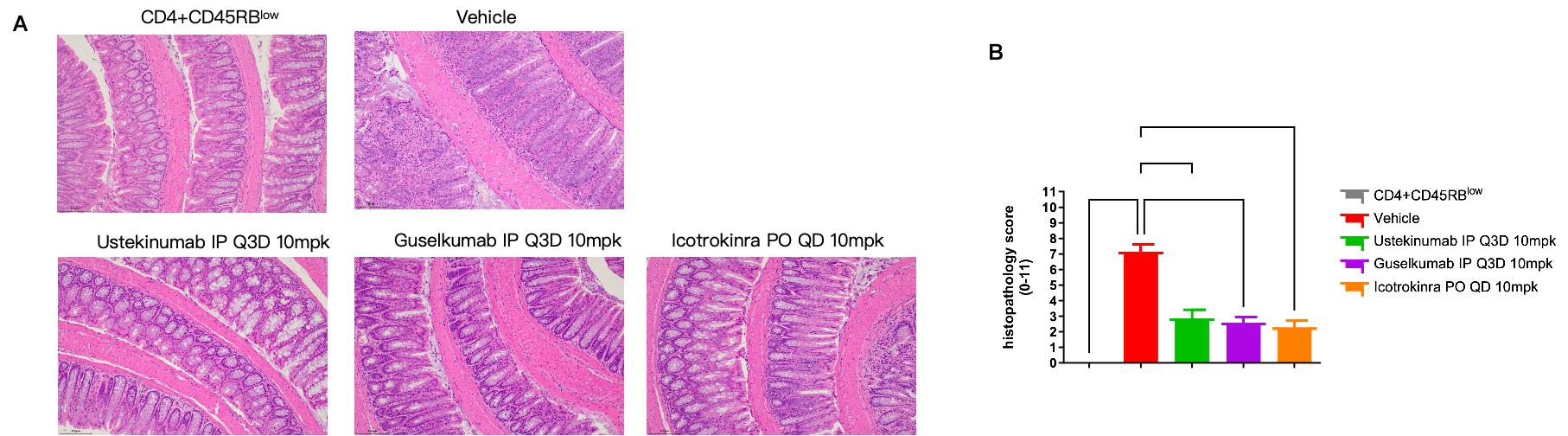
The therapeutic efficacy of Ustekinumab, Guselkumab, and Icotrokinra on T cells transfer induced colitis model in B-hIL23A/hIL12B, Rag2 KO mice. CD4+CD45RBlow T and CD4+CD45RBhigh T cells were isolated from the spleen of B-hIL23R/hIL12RB1 plus mice. B-hIL23A/hIL12B, Rag2 KO mice in group G1 were injected with CD4+CD45RBlow T cells, while B-hIL23A/hIL12B, Rag2 KO mice in groups G2-G5 were injected with CD4+CD45RBhigh T cells. The treatment groups received anti-human IL12/IL23p40 antibody Ustekinumab (10 mpk, provided by WuXi AppTec), anti-human IL23p19 antibody Guselkumab (10 mpk, provided by WuXi AppTec) and anti-human IL23R oral peptide Icotrokinra (10 mpk, provided by WuXi AppTec). (A) H&E staining of colon tissue. (B) Pathological score. Two-way ANOVA or one-way ANOVA was used for multiple comparisons, with each group compared to Vehicle. Values are expressed as mean ± SEM. *p<0.05, **p<0.01, ***p<0.001, ****p<0.0001. Note: This experiment was conducted by WuXi AppTec, T cell transfer induced chronic colitis in B-hIL23A/hIL12B, Rag2 KO mice (Donor: B-hIL23R/hIL12RB1 plus mice).
* When publishing results obtained using this animal model, please acknowledge the source as follows: The animal model [B-hIL23A/hIL12B, Rag2 KO mice] (Cat# 113953) was purchased from Biocytogen.