Description
- Background: Klotho is an anti-aging gene encoding a type I transmembrane protein with two main isoforms: α-Klotho and β-Klotho. α-Klotho is primarily expressed in kidneys, parathyroid glands, and choroid plexus, acting as a coreceptor for fibroblast growth factor 23 (FGF23) to regulate phosphate homeostasis and vitamin D metabolism. β-Klotho is mainly expressed in liver, adipose tissue, and kidneys, acting as a coreceptor for FGF15 and FGF21 to regulate lipid and energy metabolism. Klotho is related to premature aging. Klotho gene mutations or dysfunction lead to premature aging symptoms in mice, such as soft tissue calcification, atherosclerosis, skin atrophy, infertility, hypoglycemia, severe hyperphosphatemia, osteoporosis, emphysema, and shortened lifespan. Klotho knockout (KO) mice exhibit premature aging phenotypes, including growth retardation, with cessation of growth by 3 weeks of age.
- Targeting strategy: The exons 2~5 of mouse Klotho gene were knocked out in B-Klotho KO mice.
- Validation: Mouse Klotho mRNA was only detectable in wild-type C57BL/6JNifdc mice, but not in B-Klotho KO mice. Mouse Klotho protein was only detectable in wild-type C57BL/6JNifdc mice, but not in B-Klotho KO mice.
- Application: B-Klotho KO mice can be used as a model to study the pathogenesis of aging and drug efficacy research.
Targeting strategy
Gene targeting strategy for B-Klotho KO mice.The exons 2~5 of mouse Klotho gene were knocked out in B-Klotho KO mice.
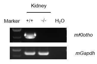
mRNA expression analysis
Strain specific analysis of Klotho mRNA expression in wild-type C57BL/6JNifdc mice and B-Klotho KO mice by RT-PCR. Kidney RNA were isolated from wild-type C57BL/6JNifdc mice (+/+) and homozygous B-Klotho KO mice (-/-), then cDNA libraries were synthesized by reverse transcription, followed by PCR with mouse Klotho primers. Mouse Klotho mRNA was only detectable in wild-type but not in homozygous B-Klotho KO mice.
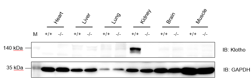
Protein expression analysis
Western blot analysis of Klotho protein expression in wild-type C57BL/6JNidc mice and homozygous B-Klotho KO mice by WB. Various tissues were collected from wild-type C57BL/6JNifdc mice (+/+) and homozygous B-Klotho KO mice (-/-), and then analyzed by western blot with anti-Klotho antibody (abcam, ab181373). 50 μg total proteins were loaded for western blotting analysis. GAPDH were detected as internal control. Klotho was detectable in kidney from C57BL/6JNifdc but not in homozygous B-Klotho KO mice.
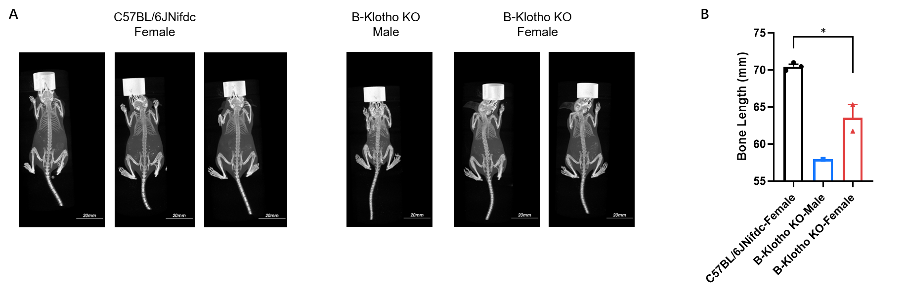
Micro-CT imaging of whole body
Whole-body micro-CT imaging of homozygous B-Klotho KO mice. (A) Representative whole-body micro-CT images of mice wild-type C57BL/6JNifdc mice (n=3, female, 5-week-old) and homozygous B-Klotho KO mice (n=1, male, 5-week-old; n=2, female, 5-week-old). (B) Quantitative comparison of body length (from oral-nasal region to tail tip). Body size of homozygous B-Klotho KO mice was smaller than wild-type C57BL/6JNifdc mice. Data are presented as mean ± SEM. *P < 0.05, **P < 0.01, ***P < 0.001. Scale bar = 20 mm.
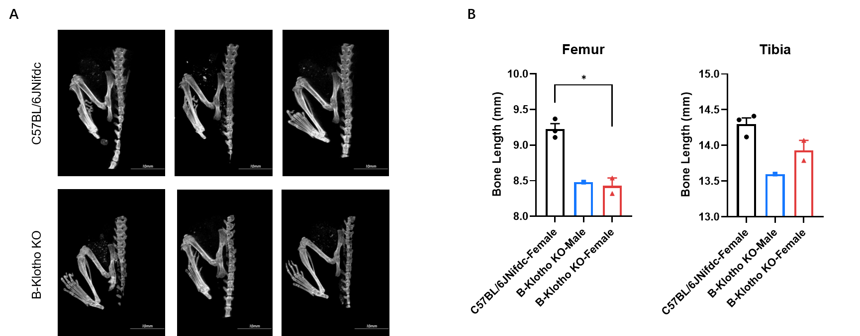
Micro-CT analysis of femur and tibia length
Micro-CT analysis of femur and tibia length in homozygous B-Klotho KO mice. (A) Representative micro-CT images of femur and tibia from wild-type C57BL/6JNifdc mice (n=3, female, 5-week-old) and homozygous B-Klotho KO mice (n=1, male, 5-week-old, left; n=2, female, 5-week-old, right). (B) Quantitative comparison of femur and tibia length. In the homozygous B-Klotho KO mice, the lengths of the femur and tibia were shortened. Data are presented as mean ± SEM. *P < 0.05, **P < 0.01, ***P < 0.001. Scale bar = 10 mm.
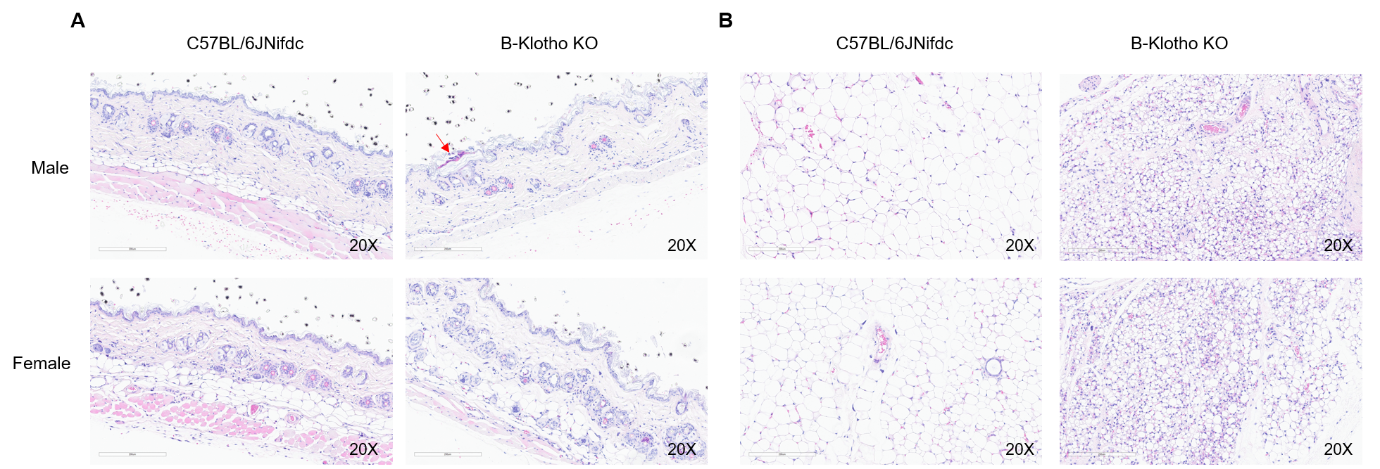
Histopathological analysis of adipocyte atrophy in B-Klotho KO mice
Histopathological analysis of dorsal skin and subcutaneous adipose tissue in homozygous B-Klotho KO mice. (A) H&E-stained sections showed skin pathological changes with punctate crusts, hyperkeratosis and subcutaneous fat layer was not visible in homozygous B-Klotho KO mice (-/-, 7-week-old, 1 male and 1 female) compared with wild-type C57BL/6JNifdc mice (+/+, 7-week-old, 1 male and 1 female). (B) H&E-stained subcutaneous adipose tissue showed adipocyte atrophy in homozygous B-Klotho KO mice compared with wild-type C57BL/6JNifdc. Scale bars: 200 µm.
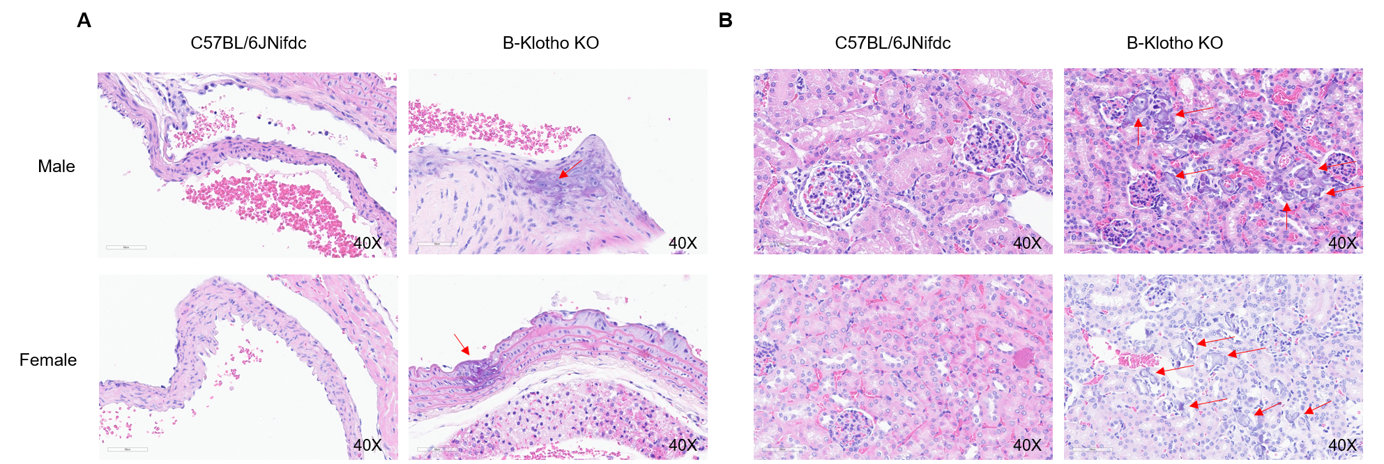
Histopathological analysis of vascular calcification in B-Klotho KO mice
Histopathological analysis of aortas and kidney tissue in homozygous B-Klotho KO mice. H&E-stained sections showed aortas (A) and kidney (B) pathological changes with calcification of the aorta and renal vasculature in homozygous B-Klotho KO mice (-/-, 7-week-old, 1 male and 1 female) compared with wild-type C57BL/6JNifdc mice (+/+, 7-week-old, 1 male and 1 female). Scale bars: 60 µm.
* When publishing results obtained using this animal model, please acknowledge the source as follows: The animal model [B-Klotho KO mice] (Cat# 113377) was purchased from Biocytogen.