B-Cep290*rd16 mice

C57BL/6JNifdc-Cep290tm1Bcgen/Bcgen • 113755

B-Cep290*rd16 mice

Product nameB-Cep290*rd16 mice
Catalog number113755
Strain nameC57BL/6JNifdc-Cep290tm1Bcgen/Bcgen
Strain backgroundC57BL/6JNifdc
NCBI gene ID80184 (Mouse)
Aliases3H11Ag, BBS14, CT87, JBTS5, LCA10, MKS4, NPHP6, POC3, SLSN6, rd16

在此页面上

  • Description
  • Targeting strategy
  • Phenotypic analysis
  • Physiological data

海报

查看全部

    发表文章

      Description

      Leber congenital amaurosis (LCA) is one of the most common inherited retinal disorders, first described in 1869 by the German ophthalmologist Theodor Leber. LCA is an autosomal recessive disease with an estimated incidence of 1/33,000 to 1/81,000 live births. Affected individuals typically present with severe visual impairment shortly after birth, accounting for approximately 20% of childhood blindness cases. Additional clinical features include photophobia, involuntary eye movements (nystagmus), extreme hyperopia, and abnormal pupillary light responses. To date, mutations in more than 20 retinal disease genes have been identified as causative for LCA. Among these, CEP290 and RPE65 represent the most frequent disease-associated genes, accounting for approximately 20% and 10% of LCA cases, respectively. The protein encoded by CEP290 localizes to the connecting cilium of photoreceptor cells. This specialized structure facilitates the transport of visual pigments and other proteins from the inner segment to the outer segment, which is essential for maintaining photoreceptor function. Since the outer segment's structural integrity depends on continuous protein and lipid supply from the inner segment, reduced CEP290 functionality ultimately leads to retinal degeneration.

      • Targeting strategy:

      The exons 37-41 of mouse Cep290 gene were knocked out in B-Cep290*rd16 mice. As a result, the expression of functional CEP290 protein was disrupt.

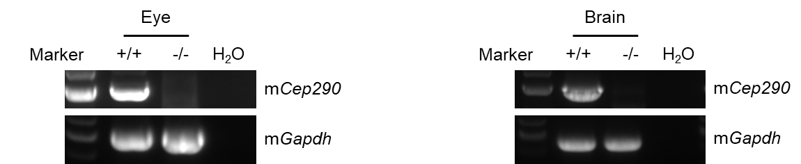
      • mRNA expression analysis:

      Mouse Cep290 mRNA was only detected in wild-type mice, but not in homozygous B-Cep290*rd16 mice.

      • Functional analysis:

      The retinal layers were disordered, and multiple layers such as the outer nuclear layer, the outer plexiform layer and the cone-rod layer disappeared in B-Cep290*rd16 mice compared to the C57BL/6JNifdc mice.

      Targeting Strategy

      Gene targeting strategy for B-Cep290*rd16 mice. The exons 37-41 of mouse Cep290 gene were knocked out in B-Cep290*rd16 mice. As a result, the expression of functional CEP290 protein was disrupt.

      mRNA expression analysis

      Strain specific analysis of Cep290 mRNA expression in wild-type C57BL/6JNifdc and B-Cep290*rd16 mice by RT-PCR. Eye and brain RNA were isolated from wild-type C57BL/6JNifdc (+/+) and homozygous B-Cep290*rd16 mice (-/-), then cDNA libraries were synthesized by reverse transcription, followed by PCR with mouse Cep290 primers. Mouse Cep290 mRNA was only detectable in wild-type mice, not in homozygous B-Cep290*rd16 mice.

      H&E staining of B-Cep290*rd16 mice

      Representative images of HE staining of wild-type C57BL/6JNifdc mice and B-Cep290*rd16 mice. Retina tissues of wild-type C57BL/6JNifdc mice (+/+) and B-Cep290*rd16 mice (-/-) (3 or 6 weeks old, male) were collected and analyzed with H&E staining. The results showed that the retinal layers were disordered, and multiple layers such as the outer nuclear layer, the outer plexiform layer and the cone-rod layer disappeared in B-Cep290*rd16 mice compared to the C57BL/6JNifdc mice. Scale bar, 100 μm.