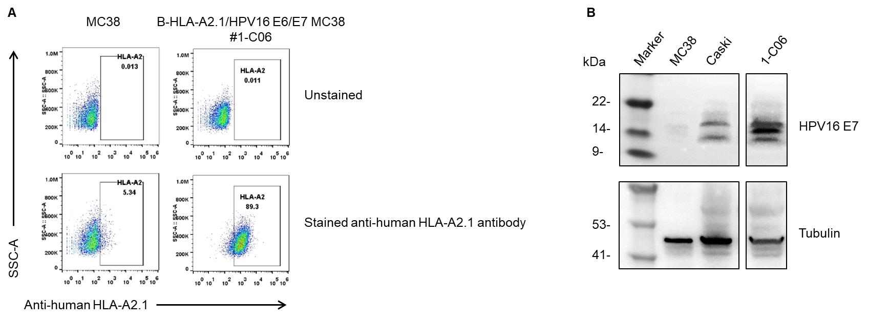
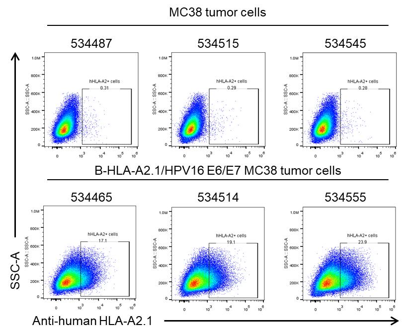
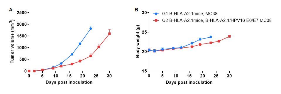
B-HLA-A2.1/HPV16 E6/E7 MC38
Catalog Number: 322401
Strain Background: C57BL/6
Aliases: IMD43; AMYLD6; MHC1D4; HLAA; HPV16 E6; HPV16 E7
---
可提供授权方案