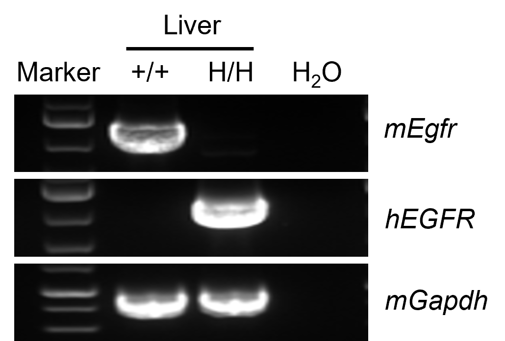
EGFR Expression by RT-PCR
- Human EGFR is specifically and correctly expressed in B-hEGFR mice.
Human EGFR was detectable in B-hEGFR mice by RT-PCR and sequencing. Liver tissues were isolated from wild-type C57BL/6 mice (+/+) and homozygous B-hEGFR mice (H/H). (A) Primers were designed to detect human and mouse EGFR. (B) Human EGFR mRNA was detectable in B-hEGFR mice, but not in wild-type mice. Sequencing of PCR products confirmed that the amplified sequences were consistent with database reference sequences.
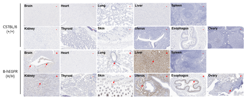
Immunohistochemical Analysis
- Human EGFR expression was detected in the brain, lung, liver, kidney, skin, uterus, esophagus, and ovary of homozygous B-hEGFR mice, but not in the heart, spleen, or thyroid. The antibodies were species-specific and did not bind to mouse EGFR in wild-type C57BL/6 mice.
- The results demonstrated that the EGFR expression profile in B-hEGFR mice closely resembles that in normal human tissues (Table 1).
Immunohistochemical analysis of organs in B-hEGFR mice. Major organs were collected from wild-type C57BL/6 mice and homozygous B-hEGFR mice (2 females, 8 weeks old), and analyzed by IHC using anti-EGFR antibody (Invitrogen, MA5-49312). The arrow indicates tissue cells with positive EGFR staining (brown). "+" and "-" indicate positive and negative tissues, respectively.
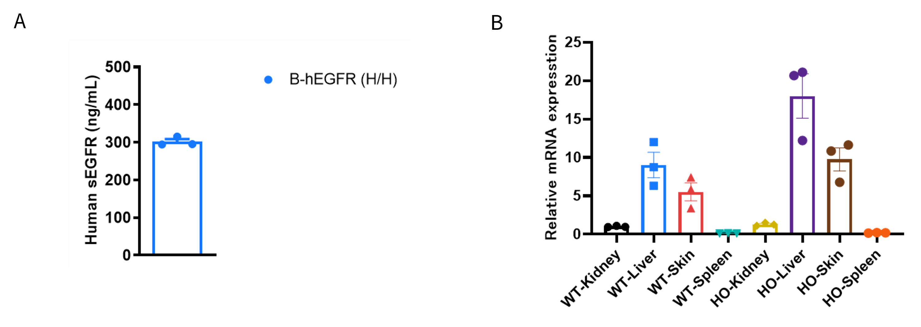
sEGFR Protein and EGFR mRNA Expression Analysis
- Approximately 300 ng/mL of human sEGFR is detectable in the plasma of B-hEGFR mice.
- The relative expression levels of Egfr in the liver and skin were higher in B-hEGFR mice than in wild-type mice.
(A) Strain specific soluble EGFR expression analysis in homozygous B-hEGFR mice by ELISA. Plasma was collected from homozygous B-hEGFR mice (H/H) (male, n=3, 7-week-old). The expression levels of human sEGFR in B-hEGFR mice were analyzed by ELISA using the anti-human EGFR kit (RD, DEGFR0). Values are expressed as mean ± SEM.
(B) qPCR analysis of EGFR expression in homozygous B-hEGFR mice. RNA was isolated from the kidney, liver, skin, and spleen of wild-type (WT, +/+) and homozygous B-hEGFR (HO, H/H) mice (female, 6 weeks old, n=3 per group). cDNA libraries were generated by reverse transcription, followed by qPCR using mouse Egfr-specific primers. The relative expression levels of Egfr in the liver and skin were higher in B-hEGFR mice than in wild-type C57BL/6 mice.
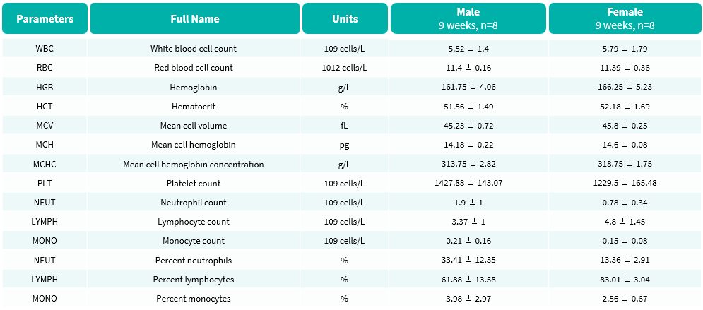
Hematology Analysis
- No significant differences were observed.
Complete blood count (CBC) of B-hEGFR Mice. Values are expressed as mean ± SD.
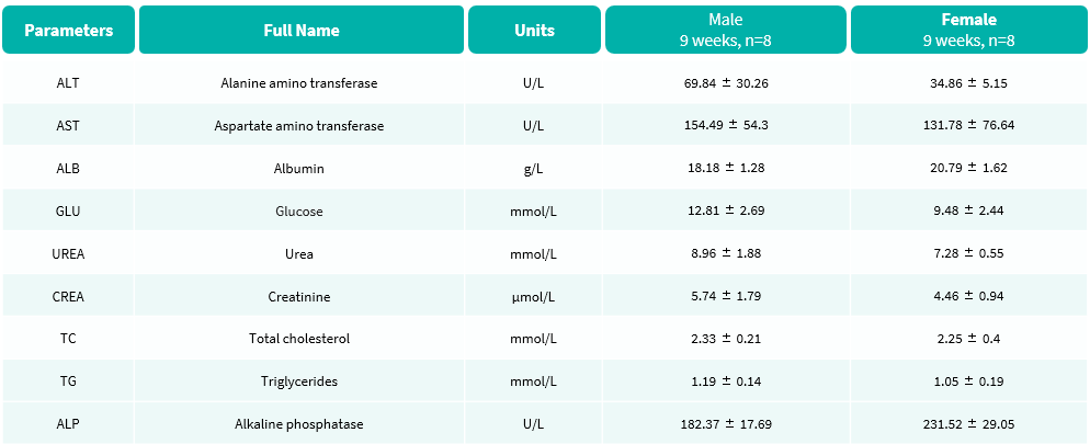
Blood Biochemical Analysis
- No significant differences were observed.
Blood biochemical parameters of B-hEGFR Mice are shown. Values are expressed as mean ± SD.
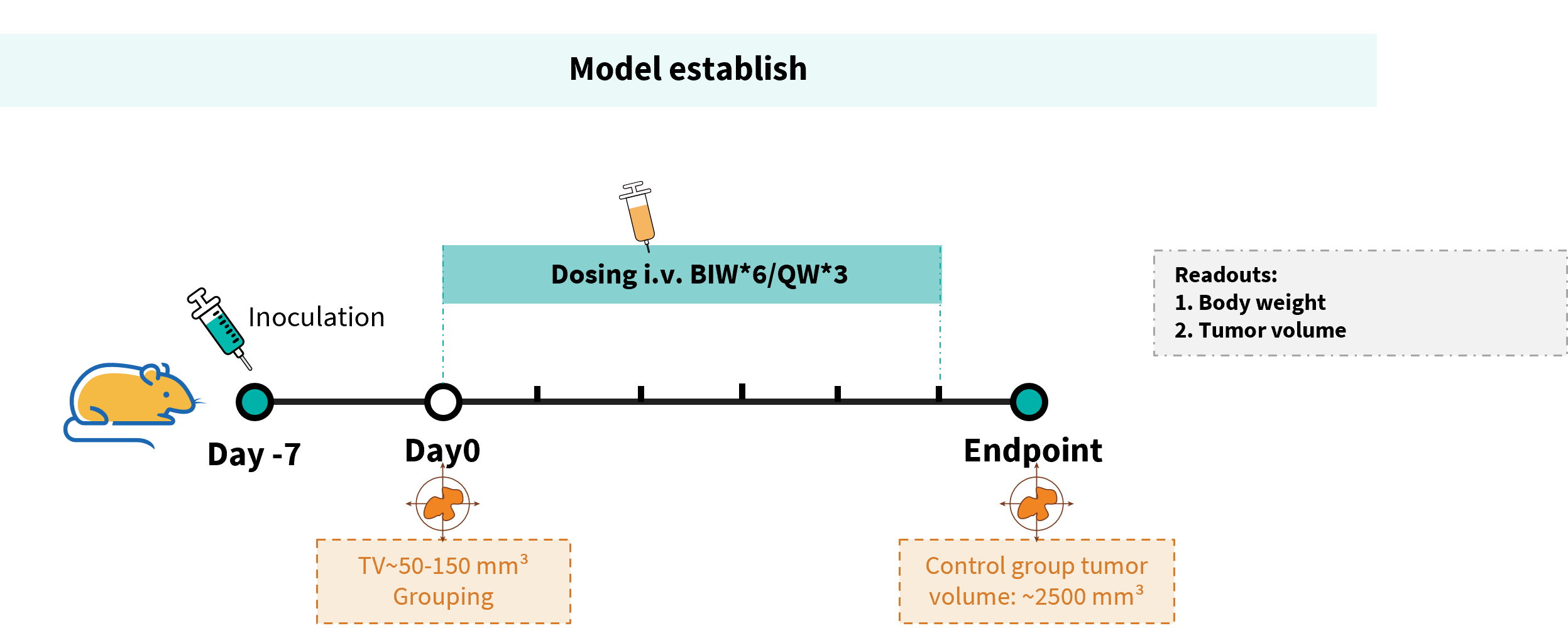
Efficacy Evaluation of Cetuximab analog-MMAE in the Treatment of the Subcutaneous B-Tg(hEGFR) MC38 Model in B-hEGFR Mice
Establishment of a B-Tg(hEGFR) MC38 model and in vivo efficacy study of Cetuximab analog-MMAE. B-Tg(hEGFR) MC38 cells were implanted subcutaneously into homozygous B-hEGFR mice (female, 6-8 weeks-old, n = 6). When the average tumor volume reached approximately 50-150 mm³, mice were randomized and subsequently administered the Cetuximab analog-MMAE (in house) via intravenous injection.
Efficacy of a Cetuximab analog-MMAE in B-hEGFR mice. (A) Tumor growth curves. (B) Body weight changes during treatment. As shown in panel A, anti-human EGFR antibody (in house) inhibited B-Tg(hEGFR) MC38 tumor growth in homozygous B-hEGFR mice, demonstrating that the B-hEGFR mice provide a powerful preclinical model for in vivo evaluation of EGFR-targeting ADC. Values are expressed as mean ± SEM.
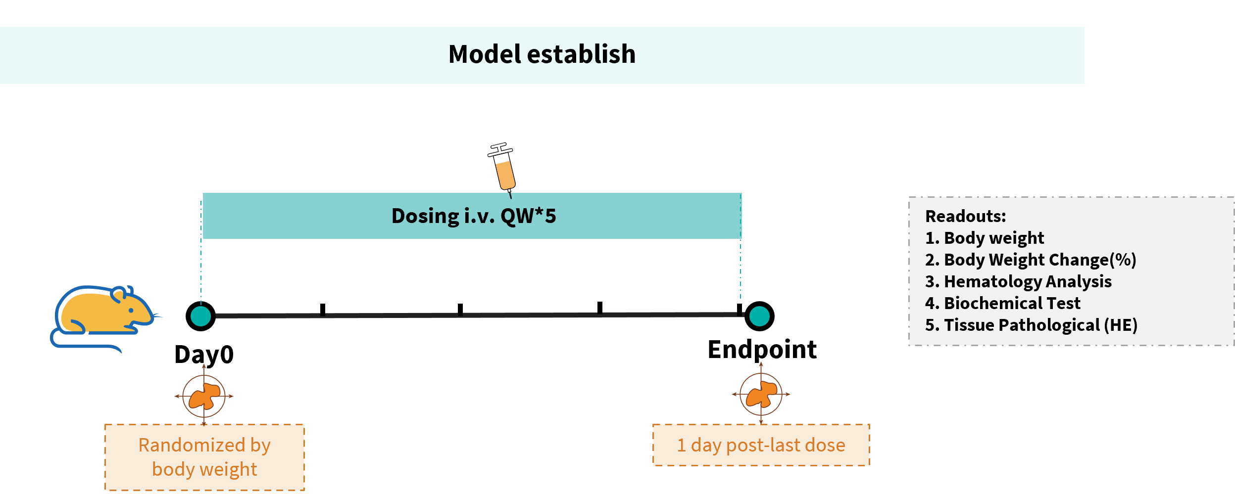
Toxicity Analysis of Anti-human EGFR Antibody in B-hEGFR Mice
Establishment of a toxicity evaluation model for EGFR-targeted antibody drugs in B-hEGFR mice. Anti-human EGFR antibody cetuximab biosimilar (Bio X Cell, 907623J2) or panitumumab (Takeda, 549661) were intravenously injected into B-hEGFR mice (male, 14-15 weeks-old, n=6). Mice were weighed twice a week, and their condition was observed daily. At the end of the experiment, blood samples were collected for complete blood count test. Additionally, tissue samples were collected from the nasal and oral skin, abdominal skin, stomach, duodenum, jejunum, ileum, cecum, colon, and rectum, and then subjected to pathological analysis.
Note: This experiment is a collaboration with the client.

Establishment of a toxicity evaluation model for EGFR-targeted antibody drugs in B-hEGFR mice. (A) Body weight and body weight changes during treatment. The results showed that 45.0 mg/kg cetuximab biosimilar and 30.0 mg/kg panitumumab resulted in significant weight loss in B-hEGFR mice. Values are expressed as mean ± SEM.
Note: This experiment is a collaboration with the client.
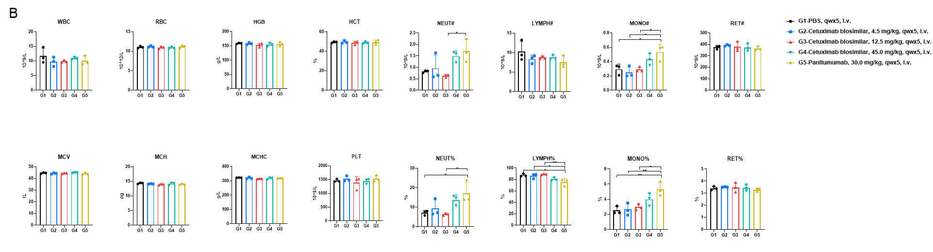
Establishment of a toxicity evaluation model for EGFR-targeted antibody drugs in B-hEGFR mice. (B) Complete blood cell count detection at the endpoint of the experiment. 45.0 mg/kg cetuximab biosimilar and 30.0 mg/kg panitumumab resulted in an increase in the number of neutrophils and monocytes, while there were no significant changes in blood parameters in the other treatment groups. The increase in neutrophils and monocytes in the peripheral blood may be induced by the activation of the immune system and mild inflammatory responses following high-dose antibody treatment. Values are expressed as mean ± SD. Significance was determined by one-way ANOVA test. *p<0.05, **p<0.01.
Note: This experiment is a collaboration with the client.
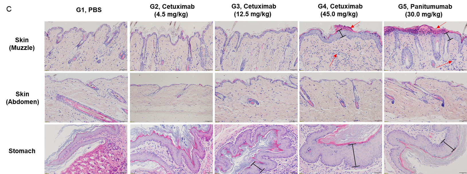
Establishment of a toxicity evaluation model for EGFR-targeted antibody drugs in B-hEGFR mice. (C) Pathological diagrams of various tissues. The histopathological examination in muzzle skin revealed article-related alteration in cetuximab biosimilar 45 mg/kg dose group (1/6, minimal), which showed squamous epithelial cells proliferation and subcutaneous infiltration of inflammatory cells. The similar article-related changes were also found in panitumumab group (5/6, minimal). Squamous epithelial cells proliferation in stomach limiting ridge were observed in mice administrated cetuximab biosimilar at the dose of 12.5 mg/kg (4/6, minimal) and 45 mg/kg groups (2/6, minimal; 4/6, slight), which were considered test-article and dose related. The similar article-related changes were also found in panitumumab group (3/6, minimal; 3/6, slight). Neither cetuximab biosimilar nor panitumumab showed any toxicity in the abdominal skin, duodenum, jejunum, ileum, cecum, colon, and rectum of mice (The data is not shown).
Note: This experiment is a collaboration with the client.
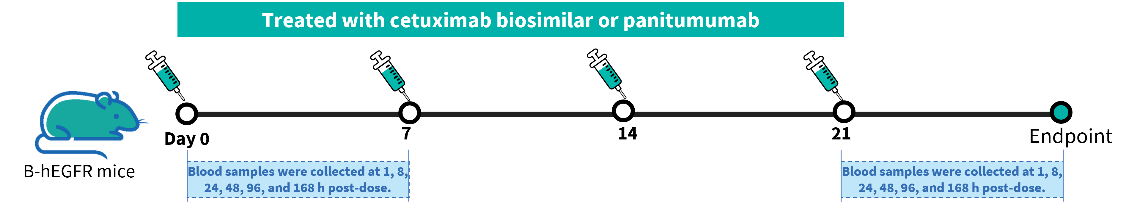
Toxicokinetic Analysis of Anti-human EGFR Antibody in B-hEGFR Mice
Toxicokinetic analysis of anti-human EGFR antibody in B-hEGFR mice. Anti-human EGFR antibody cetuximab biosimilar (Bio X Cell, 907623J2) or panitumumab (Takeda, 549661) were intravenously injected into B-hEGFR mice (male, 14-15 weeks-old, n=2). Blood samples were collected at 1h, 8h, 24h, 48h, 96h, and 168h after the first and fourth doses, and then the drug concentrations in the blood were measured.
Note: This experiment is a collaboration with the client.
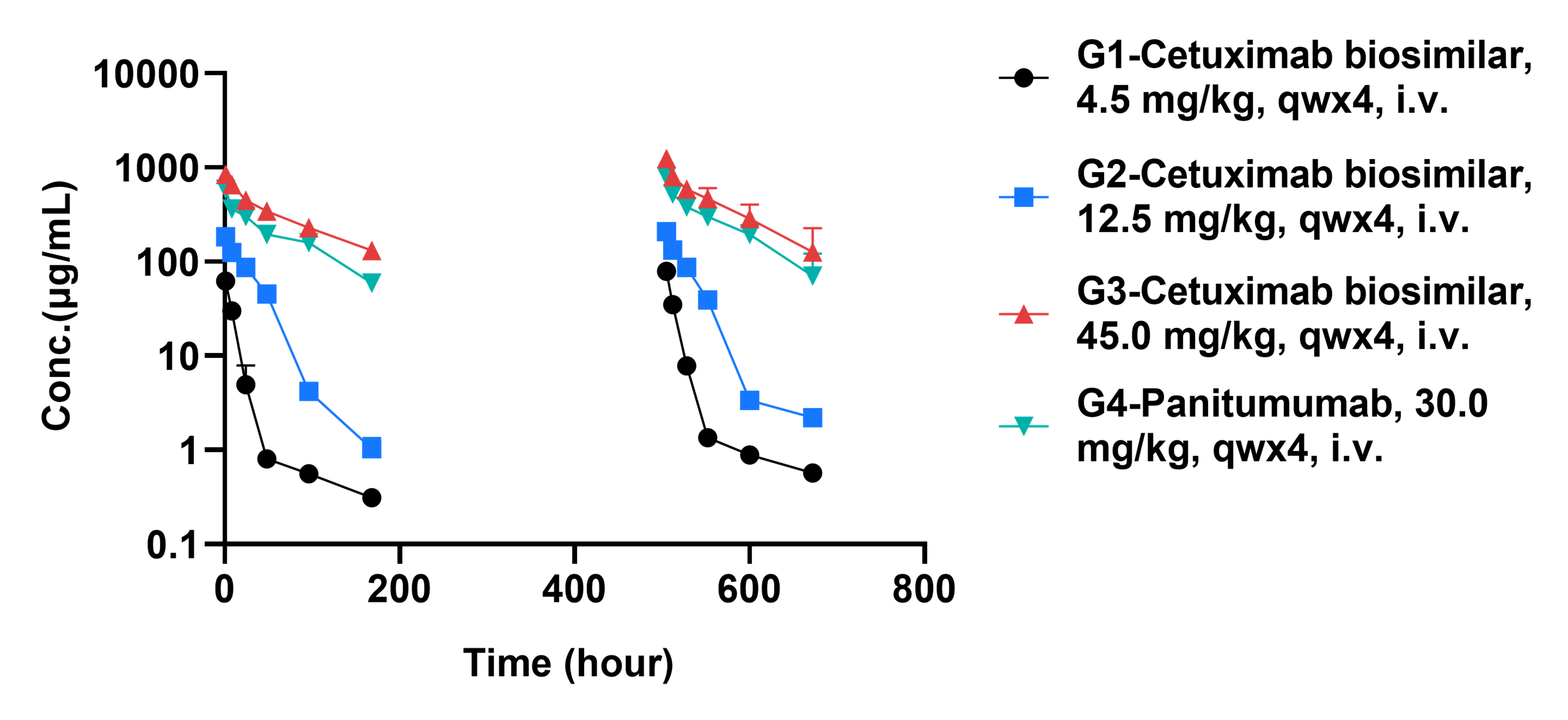
Toxicokinetic analysis of anti-human EGFR antibody in B-hEGFR mice. As shown in the figure, with the increase of dosage, the blood concentration of cetuximab biosimilar also increases. This indicates that the relationship between the blood concentration of cetuximab biosimilar and dosage is linear within the experimental dosage range. The existence of this linear relationship may be due to the absence of saturation during the absorption, distribution, metabolism, and excretion of drugs in mice.
Note: This experiment is a collaboration with the client.
* When publishing results obtained using this animal model, please acknowledge the source as follows: The animal model [B-hEGFR mice] (Cat# 120771) was purchased from Biocytogen.