
C57BL/6-Pdcd1tm3(PDCD1)Bcgen Il2tm1(IL2)Bcgen Il2ratm1(IL2RA)Bcgen ll2rbtm2(IL2RB)Bcgen ll2rgtm2(IL2RG)Bcgen ll15tm1(IL15)Bcgen ll15ratm1(IL15RA)Bcgen/Bcgen • 113319
| Product name | B-hPD-1 plus/hIL2/hIL2RA/hIL2RB/hIL2RG/hIL15/hIL15RA mice |
|---|---|
| Catalog number | 113319 |
| Strain name | C57BL/6-Pdcd1tm3(PDCD1)Bcgen Il2tm1(IL2)Bcgen Il2ratm1(IL2RA)Bcgen ll2rbtm2(IL2RB)Bcgen ll2rgtm2(IL2RG)Bcgen ll15tm1(IL15)Bcgen ll15ratm1(IL15RA)Bcgen/Bcgen |
| Strain background | C57BL/6 |
| NCBI gene ID | 5133,3558,3559,3560,3561,3600,3601 (Human) |
| Aliases | PD1; PD-1; CD279; SLEB2; hPD-1; hPD-l; hSLE1; ADMIO4; AIMTBS; IL-2; TCGF; lymphokine; p55; CD25; IL2R; IMD41; TCGFR; IDDM10; CD122; IMD63; IL15RB; P70-75; P64; CIDX; IMD4; CD132; SCIDX; IL-2RG; SCIDX1; IL-15; CD215 |
Gene targeting strategy for B-hPD-1 plus/hIL2/hIL2RA/hIL2RB/hIL2RG/hIL15/hIL15RA mice.
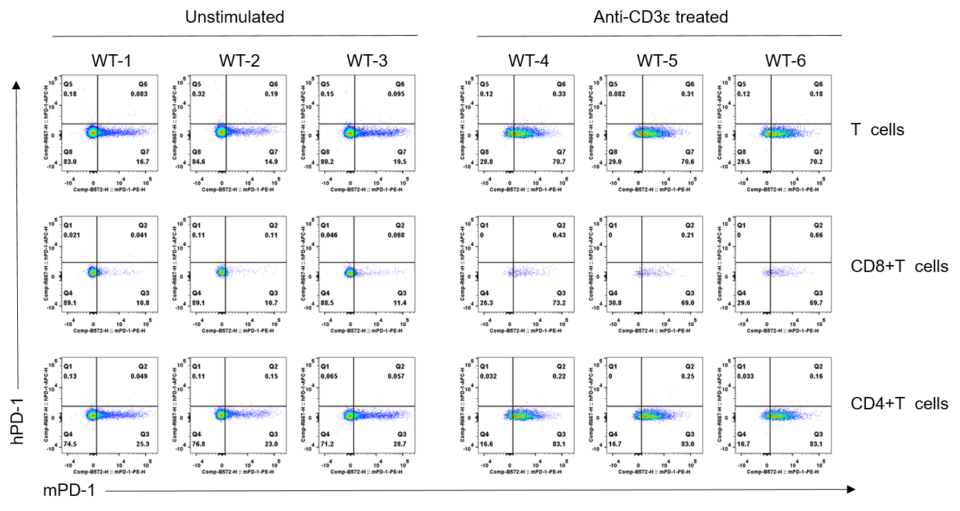
Strain specific PD-1 expression analysis in wild-type C57BL/6JNifdc mice by flow cytometry. Splenocytes were collected from wild-type C57BL/6 mice (male, 7-week-old, n=3/group) stimulated with or without anti-CD3ε antibody (7.5 μg/mice, i.p.) in vivo for 24 h, and analyzed by flow cytometry with species-specific anti-mouse PD-1 antibody (Biolegend, 109104) and anti-human PD-1 antibody (Biolegend, 329908). Mouse PD-1 was only detectable in wild-type mice.
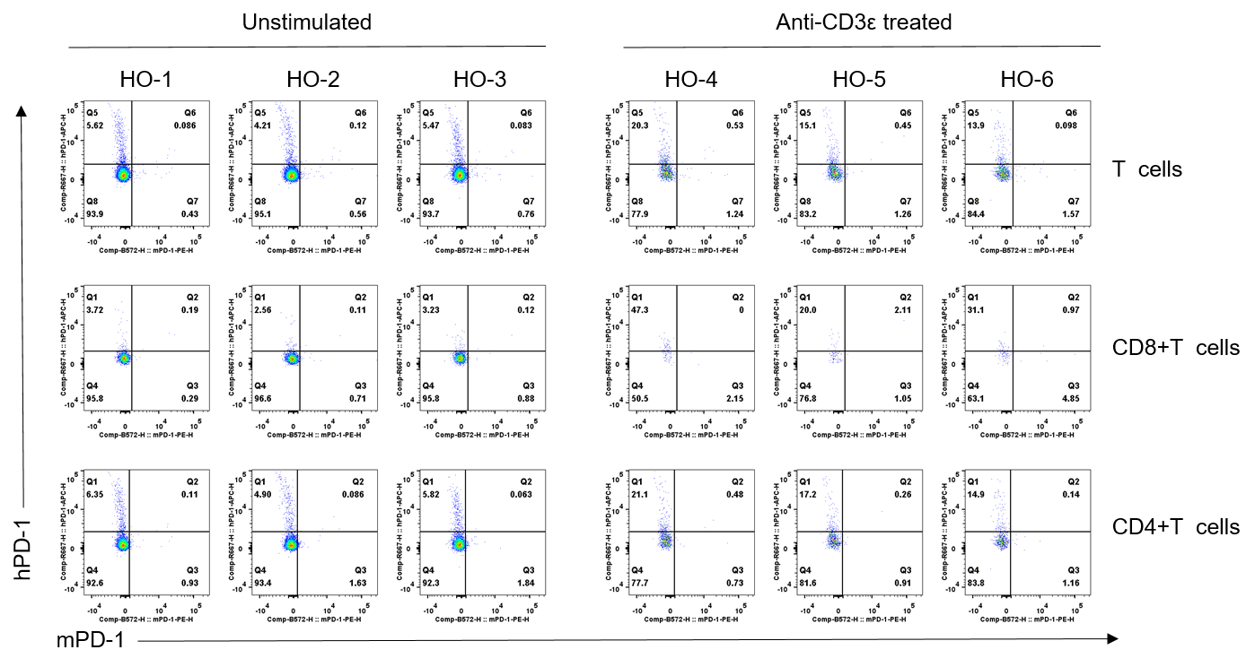
Strain specific PD-1 expression analysis in homozygous B-hPD-1 plus/hIL2RA/hIL2RB/hIL2RG/hIL15/hIL15RA mice by flow cytometry. Splenocytes were collected from homozygous B-hPD-1 plus/hIL2RA/hIL2RB/hIL2RG/hIL15/hIL15RA mice (male, 7-week-old, n=3/group) stimulated with or without anti-CD3ε antibody (7.5 μg/mice, i.p.) in vivo for 24 h, and analyzed by flow cytometry with species-specific anti-mouse PD-1 antibody (Biolegend, 109104) and anti-human PD-1 antibody (Biolegend, 329908). Human PD-1 was only detectable in homozygous B-hPD-1 plus/hIL2RA/hIL2RB/hIL2RG/hIL15/hIL15RA mice.
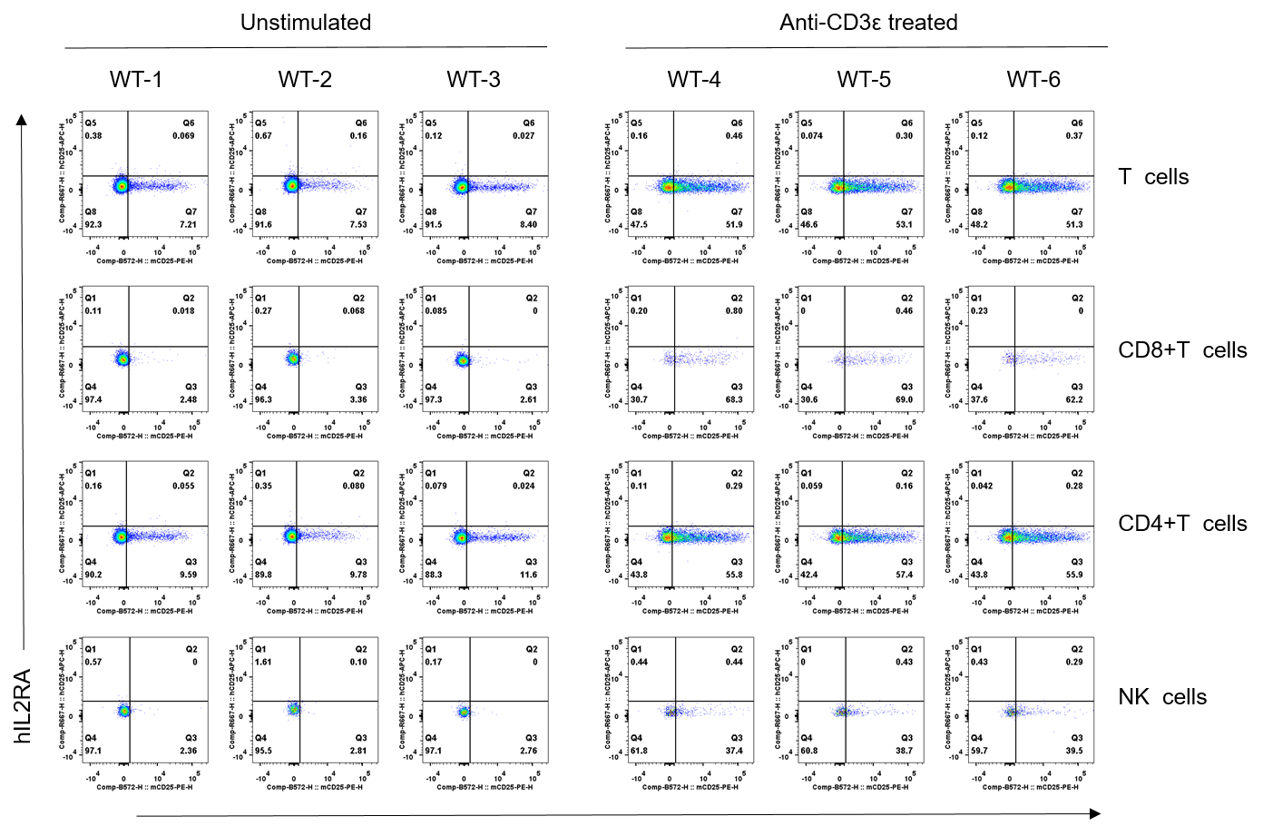
Strain specific IL2RA expression analysis in wild-type C57BL/6JNifdc mice by flow cytometry. Splenocytes were collected from wild-type C57BL/6 mice (male, 7-week-old, n=3/group) stimulated with or without anti-CD3ε antibody (7.5 μg/mice, i.p.) in vivo for 24 h, and analyzed by flow cytometry with species-specific anti-mouse IL2RA antibody (Biolegend, 102008) and anti-human IL2RA antibody (Biolegend, 302610). Mouse IL2RA was only detectable in wild-type mice.
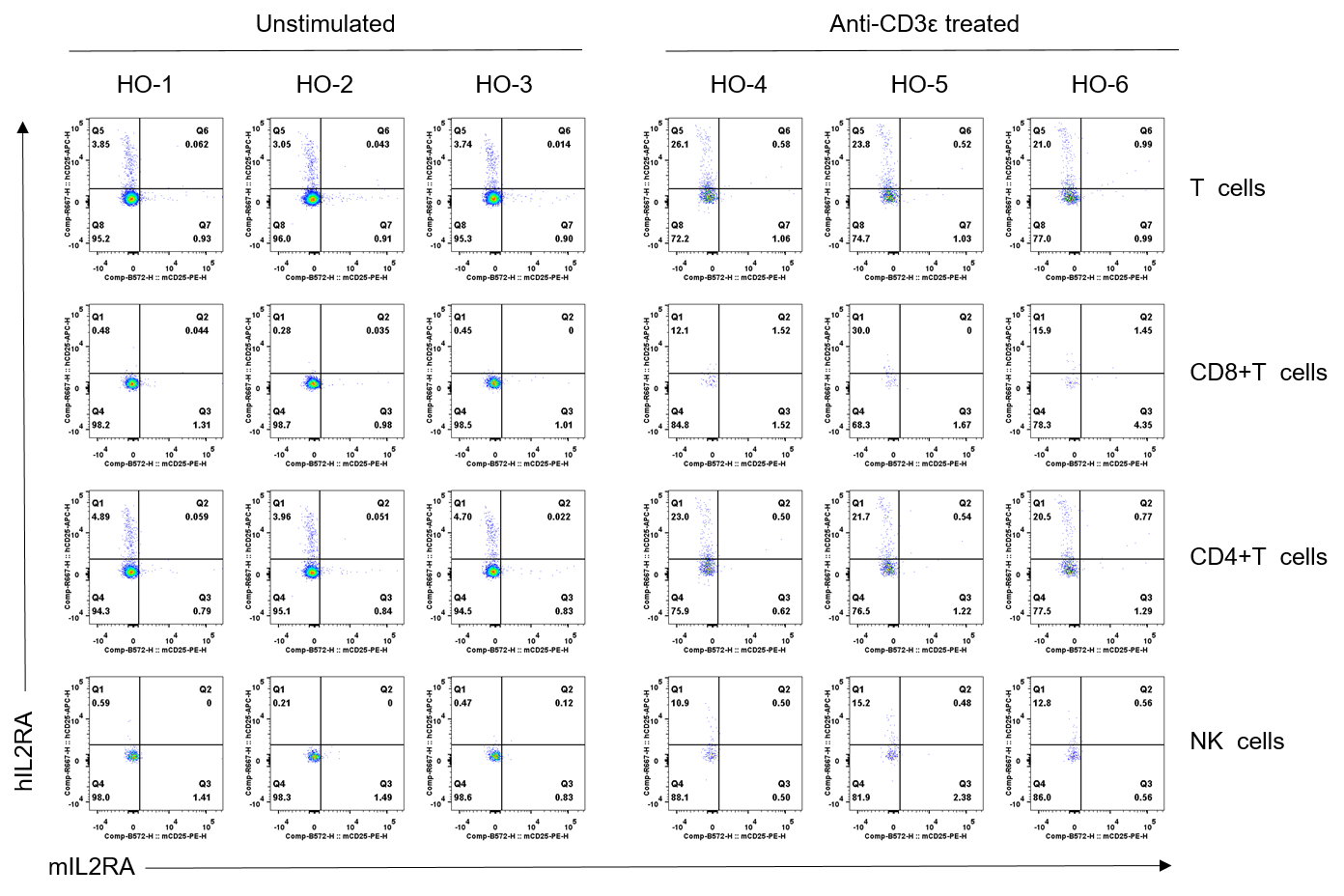
Strain specific IL2RA expression analysis in homozygous B-hPD-1 plus/hIL2RA/hIL2RB/hIL2RG/hIL15/hIL15RA mice by flow cytometry. Splenocytes were collected from homozygous B-hPD-1 plus/hIL2RA/hIL2RB/hIL2RG/hIL15/hIL15RA mice (male, 7-week-old, n=3/group) stimulated with or without anti-CD3ε antibody (7.5 μg/mice, i.p.) in vivo for 24 h, and analyzed by flow cytometry with species-specific anti-mouse IL2RA antibody (Biolegend, 102008) and anti-human IL2RA antibody (Biolegend, 302610). Human IL2RA was only detectable in homozygous B-hPD-1 plus/hIL2RA/hIL2RB/hIL2RG/hIL15/hIL15RA mice.
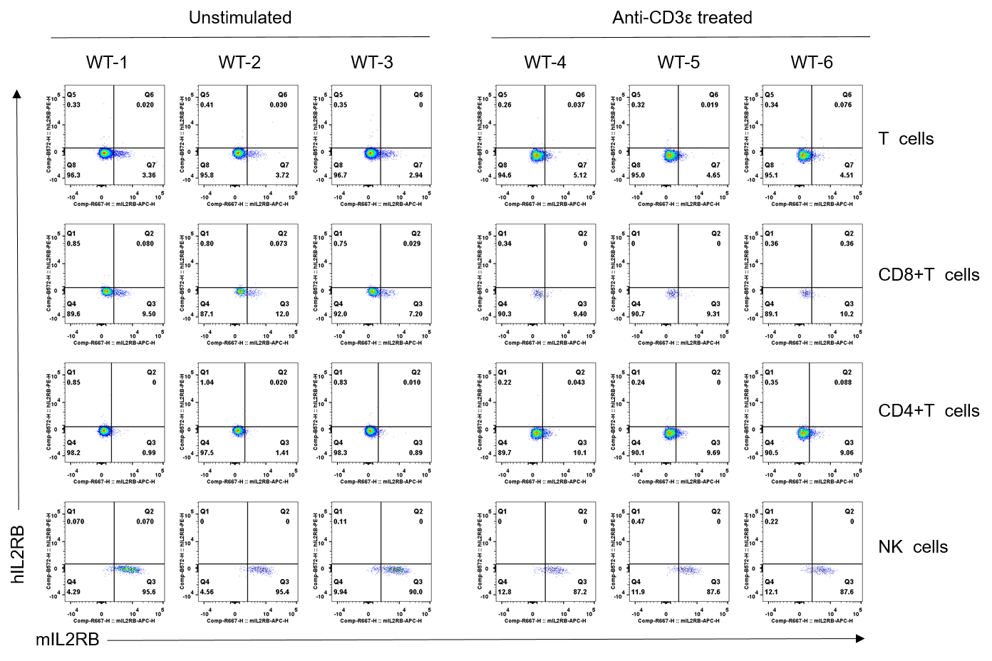
Strain specific IL2RB expression analysis in wild-type C57BL/6JNifdc mice by flow cytometry. Splenocytes were collected from wild-type C57BL/6 mice (male, 7-week-old, n=3/group) stimulated with or without anti-CD3ε antibody (7.5 μg/mice, i.p.) in vivo for 24 h, and analyzed by flow cytometry with species-specific anti-mouse IL2RB antibody (Biolegend, 105912) and anti-human IL2RB antibody (Biolegend, 339005). Mouse IL2RB was only detectable in wild-type mice.
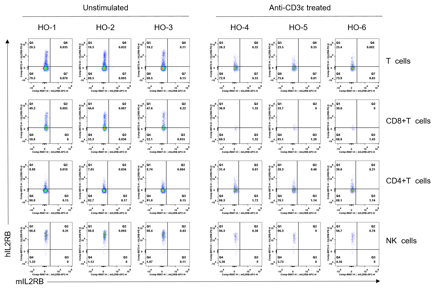
Strain specific IL2RB expression analysis in homozygous B-hPD-1 plus/hIL2RA/hIL2RB/hIL2RG/hIL15/hIL15RA mice by flow cytometry. Splenocytes were collected from homozygous B-hPD-1 plus/hIL2RA/hIL2RB/hIL2RG/hIL15/hIL15RA mice (male, 7-week-old, n=3/group) stimulated with or without anti-CD3ε antibody (7.5 μg/mice, i.p.) in vivo for 24 h, and analyzed by flow cytometry with species-specific anti-mouse IL2RB antibody (Biolegend, 105912) and anti-human IL2RB antibody (Biolegend, 339005). Human IL2RB was only detectable in homozygous B-hPD-1 plus/hIL2RA/hIL2RB/hIL2RG/hIL15/hIL15RA mice.
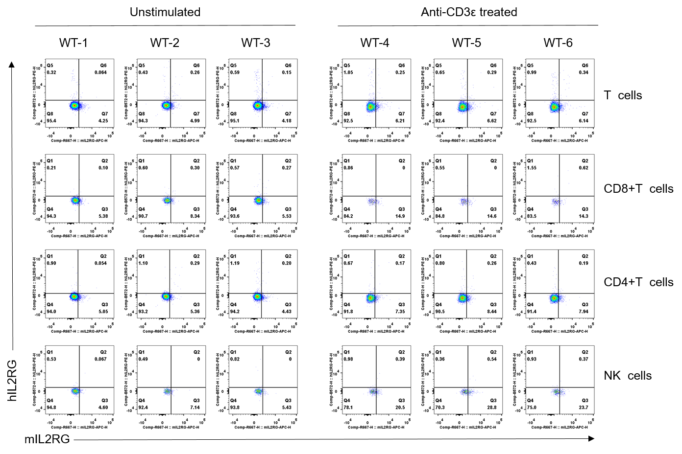
Strain specific IL2RG expression analysis in wild-type C57BL/6JNifdc mice by flow cytometry. Splenocytes were collected from wild-type C57BL/6 mice (male, 7-week-old, n=3/group) stimulated with or without anti-CD3ε antibody (7.5 μg/mice, i.p.) in vivo for 24 h, and analyzed by flow cytometry with species-specific anti-mouse IL2RG antibody (Biolegend, 132307) and anti-human IL2RB antibody (Biolegend, 338605). Mouse IL2RB was only detectable in wild-type mice.
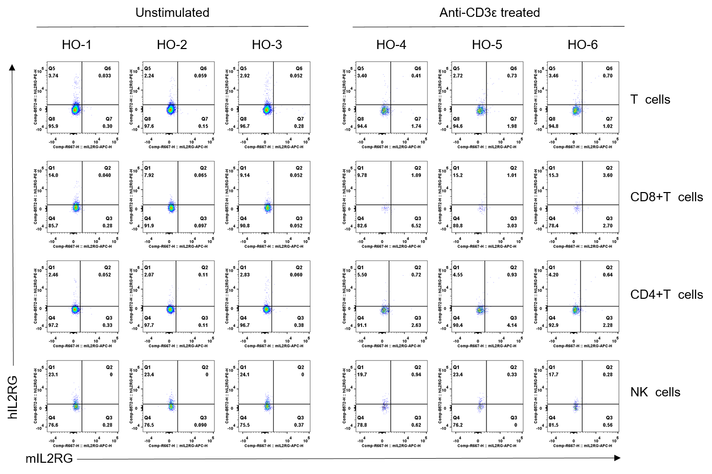
Strain specific IL2RG expression analysis in homozygous B-hPD-1 plus/hIL2RA/hIL2RB/hIL2RG/hIL15/hIL15RA mice by flow cytometry. Splenocytes were collected from homozygous B-hPD-1 plus/hIL2RA/hIL2RB/hIL2RG/hIL15/hIL15RA mice (male, 7-week-old, n=3/group) stimulated with or without anti-CD3ε antibody (7.5 μg/mice, i.p.) in vivo for 24 h, and analyzed by flow cytometry with species-specific anti-mouse IL2RG antibody (Biolegend, 132307) and anti-human IL2RG antibody (Biolegend, 338605). Human IL2RG was only detectable in homozygous B-hPD-1 plus/hIL2RA/hIL2RB/hIL2RG/hIL15/hIL15RA mice.

Strain specific IL15 expression analysis in wild-type C57BL/6JNifdc mice and homozygous B-hPD-1 plus/hIL2/hIL2RA/hIL2RB/hIL2RG/hIL15/hIL15RA mice by ELISA. Serum was collected from wild-type C57BL/6JNifdc mice and homozygous B-hPD-1 plus/hIL2/hIL2RA/hIL2RB/hIL2RG/hIL15/hIL15RA mice (Female, 6-week-old, n=3/group,) stimulated with 350mg/kg APAP in vivo for 18 hrs. Expression level of human IL15 was analyzed by ELISA (anti-human IL15 ELISA kit: R&D D1500).Human IL15 was exclusively detectable in homozygous B-hPD-1 plus/hIL2/hIL2RA/hIL2RB/hIL2RG/hIL15/hIL15RA mice. Values are expressed as mean ± SEM.
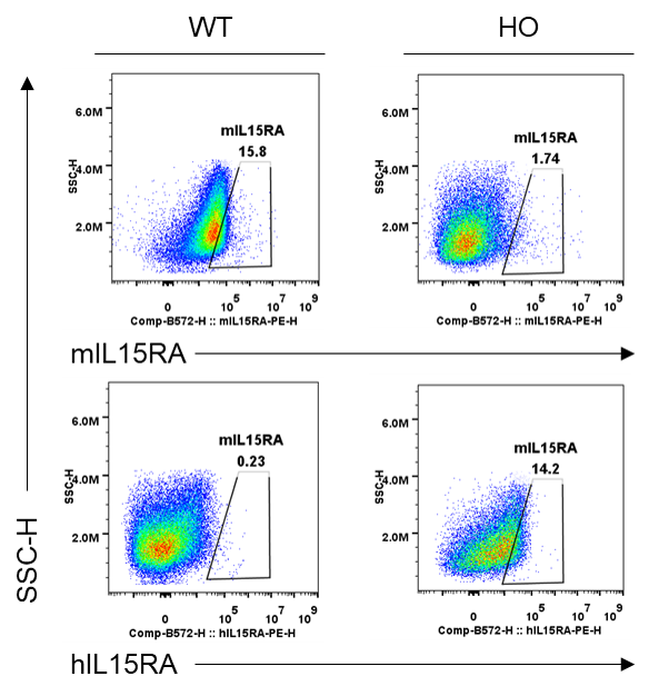
Strain specific IL15RA expression analysis in homozygous B-hPD-1 plus/hIL2/hIL2RA/hIL2RB/hIL2RG/hIL15/hIL15RA mice by flow cytometry. Bone marrow cells were collected from wild-type (WT) C57BL/6JNifdc mice and homozygous (HO) B-hPD-1 plus/hIL2/hIL2RA/hIL2RB/hIL2RG/hIL15/hIL15RA mice, and cultured in 6-well plates stimulated with GM-CSF and IL-4 for 6 days and LPS (1 μg/mL) for 18 h to induce BMDCs. Then BMDCs were collected and analyzed by flow cytometry with anti-mIL15RA antibody (BD, 568235) and anti-hIL15RA antibody (Biolegend, 330207). The mIL15RA was only detectable in wild-type mice. The hIL15RA was exclusively detectable in homozygous B-hPD-1 plus/hIL2/hIL2RA/hIL2RB/hIL2RG/hIL15/hIL15RA mice.

Frequency of leukocyte subpopulations in spleen by flow cytometry. Splenocytes were isolated from wild-type C57BL/6JNifdc mice and B-hPD-1 plus/hIL2RA/hIL2RB/hIL2RG/hIL15/hIL15RA mice (female, 8-week-old, n=3). A. Flow cytometry analysis of the splenocytes was performed to assess the frequency of leukocyte subpopulations. B. Frequency of T cell subpopulations. Frequencies of NK cells, dendritic cells, monocytes, macrophages, neutrophils and Tregs in B-hPD-1 plus/hIL2RA/hIL2RB/hIL2RG/hIL15/hIL15RA mice were similar to those in C57BL/6JNifdc mice. The frequency of T cells in B-hPD-1 plus/hIL2RA/hIL2RB/hIL2RG/hIL15/hIL15RA mice were lower than in C57BL/6JNifdc mice, while the frequency of B cells in B-hPD-1 plus/hIL2RA/hIL2RB/hIL2RG/hIL15/hIL15RA mice were higher than in C57BL/6JNifdc mice. Values are expressed as mean ± SEM. Significance was determined by two-way ANOVA test. *P < 0.05, **P < 0.01, ***p < 0.001.

Frequency of leukocyte subpopulations in blood by flow cytometry. Blood were isolated from wild-type C57BL/6JNifdc mice and B-hPD-1 plus/hIL2RA/hIL2RB/hIL2RG/hIL15/hIL15RA mice (female, 8-week-old, n=3). A. Flow cytometry analysis of the blood was performed to assess the frequency of leukocyte subpopulations. B. Frequency of T cell subpopulations. Frequencies of NK cells, dendritic cells, monocytes, macrophages, neutrophils and Tregs in B-hPD-1 plus/hIL2RA/hIL2RB/hIL2RG/hIL15/hIL15RA mice were similar to those in C57BL/6JNifdc mice. The frequency of T cells in B-hPD-1 plus/hIL2RA/hIL2RB/hIL2RG/hIL15/hIL15RA mice was lower than in C57BL/6JNifdc mice, while the frequency of B cells in B-hPD-1 plus/hIL2RA/hIL2RB/hIL2RG/hIL15/hIL15RA mice were higher than in C57BL/6JNifdc mice. Values are expressed as mean ± SEM. Significance was determined by two-way ANOVA test. *P < 0.05, **P < 0.01, ***p < 0.001.

Frequency of leukocyte subpopulations in lymph node by flow cytometry. Lymph node cells were isolated from wild-type C57BL/6JNifdc mice and B-hPD-1 plus/hIL2RA/hIL2RB/hIL2RG/hIL15/hIL15RA mice (female, 8-week-old, n=3). A. Flow cytometry analysis of the lymph node was performed to assess the frequency of leukocyte subpopulations. B. Frequency of T cell subpopulations. Frequencies of B cells, NK cells and Tregs in B-hPD-1 plus/hIL2RA/hIL2RB/hIL2RG/hIL15/hIL15RA mice were similar to those in C57BL/6JNifdc mice. Values are expressed as mean ± SEM.
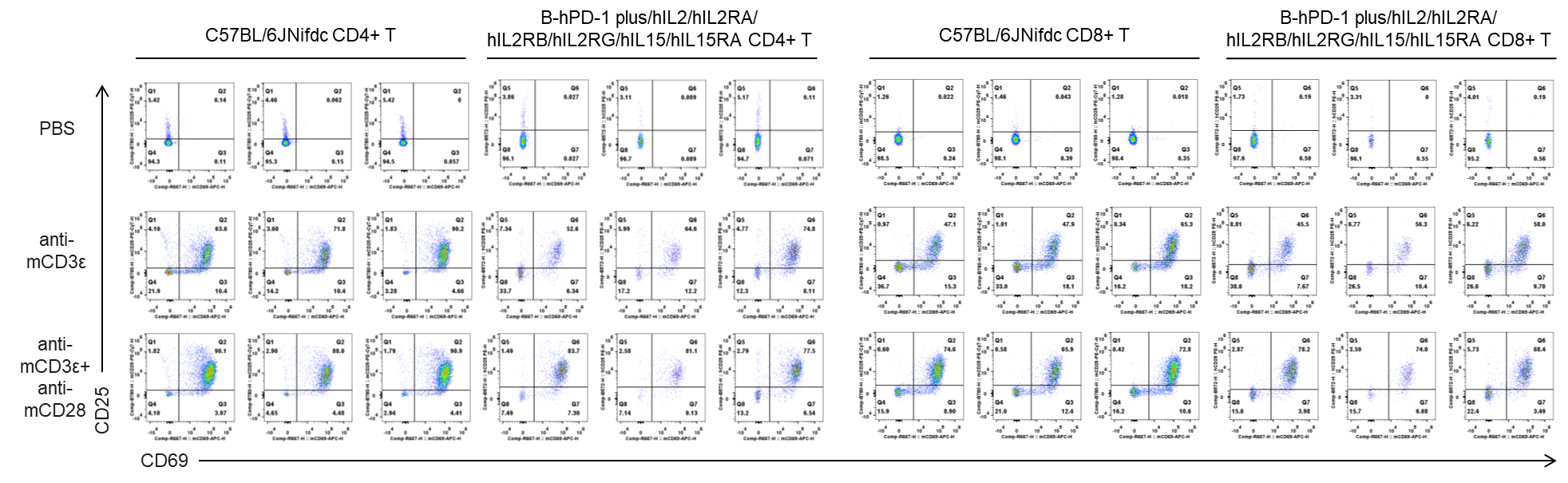
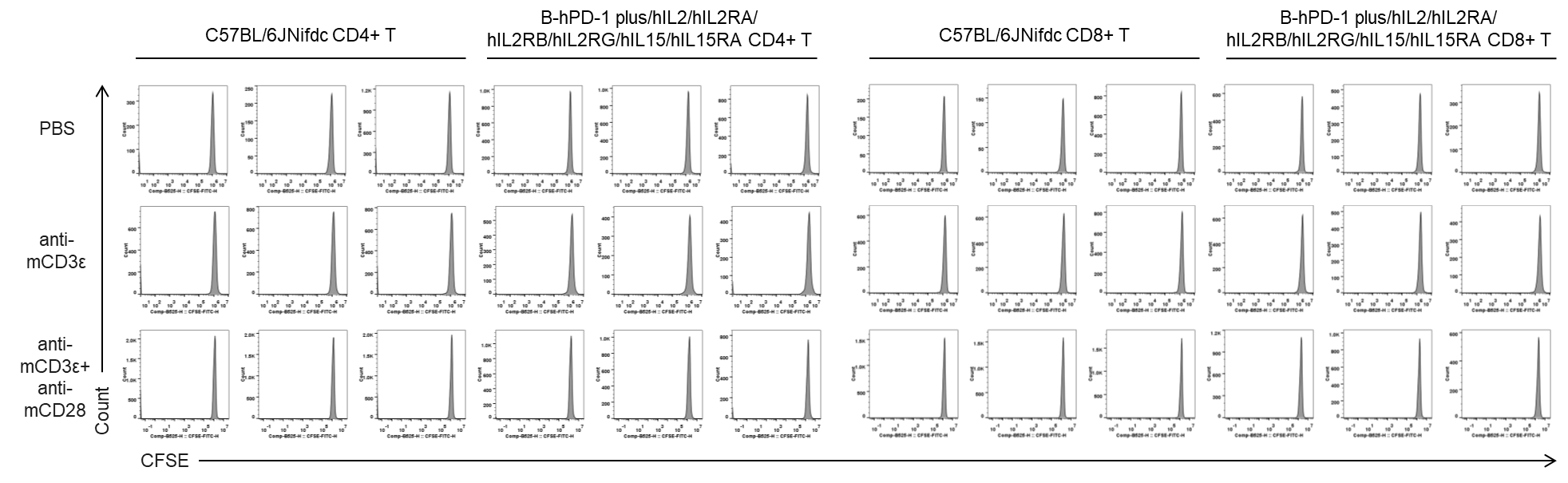
In vitro T cell activation by anti-mCD3ε antibody with or without anti-mCD28 antibody in wild-type C57BL/6JNifdc mice and homozygous humanized B-hPD-1 plus/hIL2/hIL2RA/hIL2RB/hIL2RG/hIL15/hIL15RA mice (24h). T cells were isolated from splenocytes of C57BL/6JNifdc and B-hPD-1 plus/hIL2/hIL2RA/hIL2RB/hIL2RG /hIL15/hIL15RA mice (female,13-week-old, n=3), and incubated in the presence of anti-mCD3ε antibody (2ug/ml, BioXcell, BE0001-2), with or witnout anti-mCD28 antibody (5ug/ml, BioXcell, BE0015-1) for 24h. T cell proliferation was tested by flow cytometry. T cell activation in B-hPD-1 plus/hIL2/hIL2RA/hIL2RB/hIL2RG /hIL15/hIL15RA mice was significantly up-regulated by anti-mCD3ε antibody and anti-mCD28 antibody.
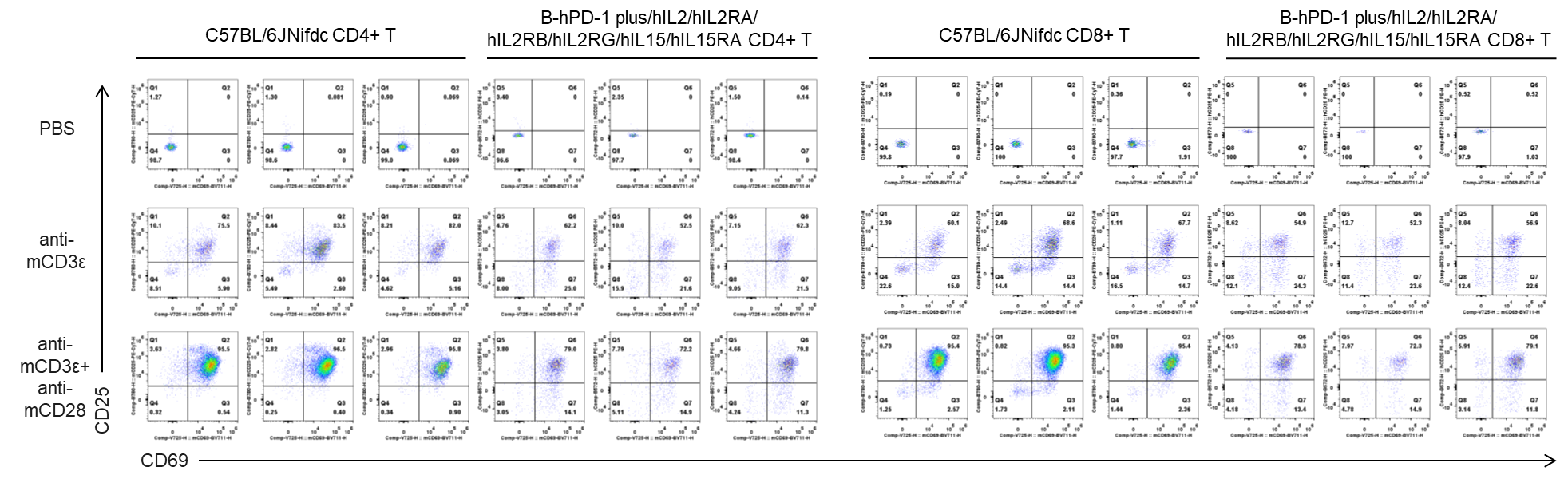
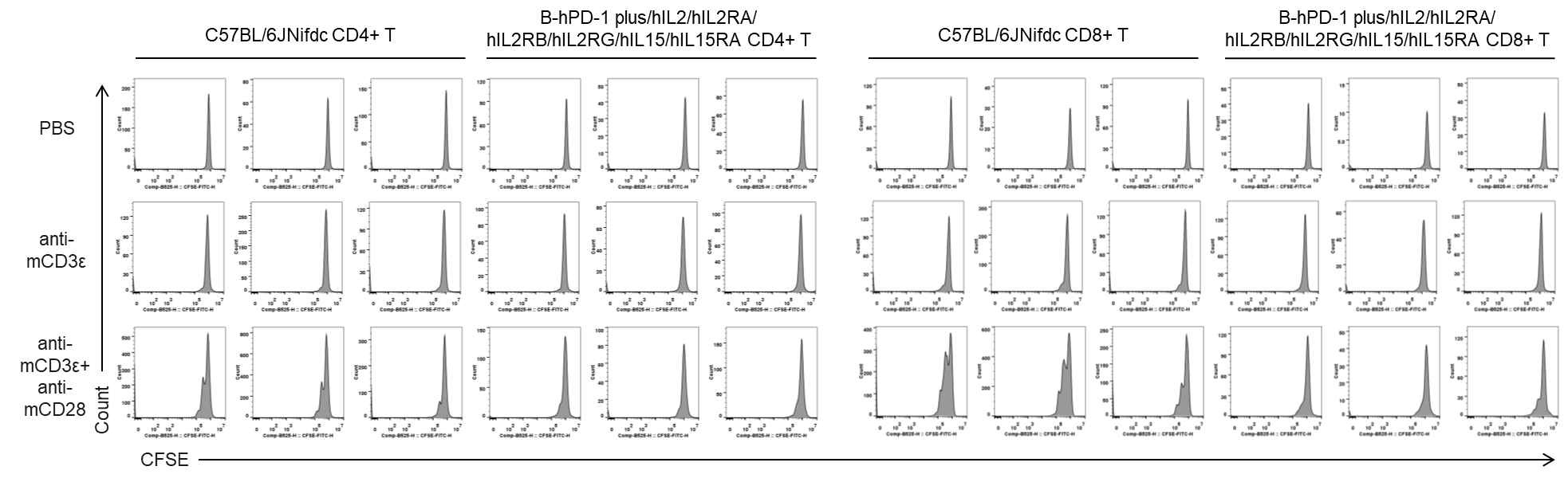
In vitro T cell activation by anti-mCD3ε antibody with or without anti-mCD28 antibody in wild-type C57BL/6JNifdc mice and homozygous humanized B-hPD-1 plus/hIL2/hIL2RA/hIL2RB/hIL2RG/hIL15/hIL15RA mice (48h). T cells were isolated from splenocytes of C57BL/6JNifdc and B-hPD-1 plus/hIL2/hIL2RA/hIL2RB/hIL2RG /hIL15/hIL15RA mice (female,13-week-old, n=3), and incubated in the presence of anti-mCD3ε antibody (2ug/ml, BioXcell, BE0001-2), with or witnout anti-mCD28 antibody (5ug/ml, BioXcell, BE0015-1) for 48h. T cell proliferation was tested by flow cytometry. T cell activation in B-hPD-1 plus/hIL2/hIL2RA/hIL2RB/hIL2RG /hIL15/hIL15RA mice was significantly up-regulated by anti-mCD3ε antibody and anti-mCD28 antibody.
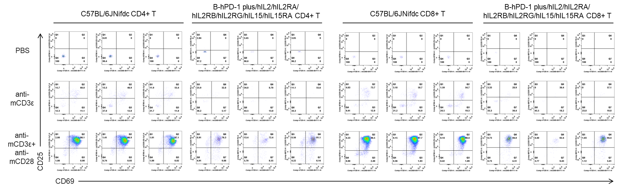
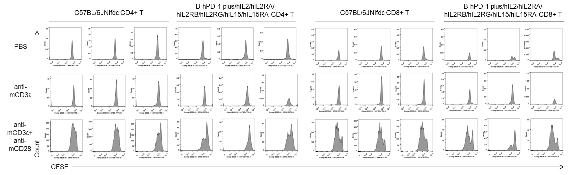
In vitro T cell activation by anti-mCD3ε antibody with or without anti-mCD28 antibody in wild-type C57BL/6JNifdc mice and homozygous humanized B-hPD-1 plus/hIL2/hIL2RA/hIL2RB/hIL2RG/hIL15/hIL15RA mice (72h). T cells were isolated from splenocytes of C57BL/6JNifdc and B-hPD-1 plus/hIL2/hIL2RA/hIL2RB/hIL2RG /hIL15/hIL15RA mice (female,13-week-old, n=3), and incubated in the presence of anti-mCD3ε antibody (2ug/ml, BioXcell, BE0001-2), with or witnout anti-mCD28 antibody (5ug/ml, BioXcell, BE0015-1) for 72h. T cell proliferation was tested by flow cytometry. T cell activation in B-hPD-1 plus/hIL2/hIL2RA/hIL2RB/hIL2RG /hIL15/hIL15RA mice was significantly up-regulated by anti-mCD3ε antibody and anti-mCD28 antibody.
Quantification of T cell proliferation in vitro by anti-CD3ε antibody with or without anti-mCD28 antibody in wild-type C57BL/6JNifdc mice and homozygous humanized B-hPD-1 plus/hIL2/hIL2RA/hIL2RB/hIL2RG/hIL15/hIL15RA mice (24h). T cells were isolated from splenocytes of C57BL/6JNifdc and B-hPD-1 plus/hIL2/hIL2RA/hIL2RB/hIL2RG/hIL15/hIL15RA mice (female,13-week-old, n=3), and incubated in the presence of anti-mCD3ε antibody (2ug/ml, BioXcell, BE0001-2), with or witnout anti-mCD28 antibody (5ug/ml, BioXcell, BE0015-1) for 24h. T cell proliferation was tested by flow cytometry.
Quantification of T cell proliferation in vitro by anti-CD3ε antibody with or without anti-mCD28 antibody in wild-type C57BL/6JNifdc mice and homozygous humanized B-hPD-1 plus/hIL2/hIL2RA/hIL2RB/hIL2RG/hIL15/hIL15RA mice (48h). T cells were isolated from splenocytes of C57BL/6JNifdc and B-hPD-1 plus/hIL2/hIL2RA/hIL2RB/hIL2RG/hIL15/hIL15RA mice (female,13-week-old, n=3), and incubated in the presence of anti-mCD3ε antibody (2ug/ml, BioXcell, BE0001-2), with or witnout anti-mCD28 antibody (5ug/ml, BioXcell, BE0015-1) for 48h. T cell proliferation was tested by flow cytometry.
Quantification of T cell proliferation in vitro by anti-CD3ε antibody with or without anti-mCD28 antibody in wild-type C57BL/6JNifdc mice and homozygous humanized B-hPD-1 plus/hIL2/hIL2RA/hIL2RB/hIL2RG/hIL15/hIL15RA mice (72h). T cells were isolated from splenocytes of C57BL/6JNifdc and B-hPD-1 plus/hIL2/hIL2RA/hIL2RB/hIL2RG/hIL15/hIL15RA mice (female,13-week-old, n=3), and incubated in the presence of anti-mCD3ε antibody (2ug/ml, BioXcell, BE0001-2), with or witnout anti-mCD28 antibody (5ug/ml, BioXcell, BE0015-1) for 72h. T cell proliferation was tested by flow cytometry.
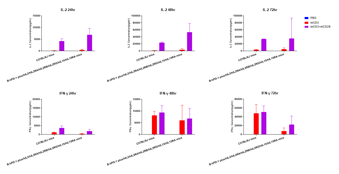
In vitro cytokine production (IFN-γ and IL-2) in B-hPD-1 plus/hIL2/hIL2RA/hIL2RB/hIL2RG/hIL15/hIL15RA mice. T cells (2×10⁵) were isolated from the splenocytes of C57BL/6JNifdc and B-hPD-1 plus/hIL2/hIL2RA/hIL2RB/hIL2RG/hIL15/hIL15RA mice (female, 13-week-old, n=3), incubated in the presence of anti-mouse CD3ε antibody (BioXCell, BE0001-1, clone 145-2C11, 2ug/ml) and anti-mCD28 antibody (BioXCell, BE0015-1, clone 37.51, 5ug/ml) for 24h, 48h and 72h. IFN-γ and IL-2 productions were then tested using ELISA method.
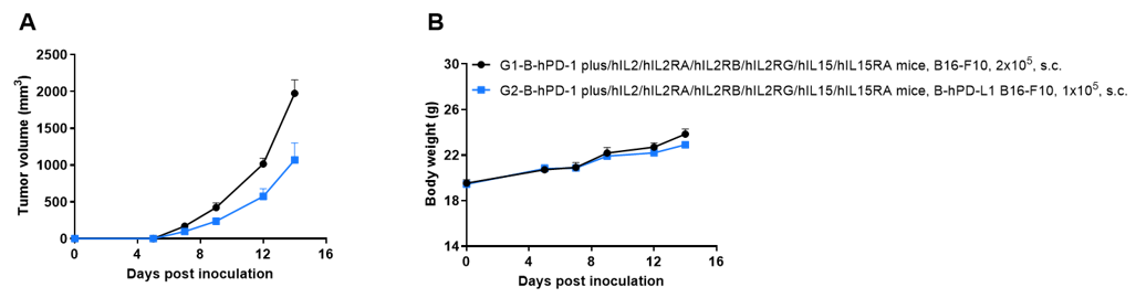
Subcutaneous tumor growth of B-hPD-L1 B16-F10 cells. B-hPD-L1 B16-F10 cells (1×10⁵) and wild-type B16-F10 cells (2×10⁵) were subcutaneously implanted into B-hPD-1 plus/hIL2/hIL2RA/hIL2RB/hIL2RG/hIL15/hIL15RA mice (female, 7-week-old, n=6). Tumor volume and body weight were measured three times a week. (A) Average tumor volume. (B) Body weight. Volume was expressed in mm3 using the formula: V=0.5 × long diameter × short diameter2. Results indicate that B-hPD-L1 B16-F10 cells were able to establish tumors in vivo and can be used for efficacy studies. Values are expressed as mean ± SEM.
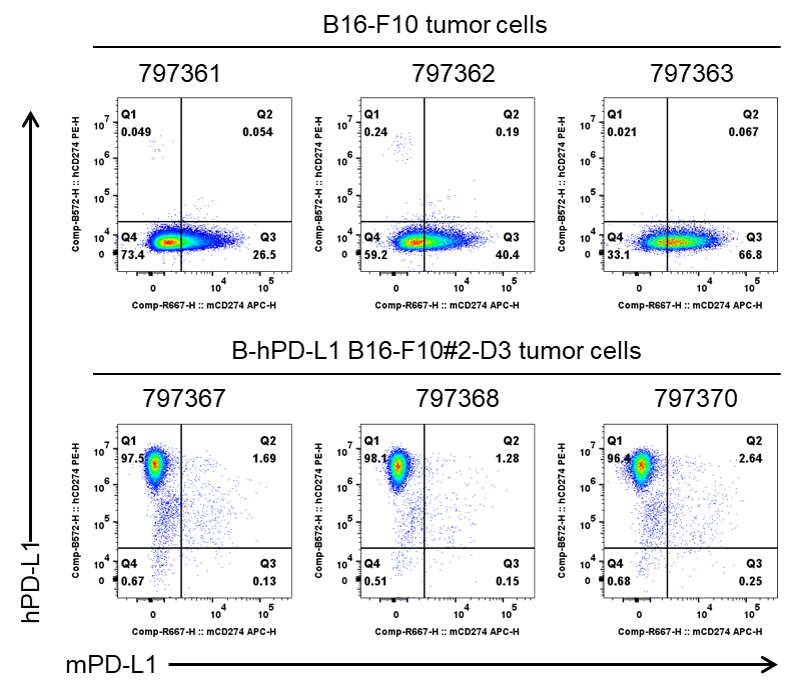
PD-L1 expression evaluated on B-hPD-L1 B16-F10 tumor cells by flow cytometry. B-hPD-L1 B16-F10 cells were subcutaneously transplanted into B-hPD-1 plus/hIL2/hIL2RA/hIL2RB/hIL2RG/hIL15/hIL15RA mice (female, 7-week-old, n=6). At the end of the experiment, tumor cells were harvested and assessed for mouse PD-L1(BioLegend, 124312) and human PD-L1(BioLegend, 329706) expression by flow cytometry. As shown, human PD-L1 was highly expressed on the surface of tumor cells. Therefore, B-hPD-L1 B16-F10 cells can be used for in vivo efficacy studies evaluating novel PD-L1 therapeutics.
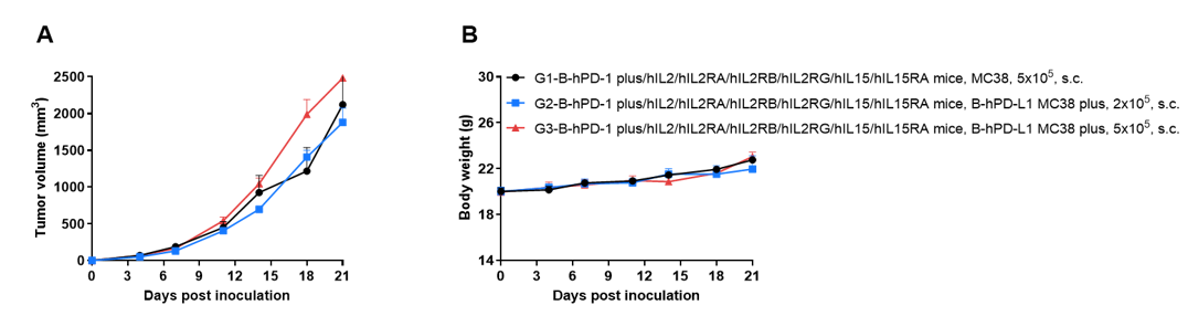
Subcutaneous tumor growth of B-hPD-L1 MC38 plus cells. B-hPD-L1 MC38 cells (2×10⁵ and 5×10⁵) and wild-type MC38 cells (5×10⁵) were subcutaneously implanted into B-hPD-1 plus/hIL2/hIL2RA/hIL2RB/hIL2RG/hIL15/hIL15RA mice (female, 9-week-old, n=6). Tumor volume and body weight were measured three times a week. (A) Average tumor volume. (B) Body weight. Volume was expressed in mm3 using the formula: V=0.5 × long diameter × short diameter2. Results indicate that B-hPD-L1 MC38 plus cells were able to establish tumors in vivo and can be used for efficacy studies. Values are expressed as mean ± SEM.
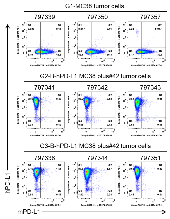
PD-L1 expression evaluated on B-hPD-L1 MC38 plus tumor cells by flow cytometry. B-hPD-L1 MC38 plus cells were subcutaneously transplanted into B-hPD-1 plus/hIL2/hIL2RA/hIL2RB/hIL2RG/hIL15/hIL15RA mice (female, 9-week-old, n=6). At the end of the experiment, tumor cells were harvested and assessed for mouse PD-L1(BioLegend, 124312) and human PD-L1(BioLegend, 329706) expression by flow cytometry. As shown, human PD-L1 was highly expressed on the surface of tumor cells. Therefore, B-hPD-L1 MC38 plus cells can be used for in vivo efficacy studies evaluating novel PD-L1 therapeutics.