B-hPD-L1/hIFNAR1/hIFNAR2 mice

C57BL/6-Cd274tm1(CD274)Bcgen Ifnar1tm1(IFNAR1)Bcgen Ifnar2tm1(IFNAR2)Bcgen/Bcgen • 114101

B-hPD-L1/hIFNAR1/hIFNAR2 mice

Catalog Number: 114101
Strain Name: C57BL/6-Cd274tm1(CD274)Bcgen Ifnar1tm1(IFNAR1)Bcgen Ifnar2tm1(IFNAR2)Bcgen/Bcgen
Strain Background: C57BL/6
NCBI gene ID: 29126,3454,3455 (Human)
Aliases: ADMIO5, B7-H, B7H1, PD-L1, PDCD1L1, PDCD1LG1, PDL1, hPD-L1; AVP, CRF2-1, IFN-R-1, IFN-alpha-REC, IFNAR, IFNBR, IFRC, IMD106; IFN-R, IFN-R-2, IFN-alpha-REC, IFNABR, IFNARB, IMD45
---
可提供授权方案

B-hPD-L1/hIFNAR1/hIFNAR2 mice

在此页面上

  • Description
  • Targeting strategy
  • Phenotypic analysis
  • Efficacy

海报

查看全部

    发表文章

      Description

      IFNAR1/IFNAR2: Key receptors to drive the transcription of interferon-stimulated genes (ISGs) for antiviral, antitumor, and immunomodulatory responses

      • Gene Information: Interferon alpha and beta receptor subunit 1 (IFNAR1) and interferon alpha and beta receptor subunit 2 (IFNAR2) are protein-coding genes located on chromosome 21q22.11. They encode type I membrane proteins that form two chains of a receptor for interferons alpha and beta.
      • Protein Expression: Both IFNAR1 and IFNAR2 are expressed on almost all nucleated cells, with notably high density found on immune cell populations such as T cells, B cells, NK cells, macrophages, and dendritic cells.
      •  Signaling Pathway: Upon binding type I interferons, IFNAR1 and IFNAR2 heterodimerize, activating the JAK1/Tyk2-dependent STAT1/STAT2 signaling pathway to drive the transcription of interferon-stimulated genes (ISGs) for antiviral, antitumor, and immunomodulatory responses.
      • Therapeutic Inhibition: By binding to the IFNAR1 subunit, anifrolumab prevents the formation of the active ternary complex with IFNAR2 and the interferon ligand, blocking all Type I IFN signaling.

      PD-1/hPD-L1: An immune-checkpoint and tumor-associated target for therapeuticintervention

      • Gene Information: PD-1 (PDCD1/CD279) is an inhibitory immune checkpointreceptor. PD-L1 (CD274/B7 H1) is a key PD-1 ligand that mediates immunesuppression in tumors and antigen presenting cell contexts.
      • Protein Expression: PD-1 is induced after antigen specific T cell stimulation and isoften sustained during chronic activation. PD-L1 can be constitutively expressed bytumor cells or induced by IFN γ and other inflammatory cues.
      • Signaling Pathway: PD-L1/PD-1 interaction initiates inhibitory signaling, includingdephosphorylation events that suppress TCR downstream signaling. This reduces Tcell activation, cytotoxicity, and tumor cell killing.
      • Therapeutic Inhibition: Anti PD-1 or anti PD-L1 monoclonal antibodies block theinhibitory interaction and restore cytotoxic T cell activity. B-hPD 1/hPD-L1 miceprovide an in vivo platform for evaluating PD-1/PD-L1 checkpoint blockade andcombination therapies.
      Targeting Strategy

      PD-L1

      • The exon 3 of mouse PD-L1 gene that encodes the IgV domain was replaced by human PD-L1 exon 3 in B- hPD-L1/hIFNAR1/hIFNAR2 mice.
      • The endogenous mouse promoter, 5′ UTR, and 3′ UTR regions are retained, allowing human PD-L1 expression to be driven by the native mouse Pd l1 promoter, while endogenous mouse Pd l1 transcription and translation are abolished.

      IFNAR1

      •  The exons 2-9 of mouse Ifnar1 gene that encode the extracellularregion were replaced by human IFNAR1 exons 2-9 in B-hPD-L1/hIFNAR1/hIFNAR2 mice.
      •  The endogenous mouse promoter, 5′ UTR, and 3′ UTR regions are retained, allowing human IFNAR1 expression to be driven by the native mouse Ifnar1 promoter, while endogenous mouse Ifnar1 transcription and translation are abolished.

      IFNAR2

      •  The chimeric CDS consists of human IFNAR2 extracellular domain and mouse Ifnar2 intracellular region was inserted into mouse ifnar2 gene locus in B-hPDL1/hIFNAR1/hIFNAR2 mice.
      •  The endogenous mouse promoter, 5′ UTR, and 3′ UTR regions are retained, allowing human IFNAR2 expression to be driven by the native mouse Ifnar2 promoter, while endogenous mouse Ifnar2 transcription and translation are abolished.
      IFNAR2 Protein Expression in Spleen

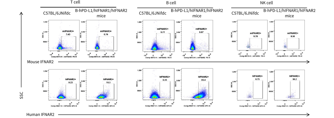
      Mouse and human IFNAR2 expression analysis in wild-type C57BL/6JNifdc and B-hPD-L1/hIFNAR1/hIFNAR2 mice by flow cytometry. Splenocytes were collected from wild type C57BL/6JNifdc and homozygous B-hPD L1/hIFNAR1/hIFNAR2 mice. Protein expression was analyzed with anti mouse IFNAR2 antibody (R&D, FAB1083P) and anti human IFNAR2 antibody (Miltenyi Biotec, 130 099 560) by flow cytometry. Mouse IFNAR2 was detectable exclusively in T cells, B cells, and NK cells of wild type mice. Human IFNAR2 was detectable in T cells, B cells, and NK cells of B-hPD L1/hIFNAR1/hIFNAR2 mice but not in wild type mice.

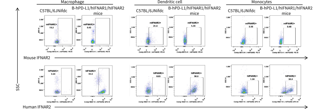
      Mouse and human IFNAR2 expression analysis in wild-type C57BL/6JNifdc and B-hPD-L1/hIFNAR1/hIFNAR2 mice by flow cytometry. Splenocytes were collected from wild type C57BL/6JNifdc and homozygous B-hPD L1/hIFNAR1/hIFNAR2 mice. Protein expression was analyzed with anti mouse IFNAR2 antibody (R&D, FAB1083P) and anti human IFNAR2 antibody (Miltenyi Biotec, 130 099 560) by flow cytometry. Mouse IFNAR2 was detectable exclusively in macrophages, monocytes, and dendritic cell of wild type mice. Human IFNAR2 was detectable in macrophages, monocytes, and dendritic cells of B-hPD-L1/hIFNAR1/hIFNAR2 mice but not in wild type mice.

      In Vivo Efficacy of Anti-Human PD-L1 Antibody and Human IFNα Protein in a Tumor Model

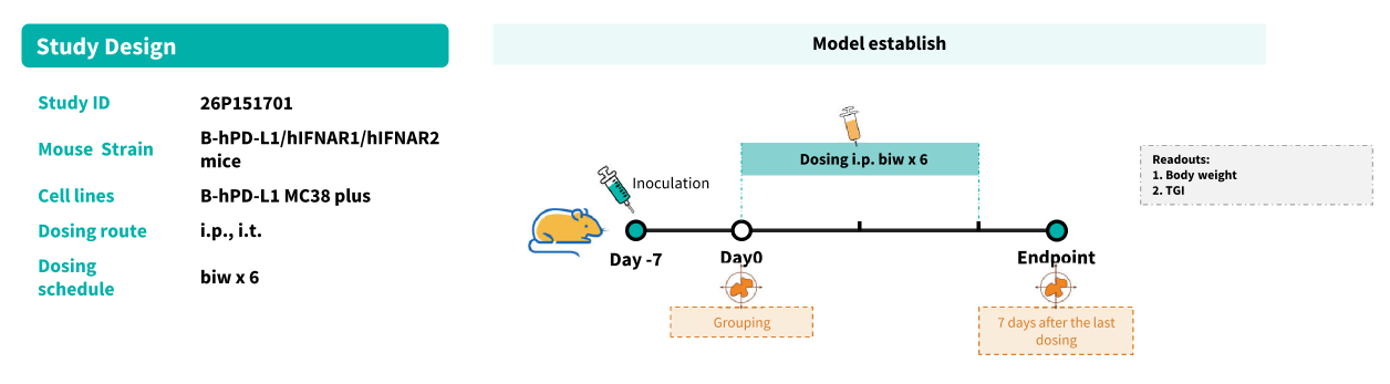
      Establishment of a B-hPD-L1 MC38 plus cell model and in vivo efficacy study of an anti-human PD-L1-specific antibody and human IFNα protein. B-hPDL1-MC38 plus cells were implanted subcutaneously into homozygous B-hPD L1/hIFNAR1/hIFNAR2 mice (female, 8 weeks old, n = 20). When the average tumor volume reached approximately 150 mm³, mice were randomized and subsequently administered the anti human PD-L1 antibody via intraperitoneal injection and human IFNα protein via intratumoral injection..

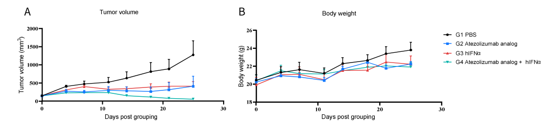
      Antitumor activity of anti-human PD-L1 antibody and human IFNα protein in B-hPD-L1/hIFNAR1/hIFNAR2 mice.
      (A) Anti human PD-L1 antibody (Atezolizumab analog, in-house) and human IFNα protein (Peginterferon alfa-2b, bought from Xiamen Amoytop Biotech Co., Ltd. )inhibited B-hPD-L1-MC38 plus tumor growth in B-hPD-L1/hIFNAR1/hIFNAR2 mice. (B) Body weight changes during treatment. As shown in figure A, single use of anti human PD-L1 antibody or human IFNα protein were efficacious in controlling tumor growth in B hPD L1/hIFNAR1/hIFNAR2 mice, and combination use of antihuman PD-L1 antibody and human IFNα protein showed better efficacy, demonstrating that the B hPD L1/hIFNAR1/hIFNAR2 mice provide a powerful preclinical model for in vivo evaluation of anti human PD-L1 antibody and human IFNα protein. Values are expressed as mean ± SEM.

      * When publishing results obtained using this animal model, please acknowledge the source as follows: The animal model [B-hPD-L1/hIFNAR1/hIFNAR2 mice] (Cat# 114101) was purchased from Biocytogen.