B-hTL1A/hDR3/hTNFA/hTNFR1/hTNFR2 mice

C57BL/6-Tnfsf15tm2(TNFSF15)Bcgen Tnfrsf25tm3(TNFRSF25)Bcgen Tnftm1(TNF)Bcgen Tnfrsf1atm1(TNFRSF1A)Bcgen Tnfrsf1btm1(TNFRSF1B)Bcgen/Bcgen • 114013

B-hTL1A/hDR3/hTNFA/hTNFR1/hTNFR2 mice

Catalog Number: 114013
Strain Name: C57BL/6-Tnfsf15tm2(TNFSF15)Bcgen Tnfrsf25tm3(TNFRSF25)Bcgen Tnftm1(TNF)Bcgen Tnfrsf1atm1(TNFRSF1A)Bcgen Tnfrsf1btm1(TNFRSF1B)Bcgen/Bcgen
Strain Background: C57BL/6
NCBI gene ID: 9966,8718,7124,7132,7133 (Human)
Aliases: TL1; TL1A; VEGI; TNLG1B; VEGI192A; DR3; TR3; DDR3; LARD; APO-3; TRAMP; WSL-1; WSL-LR; TNFRSF12; DIF; TNFA; IMD127; TNFSF2; TNLG1F; TNF-alpha; FPF; p55; p60; TBP1; TNF-R; TNFAR; TNFR1; p55-R; CD120a; TNFR55; TNFR60; TNF-R-I; TNF-R55; p75; TBPII; TNFBR; TNFR2; CD120b; TNFR1B; TNFR80; TNF-R75; p75TNFR; TNF-R-II
---
可提供授权方案
B-hTL1A/hDR3/hTNFA/hTNFR1/hTNFR2 mice

在此页面上

  • Description
  • Targeting strategy
  • Phenotypic analysis
  • Efficacy

海报

查看全部

    发表文章

      Description

      TL1A: A key inflammation cytokine in chronic intestinal inflammation and fibrosis-related diseases

      • Gene Information: TNF Superfamily Member 15 (TNFSF15, also known as TL1A) is a protein-coding gene located on chromosome 9q32. This cytokine is a ligand for receptor TNFRSF25 (also known as DR3) and  TNFRSF6B (also known as DcR3).
      • Protein Expression: TL1A is expressed in various immune cells (such as monocytes, macrophages, dendritic cells, and T cells) as well as in non-immune cells (such as synovial fibroblasts and endothelial cells). TL1A is a type II transmembrane protein that exists in either membrane-bound (mTL1A) or soluble (sTL1A) forms.
      • Signaling Pathway: TL1A competes with Death Receptor 3 (DR3) for binding, providing stimulus signals for downstream signaling pathways, thereby regulating the proliferation, activation, apoptosis of effector cells, and the production of cytokines and chemokines.
      • Therapeutic Inhibition: Blocking the interaction between TL1A and DR3 can reduce the severity of autoimmune diseases, such as the IBD model.

      TNFA: A master pathway of regulating inflammation, immune responses, and cell survival

      • Gene Information: Tumor necrosis factor (TNF) is a protein-coding gene located on chromosome 6p21.33. It encodes a multifunctional proinflammatory cytokine that belongs to the tumor necrosis factor (TNF) superfamily. TNF receptor superfamily member 1A (TNFRSF1A, TNFR1) encodes a member of the TNF receptor superfamily of proteins located on chromosome 12p13.31. TNF receptor superfamily member 1B (TNFRSF1B, TNFR2) encodes a member of the TNF receptor superfamily of proteins located on chromosome 1p36.22.
      • Protein Expression: TNF is primarily expressed by macrophages. TNF is synthesized as a membrane-bound precursor and cleaved into a soluble circulating protein. TNFR1 is expressed on almost all nucleated cells. TNFR2 is expressed on CD4⁺ T cells, CD8⁺ T cells, Tregs, endothelial cells, microglia, specific neuronal subpopulations, oligodendrocytes, cardiomyocytes, and human mesenchymal stem cells.
      • Signaling Pathway: TNF exerts its effects primarily through two receptors: TNFR1 and TNFR2. TNFR1 contains an intracellular "death domain" that can drive either cell survival and inflammation via NF-κB activation, or programmed cell death via caspase cascades (apoptosis/necroptosis). TNFR2 lacks a death domain and preferentially recruits TRAF proteins to directly promote tissue regeneration, cell survival, and localized immune suppression.
      • Therapeutic Inhibition: By blocking a central driver of the immune system's inflammatory response, adalimumab inhibits downstream inflammation and improves clinical outcomes, including reducing other inflammatory markers and reducing tissue damage in various autoimmune conditions.
      Targeting strategy

      TL1A

      • The exons 1-4 of mouse Tl1a gene that encode extracellular domain were replaced by human counterparts in B-hTL1A/hDR3/hTNFA/hTNFR1/hTNFR2 mice.
      • The genomic region of mouse Tl1a gene that encodes transmembrane domain and cytoplasmic portion was retained. The promoter, 5’UTR and 3’UTR region of the mouse gene were also retained. The TL1A expression was driven by endogenous mouse Tl1a promoter, while mouse Tl1a gene transcription and translation will be disrupted.

      DR3

      • The exons 1-10 of mouse DR3 gene that encode the whole molecule (ATG to STOP codon), including promoter, 5’UTR and 3’UTR were replaced by human counterparts in B-hTL1A/hDR3/hTNFA/hTNFR1/hTNFR2 mice.
      • The human DR3 expression was driven by human DR3 promoter, while mouse DR3 gene transcription and translation will be disrupted.

      TNFA

      • The targeted mouse Tnfa whole genomic sequences including 5'UTR, 3'UTR and coding region were replaced by human TNFA whole genomic sequences in B-hTL1A/hDR3/hTNFA/hTNFR1/hTNFR2 mice.
      • The human TNFA expression is driven by human TNFA promoter, while mouse Tnfa gene transcription and translation will be disrupted.

      TNFR1

      • The exons 2-6 of mouse Tnfr1 gene that encode extracellular domain are replaced by human counterparts in B-hTL1A/hDR3/hTNFA/hTNFR1/hTNFR2 mice. The genomic region of mouse Tnfr1 gene that encodes transmembdomain and cytoplasmic portion is retained. The promoter, 5’UTR and 3’UTR region of the mouse gene are also retained.
      • The human TNFR1 expression is driven by endogenous mouse Tnfr1 promoter, while mouse Tnfr1 gene transcription and translation will be disrupted.

      TNFR2

      • The exons 2-6 of mouse Tnfr2 gene that encode extracellular domain are replaced by human counterparts in B-hTL1A/hDR3/hTNFA/hTNFR1/hTNFR2 mice. The genomic region of mouse Tnfr2 gene that encodes transmembrane domain and cytoplasmic portion is retained. The promoter, 5’UTR and 3’UTR region of the mouse gene are also retained.
      • The human TNFR2 expression is driven by endogenous mouse Tnfr2 promoter, while mouse Tnfr2 gene transcription and translation will be disrupted.
      Soluble TL1A Protein Expression Analysis in BMDC Supernatants
      • Soluble human TL1A was exclusively detectable in homozygous B-hTL1A/hDR3/hTNFA/hTNFR1/hTNFR2 mice but not wild-type C57BL/6JNifdc mice.

      Soluble TL1A expression analysis in B-hTL1A/hDR3/hTNFA/hTNFR1/hTNFR2 mice by ELISA. Bone marrow derived dendritic cells (BMDCs) were produced by culturing the bone marrow from wild-type C57BL/6JNIfdc mice (+/+) and homozygous B-hTL1A/hDR3/hTNFA/hTNFR1/hTNFR2 mice (H/H;H/H;H/H;H/H;H/H), which were stimulated with LPS in vitro. After stimulation, the supernatants were collected and the levels of soluble TL1A were measured using the species-specific human TL1A ELISA kit. Soluble human TL1A was exclusively detectable in homozygous B-hTL1A/hDR3/hTNFA/hTNFR1/hTNFR2 mice but not wild-type C57BL/6JNifdc mice. Values are expressed as mean ± SEM. ND: not detectable.
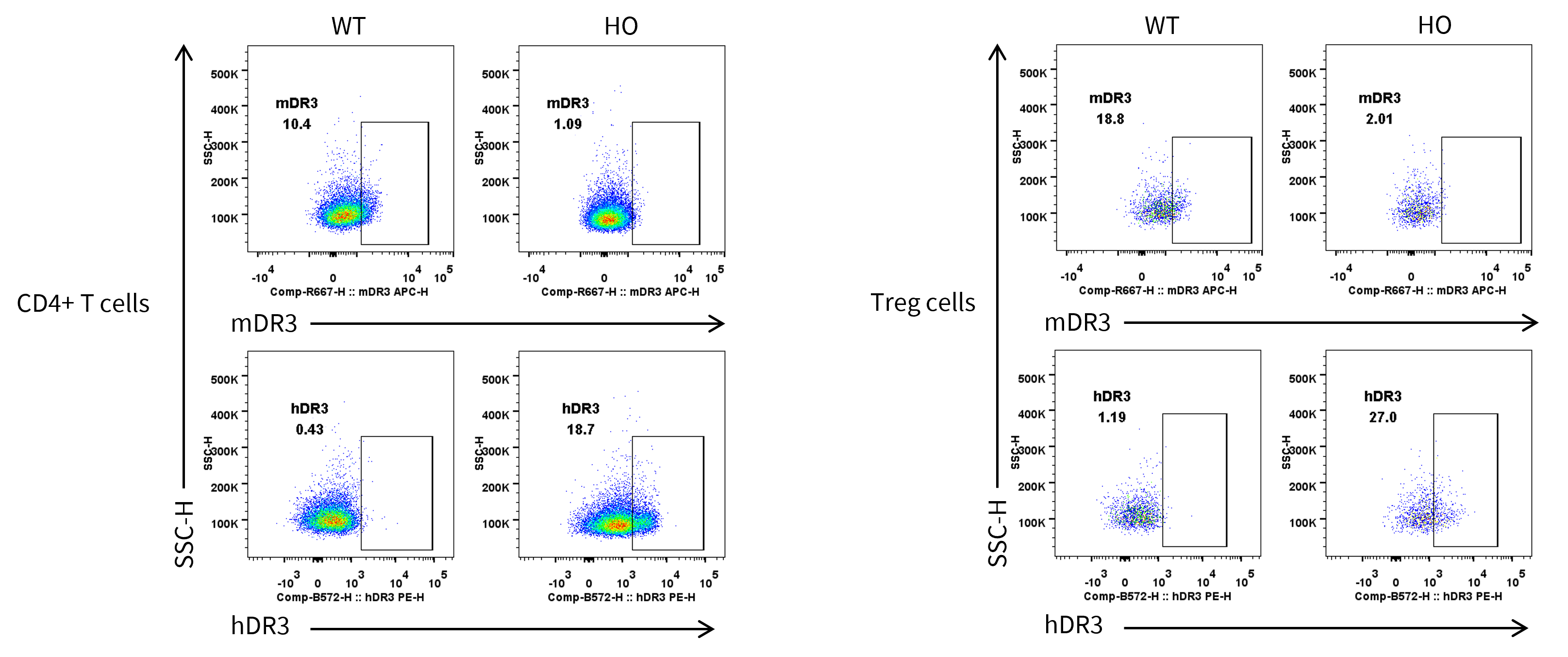
      DR3 Protein Expression Analysis in Spleen
      • Human DR3 was detectable in CD4+ T cells and Treg cells of homozygous B-hTL1A/hDR3/hTNFA/hTNFR1/hTNFR2 mice.

      Strain specific DR3 expression analysis in homozygous B-hTL1A/hDR3/hTNFA/hTNFR1/hTNFR2 mice by flow cytometry. Splenocytes were collected from wild-type C57BL/6JNifdc mice (+/+) and homozygous B-hTL1A/hDR3/hTNFA/hTNFR1/hTNFR2 mice (H/H;H/H;H/H;H/H;H/H), protein expression was analyzed with anti-mouse DR3 antibody (Biolegend, 144407) and anti-human DR3 antibody (Biolegend, 307105) by flow cytometry. Mouse DR3 was detectable in CD4+ T cells and Treg cells of wild-type C57BL/6JNifdc mice, human DR3 was detectable in CD4+ T cells and Treg cells of homozygous B-hTL1A/hDR3/hTNFA/hTNFR1/hTNFR2 mice. WT: C57BL/6JNifdc, HO: B-hTL1A/hDR3/hTNFA/hTNFR1/hTNFR2 mice.

      TNFA Protein Expression Analysis in Serum
      • Mouse TNFA was only detectable in wild-type C57BL/6JNifdc mice, and human TNFA was only detectable in homozygous B-hTL1A/hDR3/hTNFA/hTNFR1/hTNFR2 mice.

      Strain specific TNFA expression analysis in wild-type C57BL/6JNifdc mice and homozygous humanized B-hTL1A/hDR3/hTNFA/hTNFR1/hTNFR2 mice by ELISA. Serum were collected from wild-type C57BL/6JNifdc mice (+/+) and homozygous B-hTL1A/hDR3/hTNFA/hTNFR1/hTNFR2 mice (H/H;H/H;H/H;H/H;H/H), the production of TNFA in serum were assessed after 2 h of stimulation with LPS in vivo. Expression levels of mouse and human TNFA were analyzed by ELISA (Mouse TNFA ELISA Kit: Biolegend, 430904; Human TNFA ELISA Kit: Biolegend, 430204). Values are expressed as mean ± SEM.
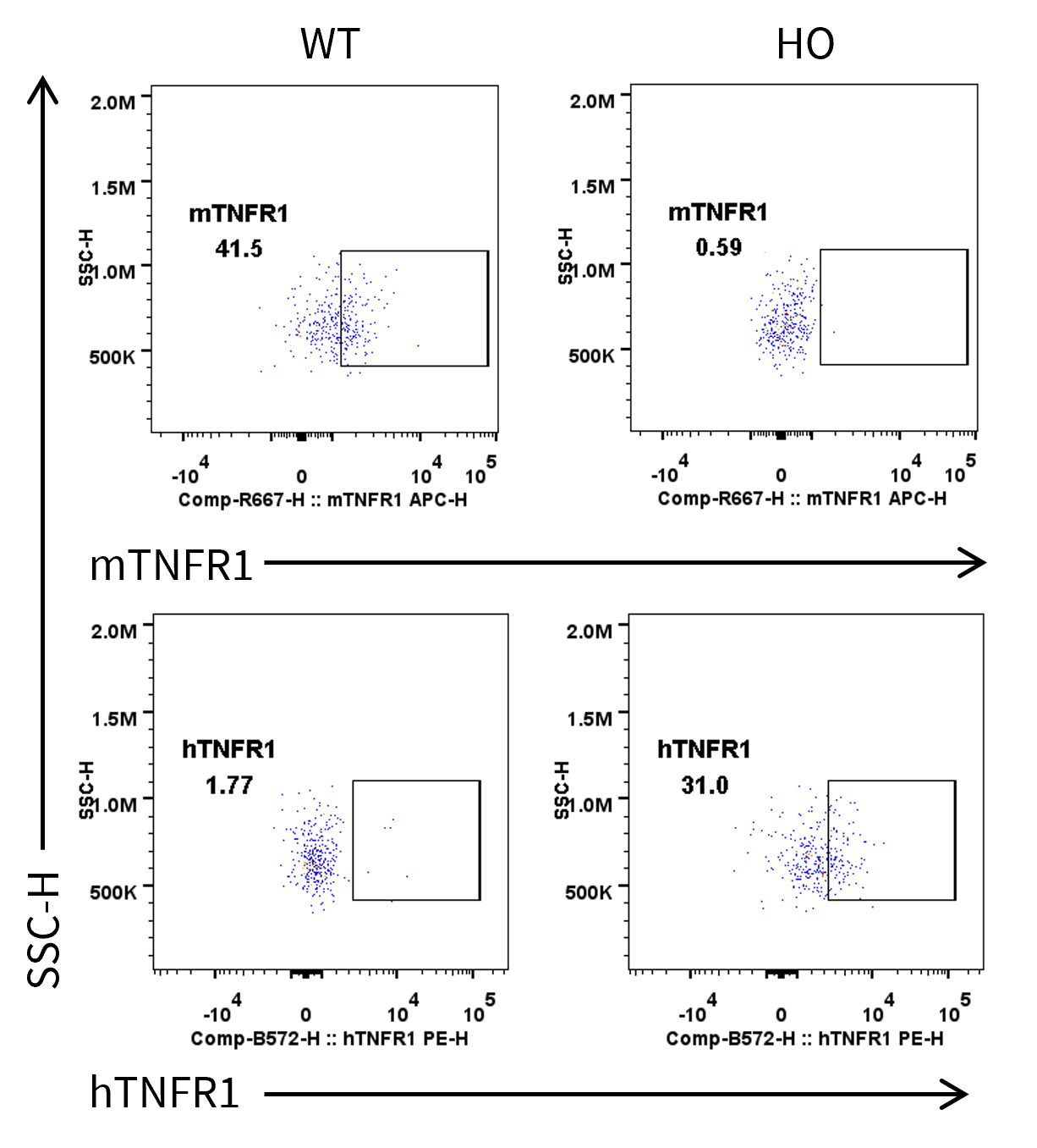
      TNFR1 Protein Expression in Spleen
      • Human TNFR1 was exclusively expressed in neutrophils of B-hTL1A/hDR3/hTNFA/hTNFR1/hTNFR2 mice.

      Strain specific TNFR1 expression analysis in homozygous B-hTL1A/hDR3/hTNFA/hTNFR1/hTNFR2 mice by flow cytometry. Splenocytes were collected from wild-type C57BL/6JNifdc mice (+/+) and homozygous B-hTL1A/hDR3/hTNFA/hTNFR1/hTNFR2 mice (H/H;H/H;H/H;H/H;H/H), and analyzed by flow cytometry with species-specific anti-TNFR1 antibody. Mouse TNFR1 was detectable in neutrophils of C57BL/6JNifdc mice. Human TNFR1 was exclusively detectable in neutrophils of homozygous B-hTL1A/hDR3/hTNFA/hTNFR1/hTNFR2 mice but not in C57BL/6JNifdc mice. WT: C57BL/6JNifdc, HO: B-hTL1A/hDR3/hTNFA/hTNFR1/hTNFR2 mice.
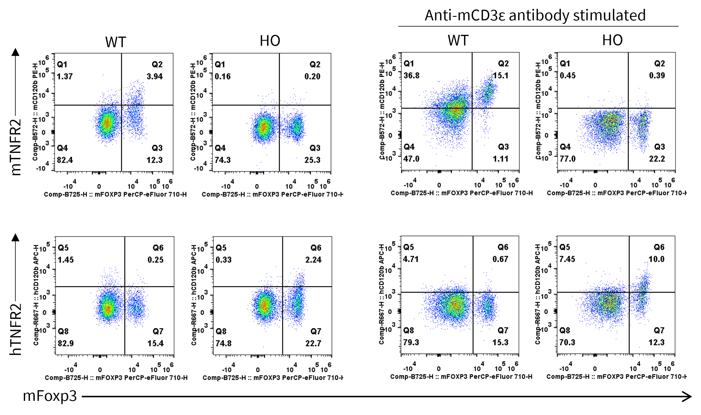
      TNFR2 Protein Expression in Spleen
      • Human TNFR2 was exclusively expressed in Treg cells of B-hTL1A/hDR3/hTNFA/hTNFR1/hTNFR2 mice.

      Strain specific TNFR2 expression analysis in homozygous B-hTL1A/hDR3/hTNFA/hTNFR1/hTNFR2 by flow cytometry. Splenocytes were collected from wild-type C57BL/6JNifdc mice (+/+) and homozygous B-hTL1A/hDR3/hTNFA/hTNFR1/hTNFR2 mice (H/H;H/H;H/H;H/H;H/H), stimulated with anti-mCD3ε antibody in vivo or not, and analyzed by flow cytometry with species-specific anti-TNFR2 antibody. Mouse TNFR2 was detectable in Treg cells of C57BL/6JNifdc mice. Human TNFR1 was exclusively detectable in Treg cells of homozygous B-hTL1A/hDR3/hTNFA/hTNFR1/hTNFR2 mice. WT: C57BL/6JNifdc, HO: B-hTL1A/hDR3/hTNFA/hTNFR1/hTNFR2 mice.

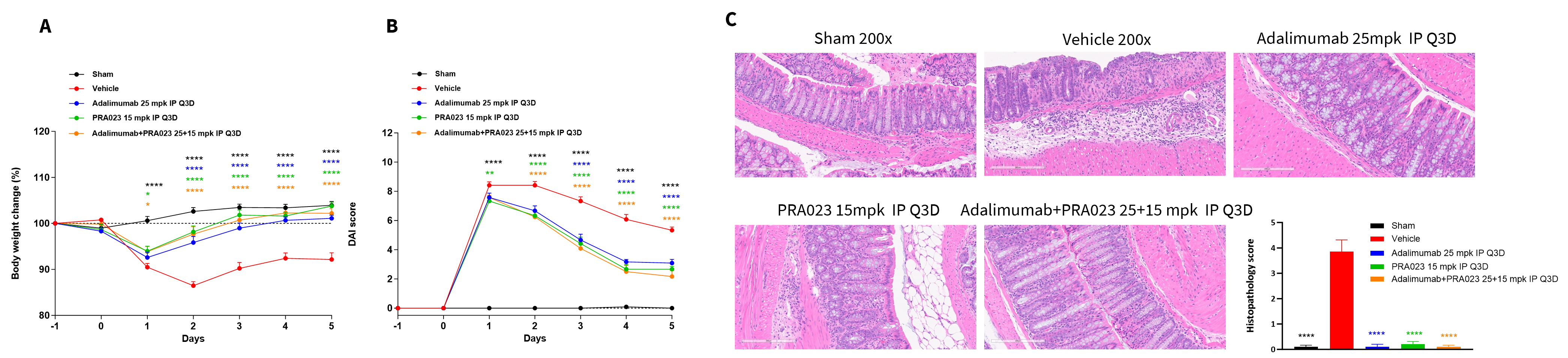
      In Vivo Efficacy of Anti-Human TL1A Antibody and Anti-Human TNFA Antibody in a TNBS Induced Acute Colitis
      • PRA023 and Adalimumab treatment efficiently improved TNBS-induced acute colitis.

      The therapeutic efficacy of anti-human TL1A antibody and anti-human TNFA antibody on the TNBS-induced acute colitis model in B-hTL1A/hDR3/hTNFA/hTNFR1/hTNFR2 mice. TNBS solution was instilled into the colon lumen of B-hTL1A/hDR3/hTNFA/hTNFR1/hTNFR2 mice. The control group (Sham) received intrarectal injections of 50% ethanol. The treatment groups received anti-human TNFA antibody Adalimumab (25 mpk, provided by WuXi AppTec), anti-human TL1A antibody PRA023 (15 mpk, provided by WuXi AppTec) alone or in combination. Body weight and DAI score were recorded daily. (A) Body weight change. (B) DAI score. (C) Pathological score. A TNBS-induced acute colitis model was established in B-hTL1A/hDR3/hTNFA/hTNFR1/hTNFR2 mice. Administration of anti-human TNFA antibody and anti-human TL1A antibody effectively improved TNBS-induced acute colitis. The results indicate that B-hTL1A/hDR3/hTNFA/hTNFR1/hTNFR2 mice are a powerful tool for evaluating in vivo efficacy of anti-human TNFα antibody and anti-human TL1A antibody. Values are expressed as mean ± SEM. *p<0.05, **p<0.01, ***p<0.001, ****p<0.0001, versus Vehicle, ANOVA.

      Note: This experiment was conducted by WuXi AppTec using B-hTL1A/hDR3/hTNFA/hTNFR1/hTNFR2 mice.

      * When publishing results obtained using this animal model, please acknowledge the source as follows: The animal model [B-hTL1A/hDR3/hTNFA/hTNFR1/hTNFR2 mice] (Cat# 114013) was purchased from Biocytogen.