B-hTNFA mice

C57BL/6-Tnftm1(TNF)Bcgen/Bcgen • 110002

B-hTNFA mice

Catalog Number: 110002
Strain Name: C57BL/6-Tnftm1(TNF)Bcgen/Bcgen
Strain Background: C57BL/6
NCBI gene ID: 7124 (Human)
Aliases: DIF; TNFA; IMD127; TNFSF2; TNLG1F; TNF-alpha
---
可提供授权方案
B-hTNFA mice

在此页面上

  • Description
  • Targeting strategy
  • Phenotypic analysis
  • Efficacy

海报

查看全部

    发表文章

      Description

      TNF: A master regulator of inflammation, immune responses, and cell survival

      • Gene Information: Tumor necrosis factor (TNF) is a protein-coding gene located on chromosome 6p21.33. It encodes a multifunctional proinflammatory cytokine that belongs to the tumor necrosis factor (TNF) superfamily.
      • Protein Expression: TNF is primarily expressed by macrophages. TNF is synthesized as a membrane-bound precursor and cleaved into a soluble circulating protein.
      • Signaling Pathway: TNF exerts its effects primarily through two receptors: TNFR1 and TNFR2.
      • Therapeutic Inhibition: By blocking a central driver of the immune system's inflammatory response, adalimumab inhibits downstream inflammation and improves clinical outcomes, including reducing other inflammatory markers and reducing tissue damage in various autoimmune conditions.
      Targeting strategy

      TNFA

      • The exons 1-4 of mouse Tnfa gene that encode signal peptide, extracellular domain, transmembrane domain and cytoplasmic region were replaced by human counterparts in B-hTNFA mice.
      • The promoter, 5’UTR and 3’UTR region of the mouse gene were replaced by human counterparts.
      • The human TNFA expression was driven by human TNFA promoter, while mouse Tnfa gene was knocked out.
      TNFA Protein Expression Analysis
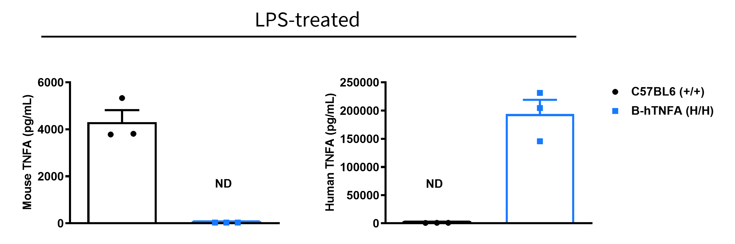
      • Mouse TNFA was detected exclusively in wild-type C57BL/6 mice.
      • Human TNFA was detected in homozygous B-hTNFA mice, but not in wild-type mice.

      Strain specific TNFA expression analysis in wild-type C57BL/6 mice and homozygous humanized B-hTNFA mice by ELISA. Serum was collected from wild-type C57BL/6 mice (+/+) and homozygous B-hTNFA mice (H/H) stimulated with LPS (20 μg/mice) in vivo for 1 hour (female, 8-week-old, n=3). Expression level of TNFA were analyzed by ELISA. Mouse TNFA was only detectable in wild-type C57BL/6 mice. Human TNFA was exclusively detectable in homozygous B-hTNFA mice. Values are expressed as mean ± SEM. ND: not detectable.
      Analysis of Leukocyte Subpopulations
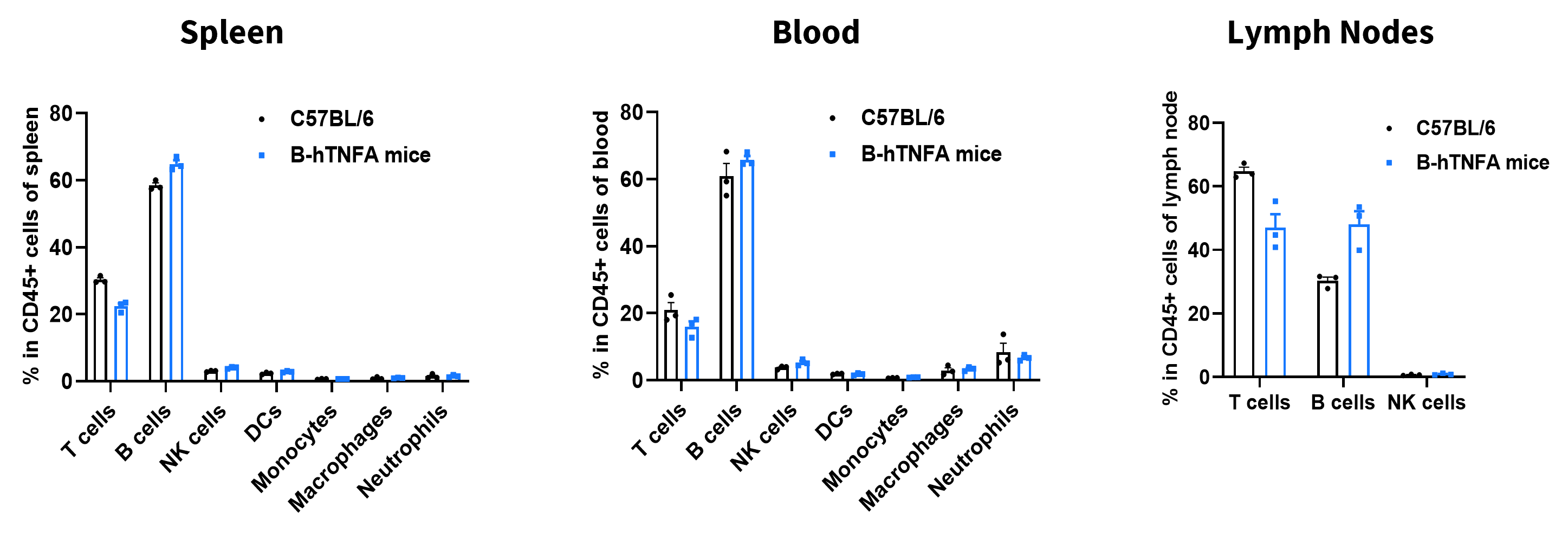
      • The frequencies of T cells, B cells, NK cells, DCs, neutrophils, monocytes, and macrophages in homozygous B-hTNFA mice were similar to those in C57BL/6 mice.
      • Humanization of TNFA does not affect normal immune cell development or splenic distribution.

      Analysis of leukocyte subpopulations by flow cytometry in immune organs and blood. Splenocytes, peripheral blood, and lymph nodes were isolated from female C57BL/6 and B-hTNFA mice plus (female, 9-week-old, n = 3). Single live cells were gated on the CD45⁺ population and analyzed by flow cytometry as indicated. Values are expressed as mean ± SEM.

      Analysis of T Cell Subpopulations
      • The proportions of CD4⁺ T cells, CD8⁺ T cells, and Tregs in homozygous B-hTNFA mice were comparable to those in C57BL/6 mice.
      • Humanization of TNFA does not affect normal T cell development, differentiation, or splenic distribution.

      Analysis of T-cell subpopulations by flow cytometry in immune organs and blood. Splenocytes, peripheral blood, and lymph nodes were isolated from C57BL/6 and B-hTNFA mice (female, 9-week-old, n = 3). Single live cells were gated on the CD3⁺ T-cell population and analyzed by flow cytometry as indicated. Values are expressed as mean ± SEM.
      Hematology Analysis
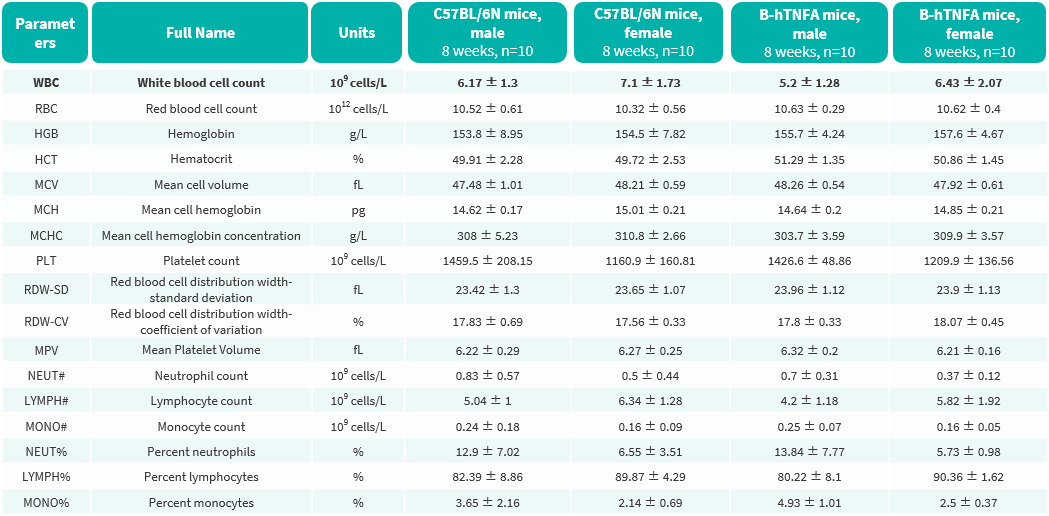
      • No significant differences were observed compared with wild-type mice.

      Complete blood count (CBC) of B-hTNFA mice. Values are expressed as mean ± SD.
      Blood Biochemical Analysis
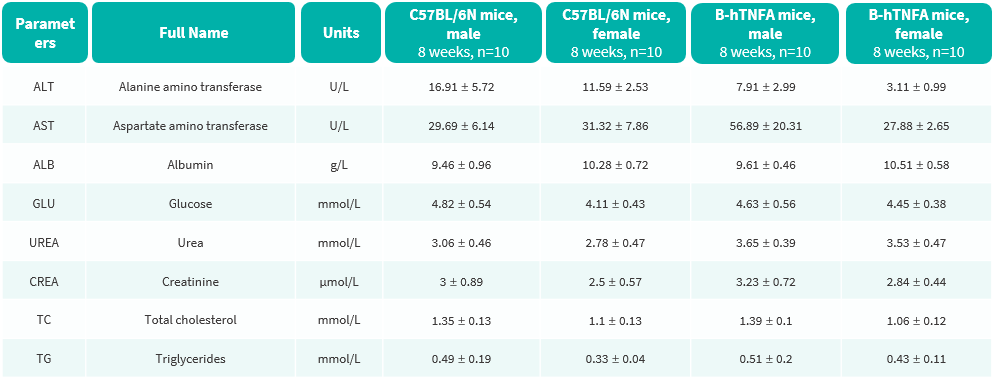
      • No significant differences were observed compared with wild-type mice.

      Blood biochemical parameters of B-hTNFA mice are shown. Values are expressed as mean ± SD.
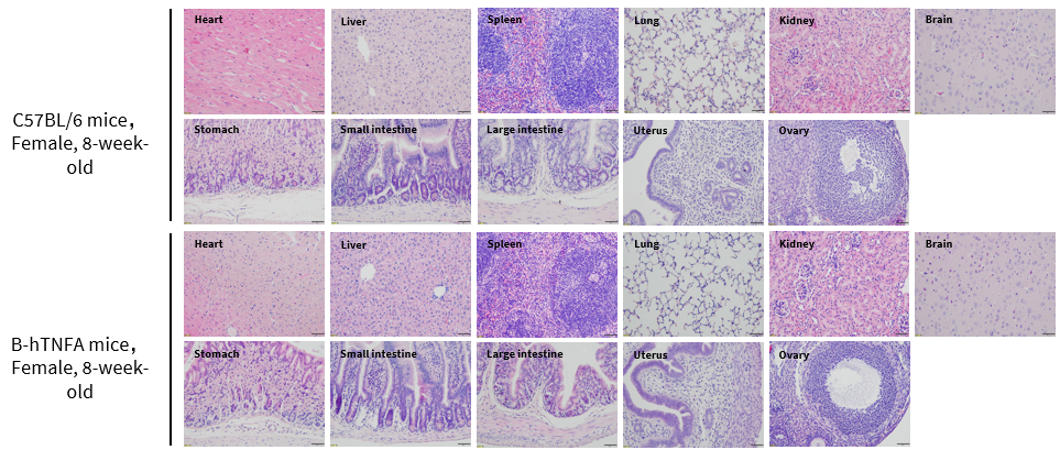
      Histopathological Analysis
      • No obvious abnormalities were observed in any organs examined (brain, heart, lung, liver, spleen, stomach, small intestine, large intestine, kidney, ovary, and uterus ).

      Histopathological analysis of organs in B-hTNFA mice. Major organs from B-hTNFA mice were collected at 8 weeks of age and analyzed by H&E staining (female, n = 10).


      In Vivo Efficacy of Anti–Human TNFA Antibody in Collagen Antibody-induced Arthritis
      • Anti-human TNFA antibody adalimumab significantly decreased clinical score in collagen antibody-induced arthritis model compared with the modelling group.

      Efficacy of anti-human TNFA antibody in B-hTNFA mice with collagen antibody induced arthritis (CAIA) model. B-hTNFA mice (female, n=10, 9-10 weeks-old) were intravenously injected with collagen antibody (mAb) on Day 0, followed by intraperitoneally injection of LPS on day 3 (A). Body weight (B) and clinical score (C) were measured on Day 0 and daily from Day 3 to Day 20 of the study. The treatment group received either anti-human TNFA antibody Adalimumab analog (in house) or methotrexate (purchased from MCE). The model group (G2 group) exhibited an arthritis phenotype after LPS injection, and its clinical score stabilized after 8 days. This indicated that the CAIA model was successfully established in B-hTNFA mice.. The clinical score of Adalimumab analog (in house) treatment group and methotrexate (purchased from MCE) were significantly lower than those of the model group (G2 group). The results indicated B-hTNFA mice provide a powerful preclinical CAIA mouse model for in vivo evaluation of anti-human TNFA antibody.

      * When publishing results obtained using this animal model, please acknowledge the source as follows: The animal model [B-hTNFA mice] (Cat# 110002) was purchased from Biocytogen.