

C57BL/6-Trem1tm1(TREM1)Bcgen/Bcgen • 111180

TREM1: A pivotal myeloid receptor target for treating autoimmune disorders and cancer.

TREM1

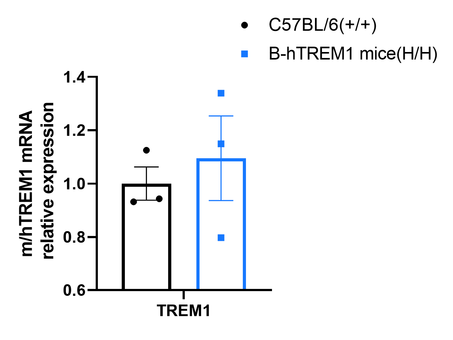
Strain specific analysis of TREM1 gene expression in wild type C57BL/6 mice and homozygous B-hTREM1 mice by RT-qPCR. Peripheral blood cell RNA was isolated from wild-type C57BL/6 mice (+/+) and homozygous B-hTREM1 mice(H/H), and then cDNA libraries were synthesized by reverse transcription, followed by qPCR with TREM1 primers.
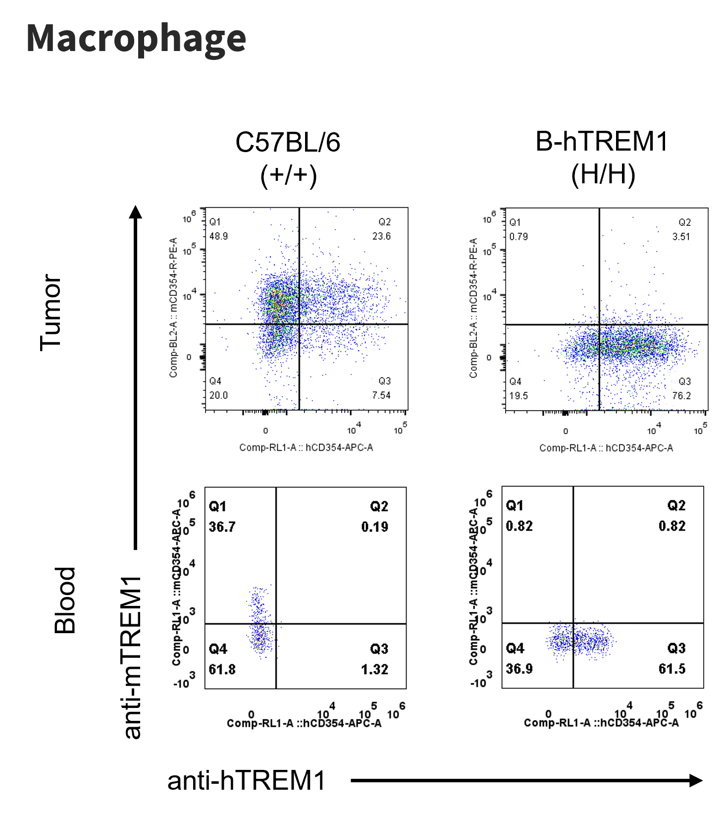
Strain specific TREM1 expression analysis in homozygous B-hTREM1 mice by flow cytometry. MC38 cells were inoculated into wild-type C57BL/6(+/+) and homozygous B-hTREM1 mice(H/H). Blood and tumor were harvested when tumor volume reached approximately 500 mm3, and analyzed by flow cytometry with species-specific anti-TREM1 antibody.
* It is not clear what population of cells was stained with specific anti-hTREM1 in tumors of C57BL/6.
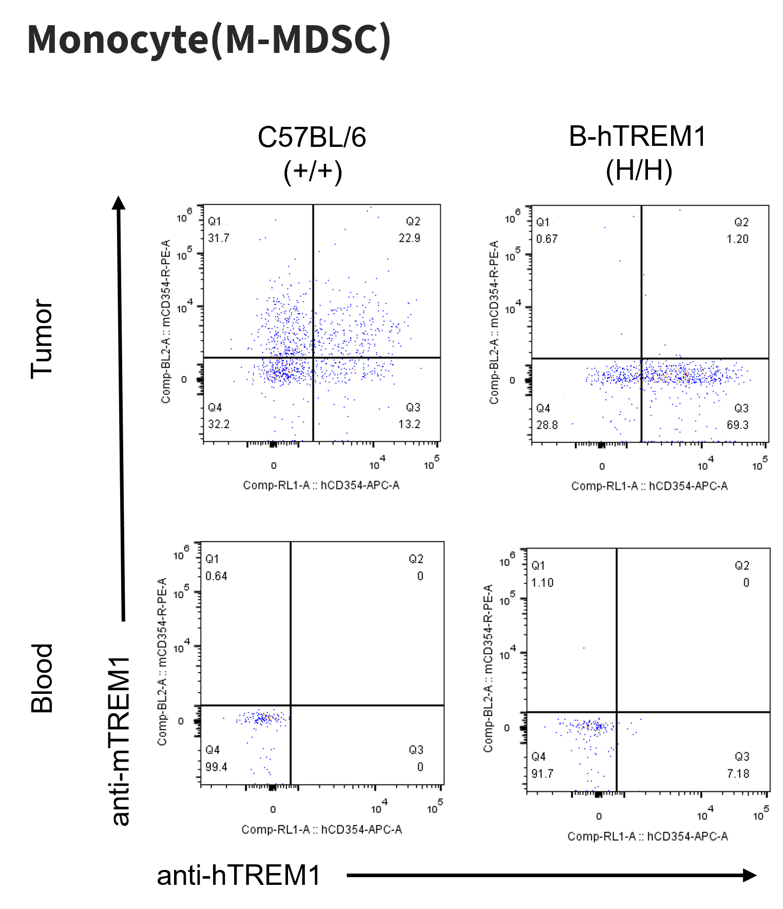
Strain specific TREM1 expression analysis in homozygous B-hTREM1 mice by flow cytometry. MC38 cells were inoculated into wild-type C57BL/6(+/+) and homozygous B-hTREM1 mice(H/H). Blood and tumor were harvested when tumor volume reached approximately 500 mm3, and analyzed by flow cytometry with species-specific anti-TREM1 antibody.
* It is not clear what population of cells was stained with specific anti-hTREM1 in tumors of C57BL/6.
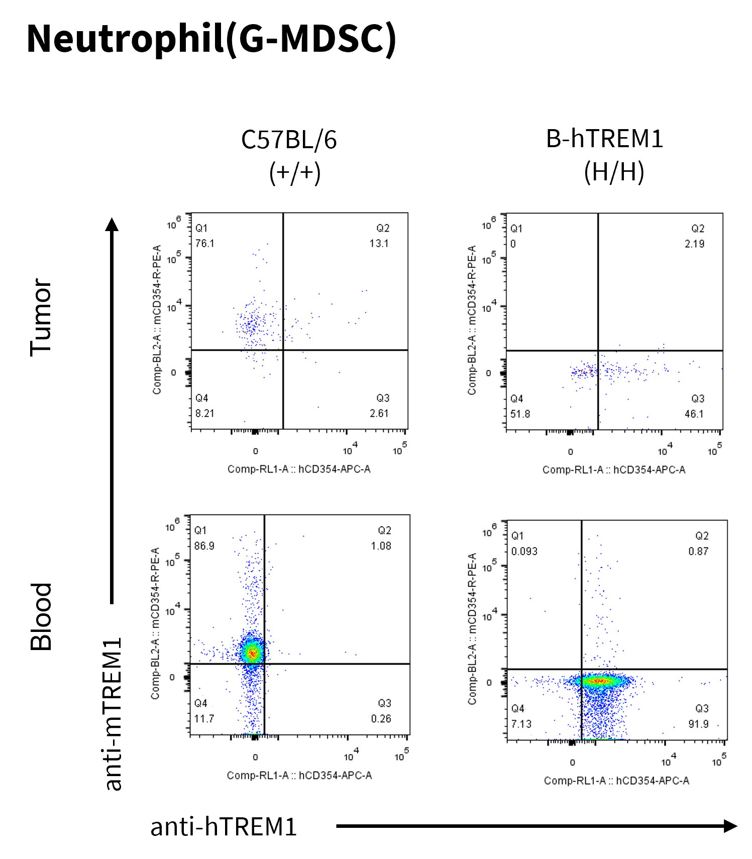
Strain specific TREM1 expression analysis in homozygous B-hTREM1 mice by flow cytometry. MC38 cells were inoculated into wild-type C57BL/6(+/+) and homozygous B-hTREM1 mice(H/H). Blood and tumor were harvested when tumor volume reached approximately 500 mm3, and analyzed by flow cytometry with species-specific anti-TREM1 antibody.
* It is not clear what population of cells was stained with specific anti-hTREM1 in tumors of C57BL/6.
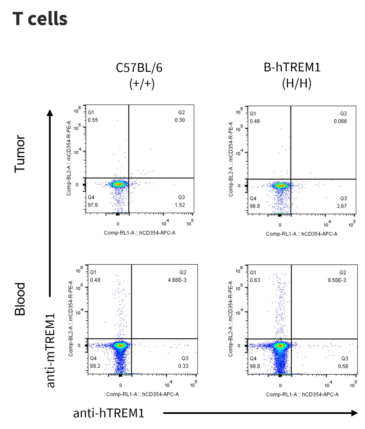
Strain specific TREM1 expression analysis in homozygous B-hTREM1 mice by flow cytometry. MC38 cells were inoculated into wild-type C57BL/6(+/+) and homozygous B-hTREM1 mice(H/H). Blood and tumor were harvested when tumor volume reached approximately 500 mm3, and analyzed by flow cytometry with species-specific anti-TREM1 antibody.
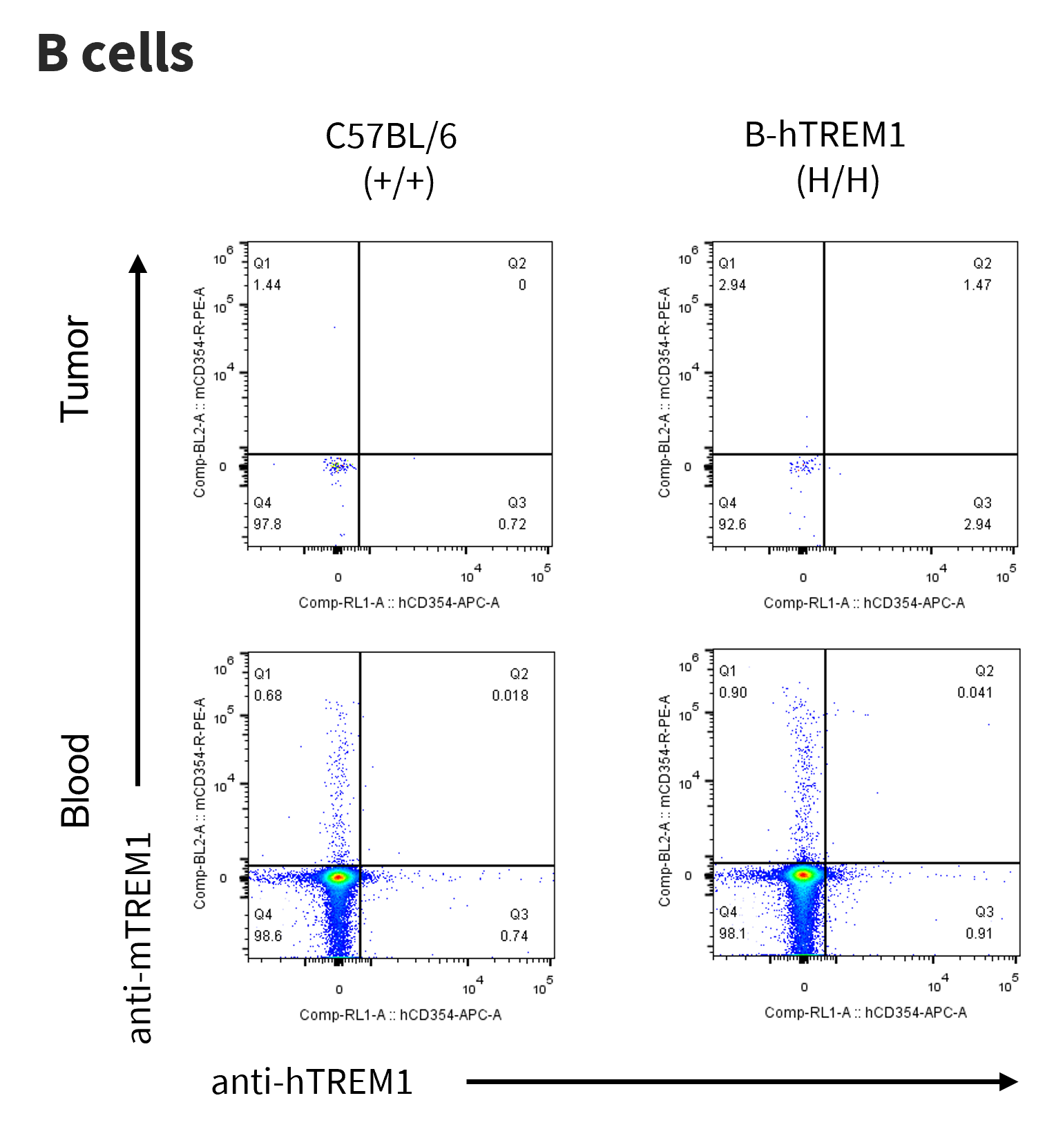
Strain specific TREM1 expression analysis in homozygous B-hTREM1 mice by flow cytometry. MC38 cells were inoculated into wild-type C57BL/6(+/+) and homozygous B-hTREM1 mice(H/H). Blood and tumor were harvested when tumor volume reached approximately 500 mm3, and analyzed by flow cytometry with species-specific anti-TREM1 antibody.
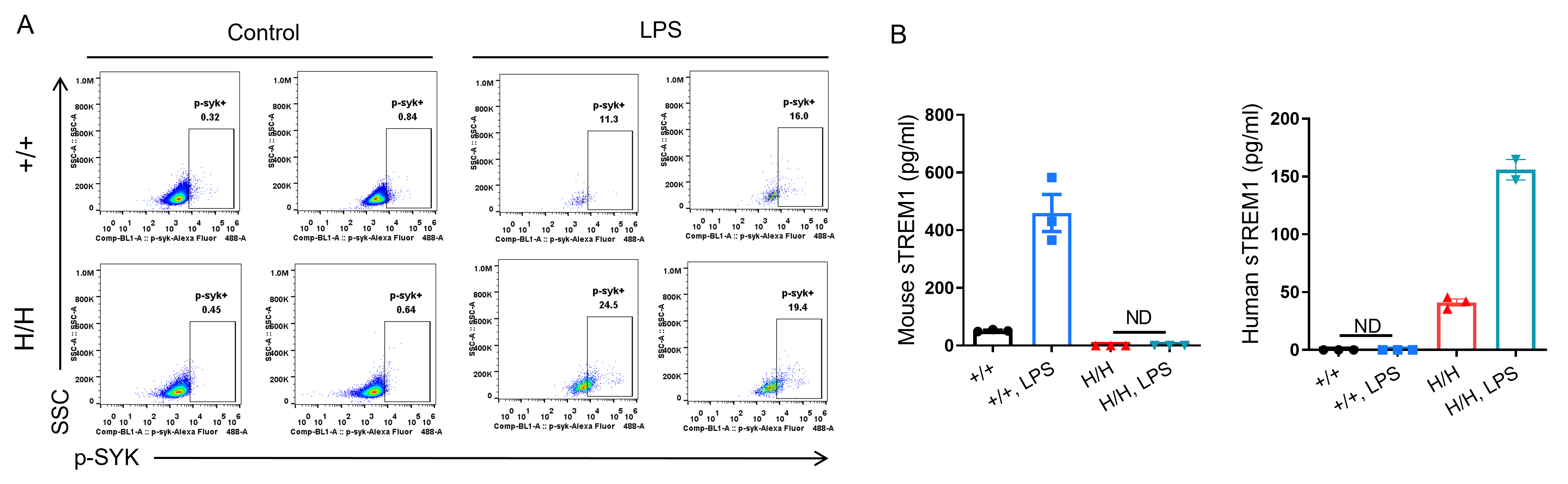
Analysis of the hTREM1 function in B-hTREM1 mice. Wild-type C57BL/6 mice(+/+) and B-hTREM1 mice(H/H) were randomly grouped and injected intraperitoneally (i.p.) with LPS from E.coli(500 ug/mouse). Serum and peritoneal macrophages were collected after LPS administration. (A) The phosphorylation of Syk. (B) the release of soluble TREM1. ND, not detected.
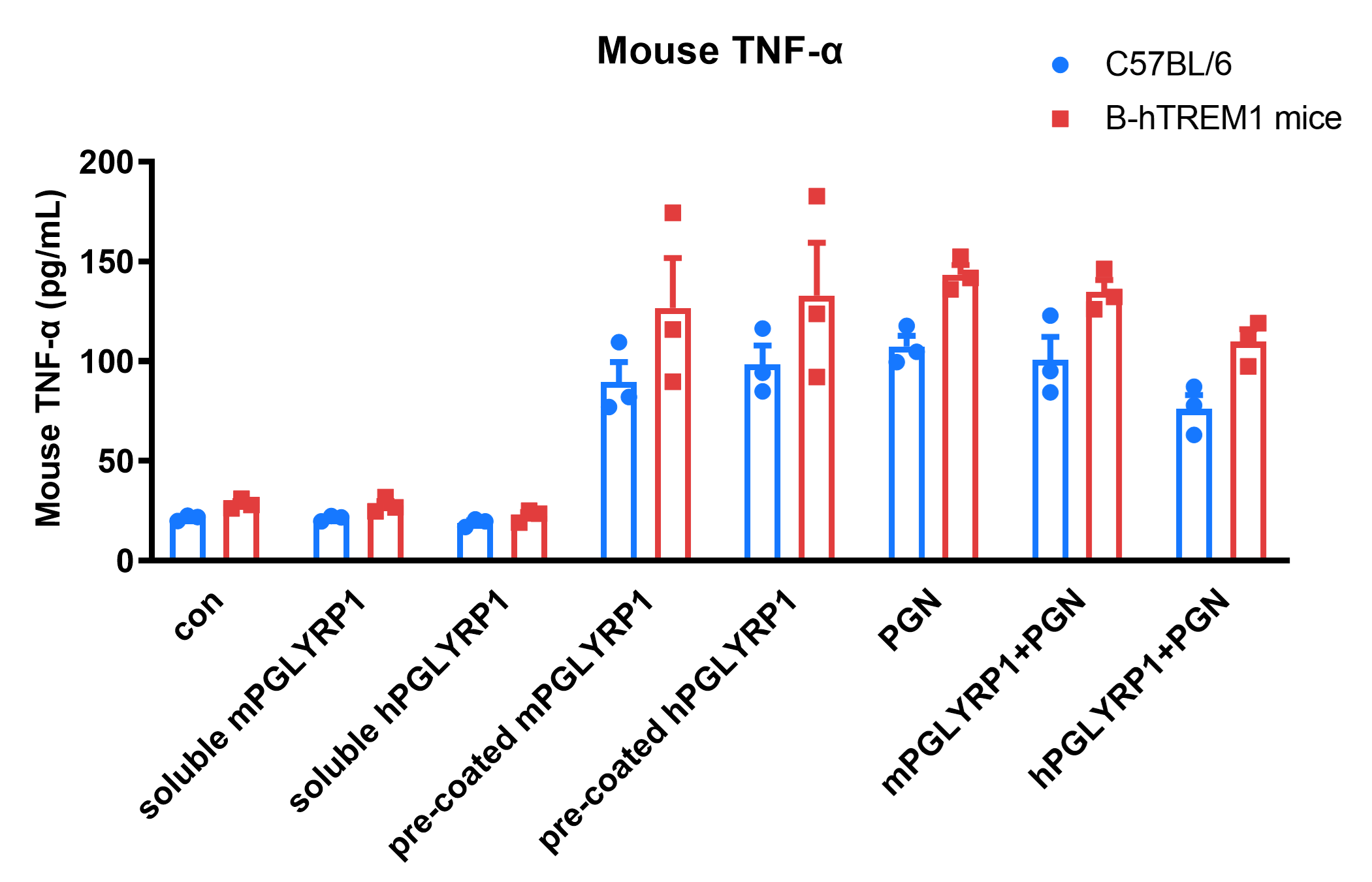
Bone marrow-derived macrophages from B-hTREM1 mice can be activated by its ligand PGLYRP1. Bone marrow-derived macrophages induced with M-CSF from wild-type C57BL/6 and B-hTREM1 mice were stimulated with m/hPGLYRP1 under the indicated conditions for 24 hours, and the levels of mouse TNF-α in the supernatant were measured. The results show that PGN alone, co-stimulation with m/hPGLYRP1 and PGN, and pre-coated m/hPGLYRP1 all significantly activated macrophages. Values are expressed as mean ± SEM. Significance was determined by two-way ANOVA test.
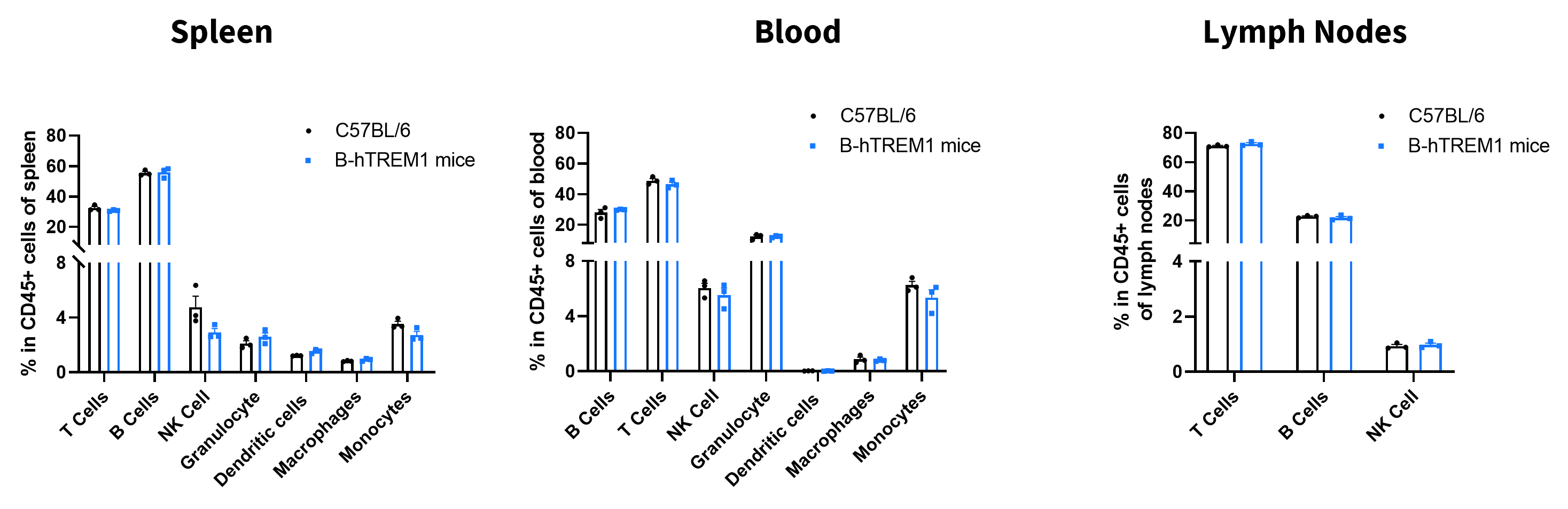
Analysis of leukocyte subpopulations by flow cytometry in immune organs and blood. Splenocytes, peripheral blood, and lymph nodes were isolated from female C57BL/6 and B-hTREM1 mice (female, 6-week-old, n = 3). Single live cells were gated on the CD45⁺ population and analyzed by flow cytometry as indicated. Values are expressed as mean ± SEM.

Analysis of T-cell subpopulations by flow cytometry in immune organs and blood. Splenocytes, peripheral blood, and lymph nodes were isolated from female C57BL/6 and B-hTREM1 mice (female, 6-week-old, n=3). Single live cells were gated on the T-cell population and analyzed by flow cytometry as indicated. Values are expressed as mean ± SEM.
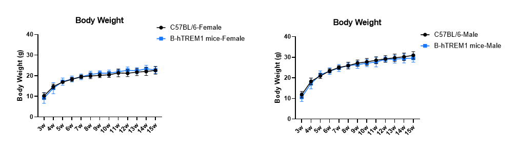
Growth curve of B-hTREM1 mice. Three-week-old mice were grouped by sex (15 males and 15 females). Body weight was measured weekly for 12 weeks on the same day each week. The minimum and maximum body weights shown in the table were calculated from the mean ± SD.
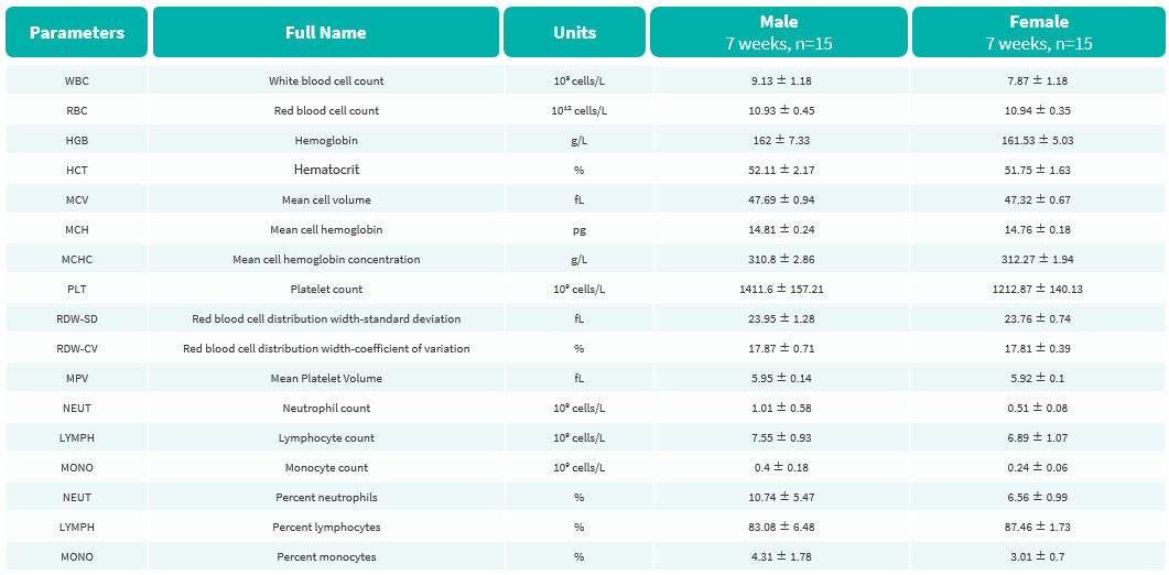
Complete blood count (CBC) of B-hTREM1 mice. Values are expressed as mean ± SD.
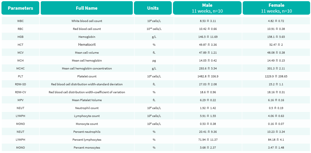
Complete blood count (CBC) of B-hTREM1 mice. Values are expressed as mean ± SD.
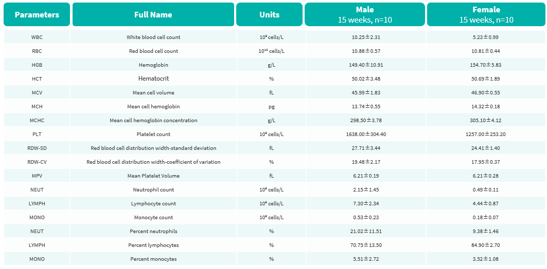
Complete blood count (CBC) of B-hTREM1 mice. Values are expressed as mean ± SD.
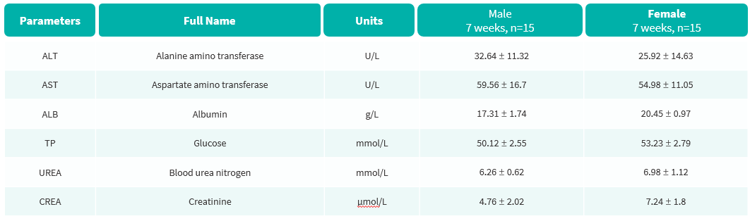
Blood biochemical parameters of B-hTREM1 mice are shown. Values are expressed as mean ± SD.
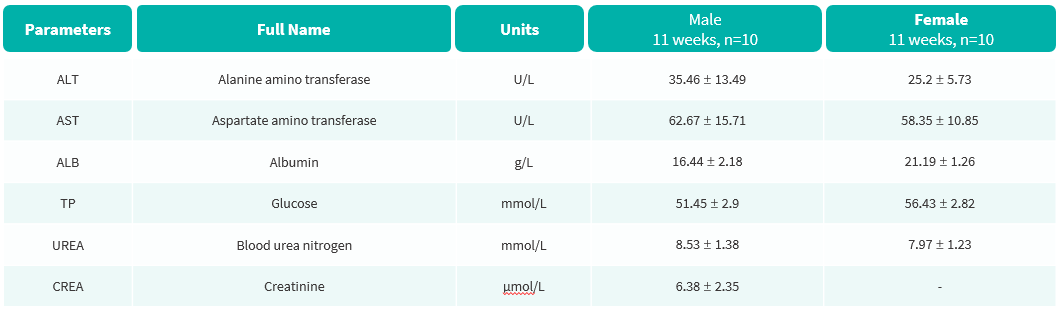
Blood biochemical parameters of B-hTREM1 mice are shown. Values are expressed as mean ± SD.
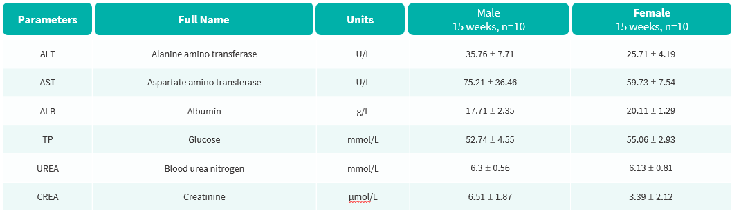
Blood biochemical parameters of B-hTREM1 mice are shown. Values are expressed as mean ± SD.
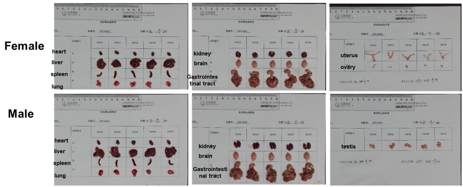
Organs of B-hTREM1 mice (7-week-old, n = 5).
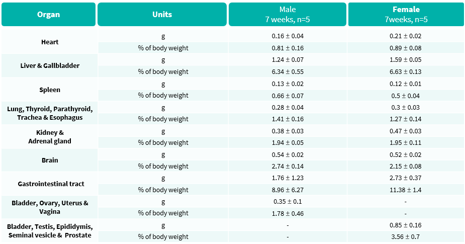
Average weights of major organs in B-hTREM1 mice.
No abnormalities were observed.
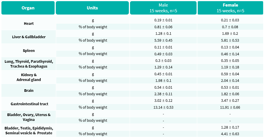
Average weights of major organs in B-hTREM1 mice.
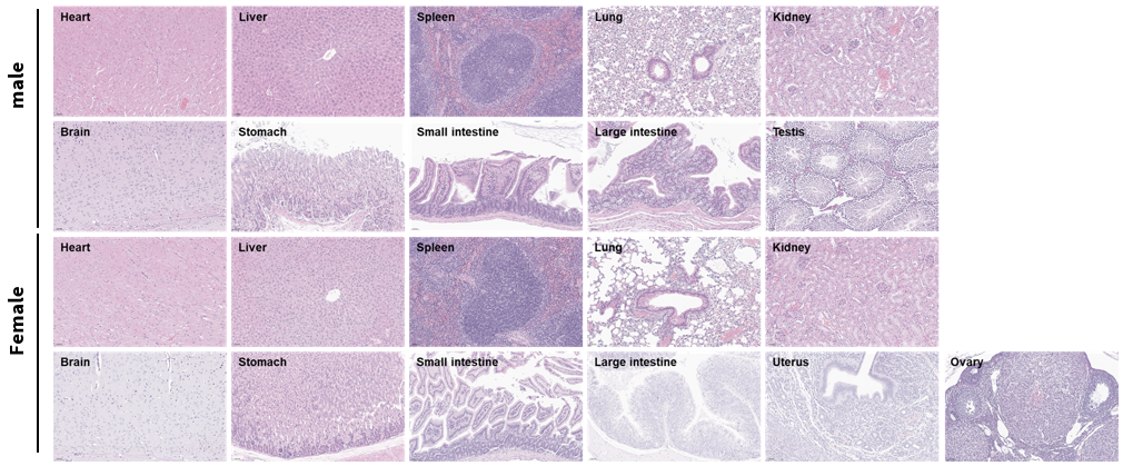
Histopathological analysis of organs in B-hTREM1 mice. Major organs from B-hTREM1 mice were collected at 15 weeks of age and analyzed by H&E staining (male, n = 5; female, n = 5).

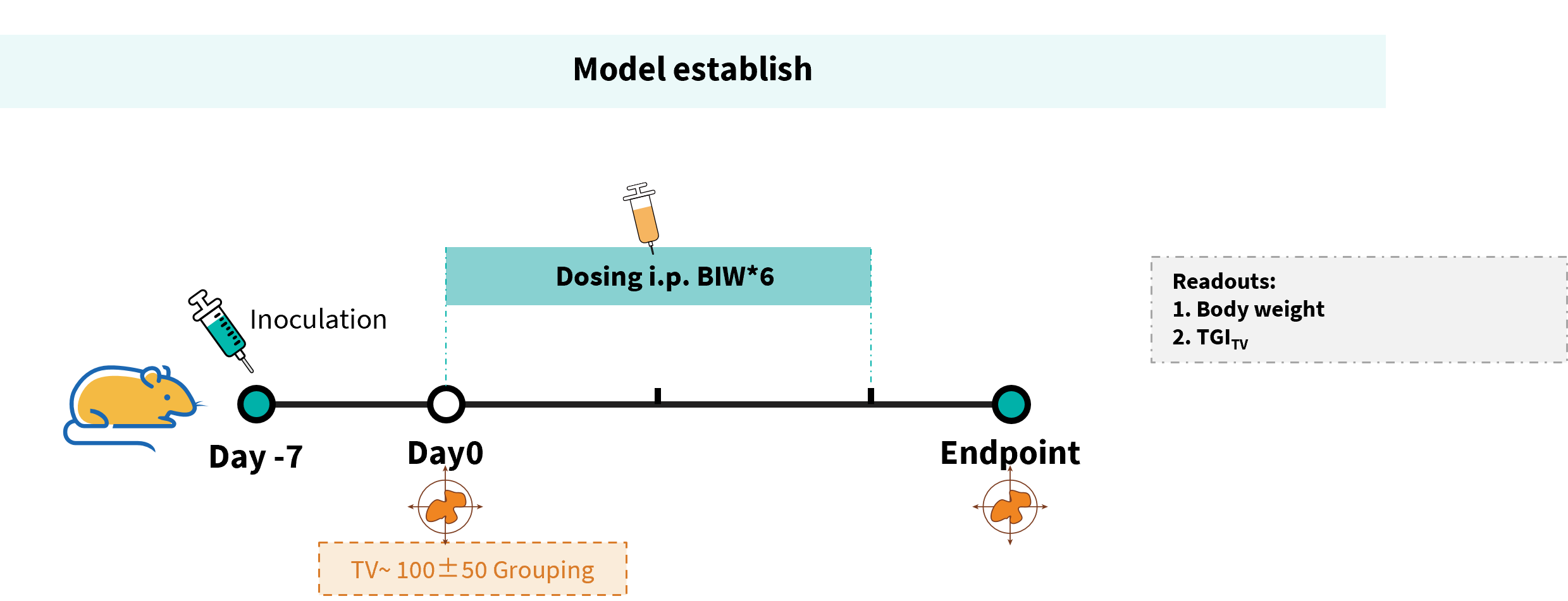
Experimental schedule for antitumor activity of anti-mouse PD-1 antibody combined with anti-human TREM1 antibody in B-hTREM1 mice. 5×10⁵ MC38 cells were subcutaneously implanted into B-hTREM1 mice (female, 9–week-old, n=5). Mice were grouped when tumor volume reached approximately 100 mm³, at which time they were injected intraperitoneally with anti-mouse PD-1 antibodies.
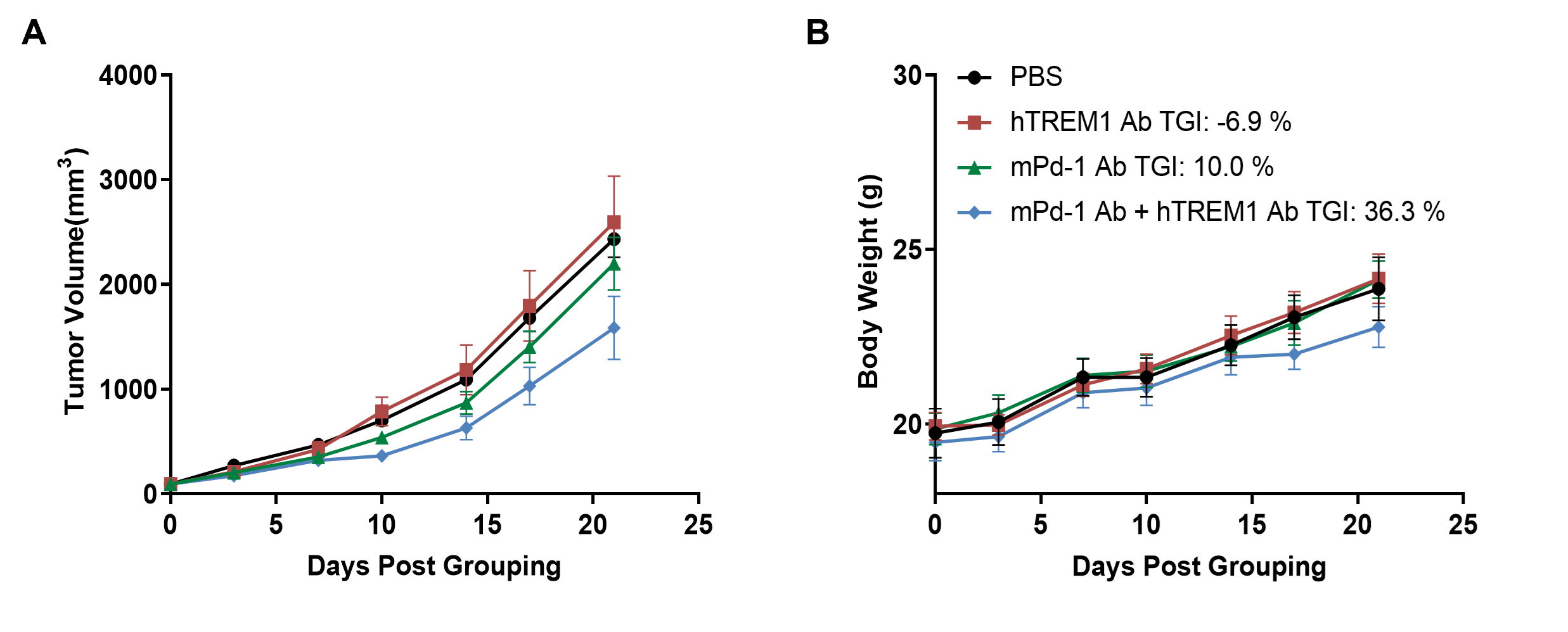
Antitumor activity of anti-mouse PD-1 antibody combined with anti-human TREM1 antibody in B-hTREM1 mice. (A) Anti-mouse PD-1 antibody combined with anti-human TREM1 antibody inhibited the growth of MC38 tumors in B-hTREM1 mice. (B) Body weight changes during treatment. Values are expressed as mean ± SEM.
