Description
TREM1: A pivotal myeloid receptor target for treating autoimmune disorders and cancer.
- Gene Information: TREM1 is a transmembrane glycoprotein belonging to the immunoglobulin superfamily. TREM1 is located on chromosome 6p21.1 and it functions as a critical activating receptor on myeloid cells, essential for amplifying inflammatory cascades and modulating immune responses across various pathological conditions.
- Protein Expression: TREM1 is predominantly expressed on the surface of myeloid cells, including circulating neutrophils, inflammatory monocytes, and tumor-associated macrophages.
- Signaling Pathway: Upon ligand engagement, TREM1 associates with the ITAM-containing adapter DAP12 to recruit SYK kinase. This initiates downstream phosphorylation cascades driving NF-κB activation, which triggers massive pro-inflammatory cytokine release in autoimmunity while amplifying myeloid-driven suppressive networks in cancer.
- Therapeutic Targeting: TREM1 is a key target for therapeutic antagonists. In autoimmune diseases, blocking TREM1 attenuates pathological hyperinflammation and tissue damage. In oncology, neutralizing TREM1 reprograms tumor-associated macrophages to relieve immunosuppression, reinvigorating robust anti-tumor responses and enhancing immunotherapy efficacy.
IL23: A key inflammation cytokine in the inflammatory response driven by Th17 cells
- Gene Information: IL-23 is a heterodimeric cytokine composed of p19 and p40 subunits, and is encoded by interleukin-23 subunit alpha (IL23A) and interleukin-12 subunit beta (IL12B) respectively. IL23A is located on chromosome 12q13.3, IL12B is located on chromosome 5q33.3.
- Protein Expression: Mature, biologically active IL-23 protein is secreted only when a cell expresses both the p19 and p40 subunits and assembles them correctly. IL-23 is mainly produced by activated antigen-presenting cells, such as macrophages and dendritic cells, as well as damaged keratinocytes or intestinal epithelial cells.
- Signaling Pathway: IL23 binds to the IL23R/IL12RB1 receptor complex to trigger JAK-STAT, p38 MAPK, and NF-κB signaling. This initiates the transcription of downstream pro-inflammatory factors, such as IL-17A, IL-17F, IL-22 and RORγt, thereby promoting the differentiation and maintenance of pathogenic Th17 cells and exacerbating chronic tissue inflammation.
- Therapeutic Inhibition: Blocking the interaction between IL23 and IL23 receptor can reduce the severity of autoimmune diseases, such as the IBD model and psoriasis.
Targeting strategy
TREM1
- The exons 1-4 of mouse Trem1 gene that encode signal peptide and extracellular domain are replaced by human counterparts in B-hTREM1/hPGLYRP1/hIL23A/hIL12B mice.
- The genomic region of mouse Trem1 gene that encodes transmembrane domain and cytoplasmic portion is retained. The promoter, 5’UTR and 3’UTR region of the mouse gene are also retained. The human TREM1 expression is driven by endogenous mouse Trem1 promoter, while mouse Trem1 gene transcription and translation will be disrupted.
PGLYRP1
- The exons 1-3 of mouse Pglyrp1 gene that encode the whole molecule (ATG to STOP codon), including 3’UTR were replaced by human counterparts in B-hTREM1/hPGLYRP1/hIL23A/hIL12B mice.
- The promoter and 5’UTR region of the mouse gene are retained. The human PGLYRP1 expression is driven by endogenous mouse Pglyrp1 promoter, while mouse Pglyrp1 gene transcription and translation will be disrupted.
IL23A
- The exons 1-4 of mouse Il23a gene that encode the whole molecule (ATG to STOP codon) were replaced by human counterparts in B-hTREM1/hPGLYRP1/hIL23A/hIL12B mice.
- The promoter, 5’UTR and 3’UTR region of the mouse gene were retained. The human IL23A expression was driven by endogenous mouse Il23a promoter, while mouse Il23a gene transcription and translation will be disrupted.
IL12B
- The exons 2-8 of mouse Il12b gene that encode the whole molecule (ATG to STOP codon), including 3’UTR were replaced by human counterparts in B-hTREM1/hPGLYRP1/hIL23A/hIL12B mice.
- The promoter and 5’UTR region of the mouse gene were retained. The human IL12B expression was driven by endogenous mouse Il12b promoter, while mouse Il12b gene transcription and translation will be disrupted.
Note: B-hTREM1/hPGLYRP1/hIL23A/hIL12B mice were obtained by mating B-hTREM1/hPGLYRP1 mice (113124) and B-hIL23A/hIL12B mice (120553).
TREM1 Protein Expression Analysis in Spleen
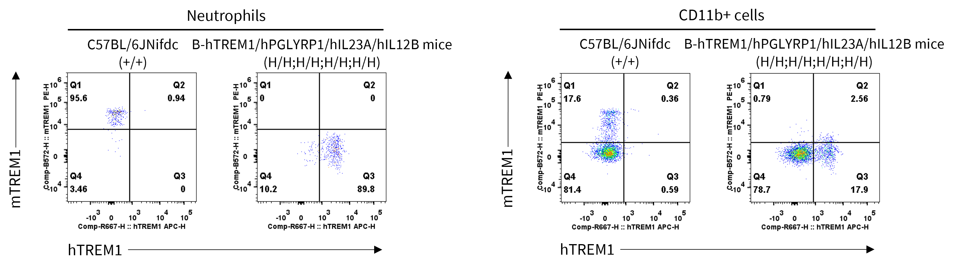
- Human TREM1 were exclusively detectable in neutrophils and CD11b+ cells of homozygous B-hTREM1/hPGLYRP1/hIL23A/hIL12B mice but not wild-type C57BL/6JNifdc mice.
Strain specific TREM1 expression analysis in homozygous B-hTREM1/hPGLYRP1/hIL23A/hIL12B mice by flow cytometry. Splenocytes were collected from wild-type C57BL/6JNifdc mice (+/+) and homozygous B-hTREM1/hPGLYRP1/hIL23A/hIL12B mice (H/H;H/H;H/H;H/H), and analyzed by flow cytometry with species-specific anti-mouse TREM1 antibody (Mouse TREM-1 PE-conjugated Antibody, R&D, FAB1187P) and anti-human TREM1 antibody (APC anti-human CD354 (TREM-1) Antibody, Biolegend, 314909). Mouse TREM1 was only detectable in neutrophils and CD11b+ cells of wild-type C57BL/6JNifdc mice. Human TREM1 was exclusively detectable in neutrophils and CD11b+ cells of homozygous B-hTREM1/hPGLYRP1/hIL23A/hIL12B mice but not in wild-type mice.
Soluble TREM1 Protein Expression Analysis in Serum
- Human soluble TREM1 was exclusively detectable in homozygous B-hTREM1/hPGLYRP1/hIL23A/hIL12B mice.
Strain specific TREM1 expression analysis in homozygous B-hTREM1/hPGLYRP1/hIL23A/hIL12B mice by ELISA. Serum was collected from wild-type C57BL/6JNifdc (+/+) and homozygous B-hTREM1/hPGLYRP1/hIL23A/hIL12B mice (H/H;H/H;H/H;H/H) stimulated with 20 μg/mouse LPS in vivo for 2 hrs. Expression level of mouse and human TREM1 were analyzed by ELISA (anti-mouse TREM1 ELISA kit: R&D, MTRM10; anti-human TREM1 ELISA kit: R&D, DTRM10C). Mouse TREM1 was only detectable in wild-type C57BL/6JNifdc mice. Human TREM1 was exclusively detectable in homozygous B-hTREM1/hPGLYRP1/hIL23A/hIL12B mice, which indicating that mouse matrix metalloproteinase (MMP) was capable of proteolytically cleaving human TREM1, and inducing the release of soluble TREM1. Values are expressed as mean ± SEM. ND: not detectable.
PGLYRP1 Protein Expression Analysis in Serum
- Human PGLYRP1 was exclusively detectable in homozygous B-hTREM1/hPGLYRP1/hIL23A/hIL12B mice.
Strain specific PGLYRP1 expression analysis in homozygous B-hTREM1/hPGLYRP1/hIL23A/hIL12B mice by ELISA. Serum was collected from wild-type C57BL/6JNifdc (+/+) and homozygous B-hTREM1hPGLYRP1hIL23AhIL12B mice (H/H;H/H;H/H;H/H) stimulated with or without LPS in vivo for 2 hrs. Expression level of mouse and human PGLYRP1 were analyzed by ELISA (anti-mouse PGLYRP1 ELISA kit: OriGene, EA102442; anti-human PGLYRP1 ELISA kit: R&D, DY2590). Mouse PGLYRP1 was only detectable in wild-type C57BL/6JNifdc mice. Human PGLYRP1 was exclusively detectable in homozygous B-hTREM1/hPGLYRP1/hIL23A/hIL12B mice. Values are expressed as mean ± SEM.
IL23 Protein Expression Analysis in BMDC Supernatants
- Human IL23 was exclusively detectable in homozygous B-hTREM1/hPGLYRP1/hIL23A/hIL12B mice but not wild-type C57BL/6JNifdc mice.
Strain specific IL23 expression analysis in homozygous B-hTREM1/hPGLYRP1/hIL23A/hIL12B mice by ELISA. Bone marrow derived dendritic cells (BMDCs) were produced by culturing the bone marrow from wild-type C57BL/6JNifdc (+/+) and homozygous B-hTREM1/hPGLYRP1/hIL23A/hIL12B mice (H/H;H/H;H/H;H/H), which were stimulated with LPS in vitro. After stimulation, the supernatants were collected and the levels of mouse and human IL23 were analyzed by ELISA (Mouse IL-23 Quantikine ELISA Kit, R&D, M2300; Human IL-23 Quantikine ELISA Kit, R&D, D2300B). Mouse IL23 was only detectable in wild-type C57BL/6JNifdc mice. Human IL23 was exclusively detectable in B-hTREM1/hPGLYRP1/hIL23A/hIL12B mice. Values are expressed as mean ± SEM. ND: not detectable.
* When publishing results obtained using this animal model, please acknowledge the source as follows: The animal model [B-hTREM1/hPGLYRP1/hIL23A/hIL12B mice] (Cat# 114307) was purchased from Biocytogen.