Description
DMD: Mutations in this gene lead to severe, progressive muscle-wastingdisorders.
- Gene Information: Human Dystrophin protein is encoded by DMD gene. Human DMD gene is one of the largest human genes, located on the short arm of X chromosome.
- Protein Expression: Dystrophin is a large protein with a molecular weight of approximately 427 kDa. Dystrophin is primarily expressed on the inner surface of the sarcolemma in skeletal, cardiac, and smooth muscle tissues. It is also expressed at lower levels in specific neurons within the brain.
- Signaling Pathway: During muscle contraction and relaxation, the sarcolemma experiences severe mechanical stress. Dystrophin links the intracellular actin cytoskeleton to the extracellular matrix. In the absence of dystrophin, muscle tissue becomes vulnerable to contraction-induced damage.
- Therapeutic Application: Genetic interventions including micro-dystrophin delivery represent a promising approach for the treatment of Duchenne Muscular Dystrophin.
TFR1 MOA: Target for Drug Delivery Across the Blood-Brain Barrier
- Primary role: TFR1 binds transferrin (TF) to mediate cellular iron uptake via endocytosis.
- Binding: At physiological pH, TFR1 binds TF-Fe²⁺ to form the TF–TFR1 complex.
- Endocytosis: The complex undergoes clathrin-mediated endocytosis and enters endosomes.
- Iron release: Endosomal acidification induces conformational changes, releasing iron ions.
- Recycling: TFR1 is recycled to the cell surface through the Golgi, completing the iron transport cycle.
Targeting strategy
DMD
- The exon 51, exon 52 and exon 53 of mouse Dmd are replaced by exon 51 and exon 53 of human DMD in B-hDMD(exon51 53, del52) mice.
- The human exon 51 is flanked by ~2kb of human intron 50 and human intron 51 sequences, while human exon 53 is flanked by ~2kb of human intron 52 and human intron 53 sequences.
TFR1
- The exons 4 19 of mouse Tfr1 gene that encode extracellular domain are replaced by human counterparts in B-hDMD(exon51 53, del52)/hTFR1 mice. The genomic region of mouse Tfr1 gene that encodes cytoplasmic portion is retained.
- The promoter, 5’UTR and 3’UTR of the mouse gene are also retained. The chimeric TFR1 expression is driven by endogenous mouse Tfr1 promoter, while mouse Tfr1 gene transcription and translation will be disrupted.
Note: B-hDMD(exon51-53, del52)/hTFR1 mice were obtained by mating B-hDMD(exon51-53, del52) mice (114172) and B-hTFR1 mice (110861).
Behavioral Performance Analysis
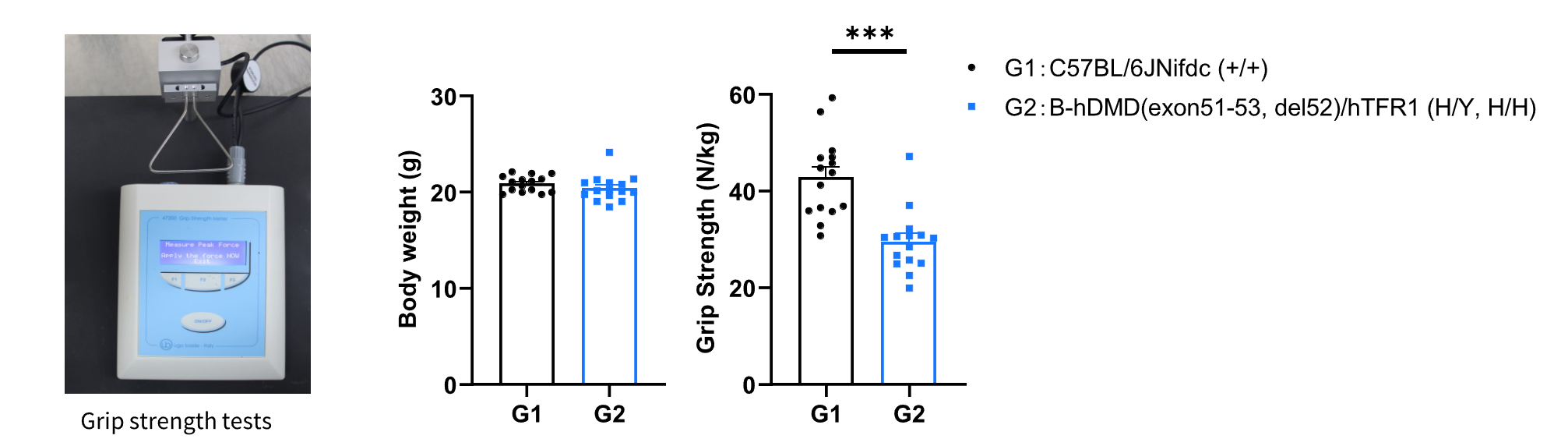
Behavioral performance in wild-type C57BL/6JNifdc and hemizygous B-hDMD(exon51-53, del52)/hTFR1 mice. Grip strength tests were conducted to assay the behavioral performance in wild-type C57BL/6JNifdc and hemizygous B-hDMD(exon51-53, del52)/hTFR1 mice (male, 6-week-old, n=15). Grip strength produced by forelimb in hemizygous B-hDMD(exon51-53, del52)/hTFR1 mice was significant weaker than that in wild-type control mice. All grip strength measurements are normalized to the individual animal’s body weight. Values are expressed as mean ± SEM. Significance was determined by unpaired t test. *P < 0.05, P < 0.01, ***P < 0.001.
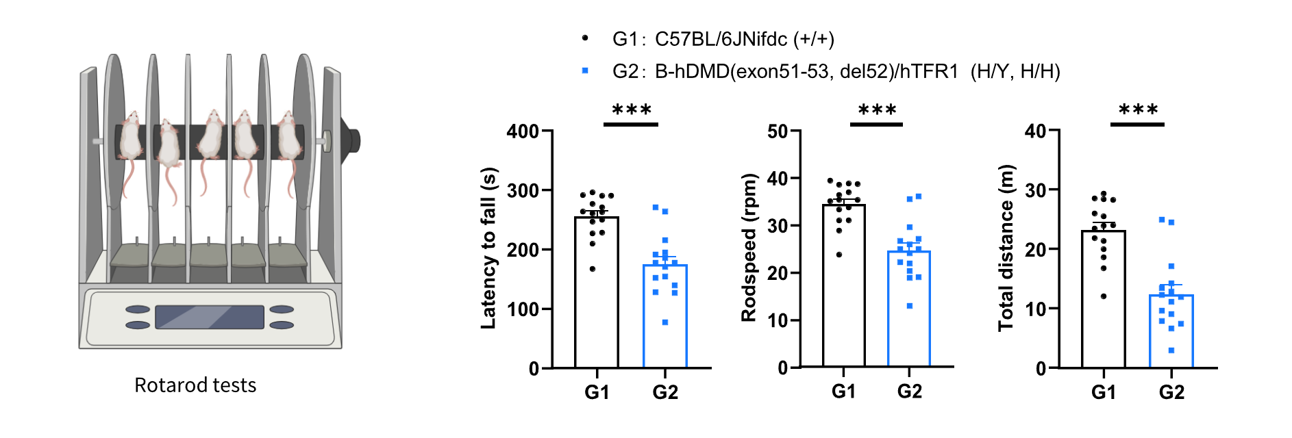
Behavioral performance in wild-type C57BL/6JNifdc and hemizygous B-hDMD(exon51-53, del52)/hTFR1 mice. Rotarod tests were conducted to assay the behavioral performance in wild-type C57BL/6JNifdc and hemizygous B-hDMD(exon51-53, del52)/hTFR1 mice (male, 6-week-old, n=15). Rotarod tests were performed to assay the motor coordination. The latency to fall, rod speed and total distance were significantly decreased in hemizygous B-hDMD(exon51-53, del52)/hTFR1 mice, showing the impairment of motor coordination and balance. Values are expressed as mean ± SEM. Significance was determined by unpaired t test. *P < 0.05, P < 0.01, ***P < 0.001.
* When publishing results obtained using this animal model, please acknowledge the source as follows: The animal model [B-hDMD(exon51-53, del52)/hTFR1 mice] (Cat# 115038) was purchased from Biocytogen.