Description
- Human uPAR (PLAUR/CD87) is a compelling therapeutic target with broad relevance in oncology, inflammation, fibrosis, and age-associated diseases. uPAR is a GPI-anchored cell-surface glycoprotein composed of three extracellular LU domains (DI–DIII) and lacks both a transmembrane region and an intracellular signaling domain. This unique structure enables uPAR to function as a cell-surface signaling hub through interactions with multiple partner molecules.
- uPAR is primarily expressed on myeloid and inflammatory cells, including monocytes, macrophages, neutrophils, and dendritic cells, and is frequently upregulated on tumor cells, stromal cells, and angiogenic endothelial cells in diseased tissues. Its canonical ligand, urokinase-type plasminogen activator (uPA), recruits proteolytic activity to the cell surface and promotes plasmin generation and extracellular matrix remodeling. In addition, uPAR interacts with vitronectin, integrins, EGFR, FPR1, and LRP1, linking extracellular proteolysis to pathways controlling cell adhesion, migration, invasion, proliferation, survival, and inflammatory responses.
- Physiologically, uPAR contributes to tissue remodeling, wound repair, immune cell trafficking, and host defense. Pathologically, it is strongly associated with tumor invasion, metastasis, angiogenesis, stromal remodeling, chronic inflammation, and fibrosis, making it highly attractive for translational research and therapeutic intervention.
- A wide range of uPAR-targeted modalities are under development, including monoclonal antibodies, radiopharmaceuticals, peptide-based theranostics, ADCs, CAR-T cells, and small-molecule or peptide antagonists. While no uPAR-targeted therapy has yet been approved, the field is advancing rapidly, with radiotheranostic programs emerging as a leading clinical direction in glioblastoma and other uPAR-positive solid tumors.
- In B-NDG hUPAR mice, the exons 2-7 of mouse uPAR gene that encode the extracellular domain and propeptide were replaced by human counterparts in B-NDG hUPAR mice. The promoter, 5’UTR, signal peptide, and 3’UTR region of the mouse gene were retained. The humanized UPAR expression was driven by endogenous mouse uPAR promoter, while mouse uPAR gene transcription and translation will be disrupted.
- Mouse UPAR was only detectable on the monocytes/macrophages of the peritoneal lavage fluid in wild-type B-NDG mice. Human UPAR was detectable on the monocytes/macrophages of the peritoneal lavage fluid and bone marrow, and the neutrophils of bone marrow in homozygous B-NDG hUPAR mice.
- Application: B-NDG hUPAR mice enable in vivo efficacy and safety evaluation of UPAR-targeted therapeutics (e.g., monoclonal antibodies, radiopharmaceuticals, ADCs, and cell therapies) with on-target/off-tumor assessment.
Targeting Strategy
Gene targeting strategy for B-NDG hUPAR mice. The exons 2-7 of mouse uPAR gene that encode the extracellular domain and propeptide were replaced by human counterparts in B-NDG hUPAR mice. The promoter, 5’UTR, signal peptide, and 3’UTR region of the mouse gene were retained. The humanized UPAR expression was driven by endogenous mouse uPAR promoter, while mouse uPAR gene transcription and translation will be disrupted.
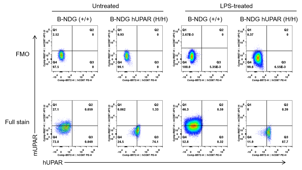
Protein Expression Analysis in Peritoneal Lavage Fluid
Strain specific UPAR expression analysis in wild-type B-NDG mice (+/+) and homozygous humanized B-NDG hUPAR mice (H/H) by flow cytometry. Peritoneal lavage fluid was collected from wild-type B-NDG mice (male, 6 weeks old, n=1) and homozygous B-NDG hUPAR mice (male, 14 weeks old, n=1) stimulated with LPS (50 ng, i.p.) in vivo for 3 hrs. Protein expression was analyzed with anti-mouse UPAR antibody (Miltenyi biotec, 130-109-898) and anti-human UPAR antibody (Miltenyi biotec, 130-114-850) by flow cytometry. Mouse UPAR was only detectable on the monocytes/macrophages of the peritoneal lavage fluid in wild-type B-NDG mice. Human UPAR was exclusively detectable on the monocytes/macrophages of the peritoneal lavage fluid in homozygous B-NDG hUPAR mice, but not in wild-type B-NDG mice.
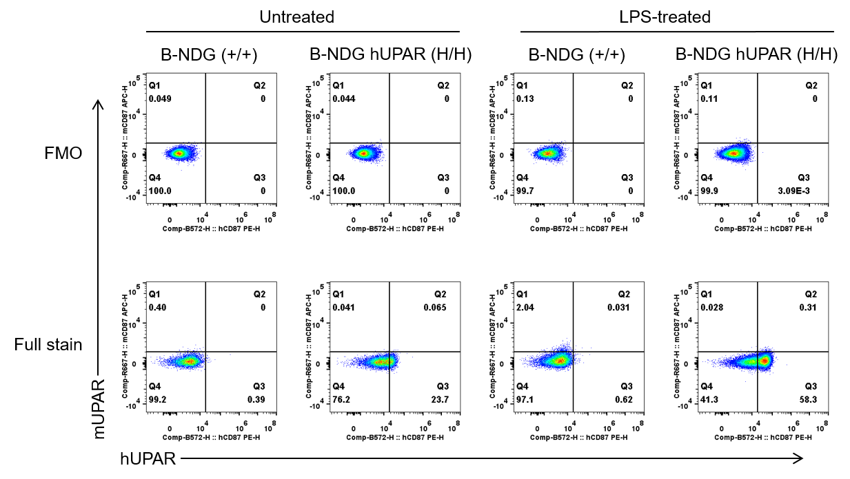
Protein Expression Analysis on Monocytes/Macrophages of Bone Marrow
Strain specific UPAR expression analysis in wild-type B-NDG mice (+/+) and homozygous humanized B-NDG hUPAR mice (H/H) by flow cytometry. Bone marrow was collected from wild-type B-NDG mice (male, 6 weeks old, n=1) and homozygous B-NDG hUPAR mice (male, 14 weeks old, n=1) stimulated with LPS (50 ng, i.p.) in vivo for 3 hrs. Protein expression was analyzed with anti-mouse UPAR antibody (Miltenyi biotec, 130-109-898) and anti-human UPAR antibody (Miltenyi biotec, 130-114-850) by flow cytometry. Mouse UPAR was not detectable on the monocytes/macrophages of the bone marrow in wild-type B-NDG mice. Human UPAR was exclusively detectable on the monocytes/macrophages of the bone marrow in homozygous B-NDG hUPAR mice, but not in wild-type B-NDG mice.
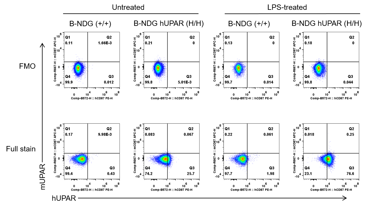
Protein Expression Analysis on Neutrophils of Bone Marrow
Strain specific UPAR expression analysis in wild-type B-NDG mice (+/+) and homozygous humanized B-NDG hUPAR mice (H/H) by flow cytometry. Bone marrow was collected from wild-type B-NDG mice (male, 6 weeks old, n=1) and homozygous B-NDG hUPAR mice (male, 14 weeks old, n=1) stimulated with LPS (50 ng, i.p.) in vivo for 3 hrs. Protein expression was analyzed with anti-mouse UPAR antibody (Miltenyi biotec, 130-109-898) and anti-human UPAR antibody (Miltenyi biotec, 130-114-850) by flow cytometry. Mouse UPAR was not detectable on the neutrophils of the bone marrow in wild-type B-NDG mice. Human UPAR was exclusively detectable on the neutrophils of the bone marrow in homozygous B-NDG hUPAR mice, but not in wild-type B-NDG mice.
* When publishing results obtained using this animal model, please acknowledge the source as follows: The animal model [B-NDG hUPAR mice] (Cat# 113376) was purchased from Biocytogen.