B-hCD38 mice

C57BL/6N-Cd38tm3(CD38)Bcgen/Bcgen • 110046

B-hCD38 mice

Catalog Number: 110046
Strain Name: C57BL/6N-Cd38tm3(CD38)Bcgen/Bcgen
Strain Background: C57BL/6N
NCBI gene ID: 952 (Human)
Aliases: ADPRC1; cADPR1; ADPRC 1
---
可提供授权方案
B-hCD38 mice

在此页面上

  • General information
  • Description
  • Targeting strategy
  • Phenotypic analysis
  • Efficacy
  • Physiological data
  • FAQ section

海报

查看全部

    发表文章

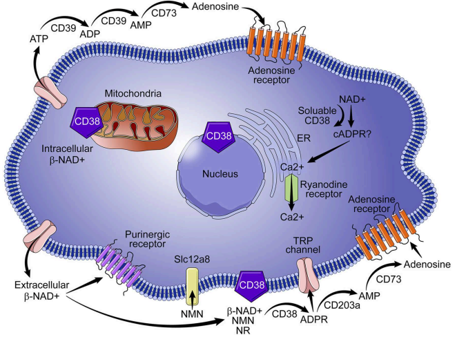
      CD38 MOA: Targeted Receptor-mediated Cellular Cytotoxicity and Ectoenzymatic Inhibition

      CD38: A multifunctional ectoenzyme regulating calcium signaling and immunity. 

      • Gene Information: Located on chromosome 4p15, the CD38 gene encodes a highly conserved transmembrane glycoprotein that functions as both a receptor and a versatile ectoenzyme.
      • Protein Expression: CD38 is highly expressed on plasma cells, multiple myeloma cells, and chronically activated T, B, and natural killer immune cells. 
      • Signaling Pathway: It synthesizes cADPR and NAADP, mobilizing intracellular calcium (Ca2+) to regulate cellular activation, proliferation, and complex metabolic signaling networks. 
      • Therapeutic Inhibition: Monoclonal antibodies like daratumumab target CD38, inducing direct tumor cell apoptosis, ADCC, CDC, and alleviating immunosuppression within the microenvironment.
      Description

      CD38 is a 42-kDa glycoprotein, also known as T10. It is an ADP-ribosyl hydrolase expressed on B cells, NK cells, and a subset of T cells, as well as in the brain, muscle, and kidney. In mice, CD38 expression is downregulated on germinal center B cells and plasma cells, whereas this downregulation is not observed in humans. By functioning as both a cyclase and a hydrolase, CD38 mediates lymphocyte activation, adhesion, and the metabolism of cADPR and NAADP. CD31 also serves as a known ligand for CD38.

      NAD⁺ is broken down into byproducts that circulate in the bone marrow plasma within the myeloma niche, accumulating various amounts of ADO. Most ADO is taken up by purinergic cell receptors (ADORs) expressed by bone cells or immune cells within the niche. The outcome is either suppression of the anti-tumor activity of immune cells (Teff, NK cells, TAMs) or an increase in regulatory T cells (Tregs), mesenchymal-derived stromal cells (MDSCs), or dendritic cells (DCs), all of which suppress immune activity against tumors.

      Increased expression of CD38 is an unfavorable diagnostic marker in chronic lymphocytic leukemia and is associated with enhanced disease progression. CD38 is also the therapeutic target of daratumumab (Darzalex), which is approved for the treatment of multiple myeloma.

      Key Advantages

      • Validated Humanized Target Expression–Successful detection of human CD38 mRNA and protein ensures reliable physiological expression for targeted therapeutic testing.
      • Proven Clinical Antibody Binding-Demonstrated binding to Daratumumab and Isatuximab analogs confirms structurally accurate and clinically relevant human epitopes.
      • Robust In Vivo Efficacy-Demonstrated tumor inhibition by Daratumumab analog validates the model's predictive power for in vivo therapeutic testing.

      Validation

      • Anti-CD38 Antibody Screening-Evaluate and screen novel human-targeted CD38 monoclonal antibodies, or antibody-drug conjugates (ADCs) in vivo.
      • In Vivo Efficacy Evaluation-Assess the therapeutic tumor-inhibiting potential of candidate drugs using clinically relevant hematological malignancy models.
      • Pharmacokinetic/Pharmacodynamic Profiling-Analyze drug clearance, target engagement, and receptor internalization kinetics of CD38-targeted therapeutics in vivo.

      Application

      • In vivo efficacy testing of anti-human CD38 therapeutic antibodies (e.g., daratumumab).
      • ADCC, CDC, and Fc-engineering–based functional characterization.
      • In vivo tumor challenge and metastasis models using human CD38-expressing tumor lines.
      • Biomarker validation for CD38-mediated immunosuppression and T-cell regulation.
      • Safety and tolerability studies for CD38-targeting immunotherapies.
      • Immune-cell binding, occupancy, and receptor engagement studies.

      Targeting strategy
      • A CDS that encodes human CD38 partial extracellular domain, followed by mouse 3’UTR-STOP was inserted at exon 2 of the mouse Cd38 gene to replace part of exon 2 and all of exon 3 of mouse Cd38 gene. The genomic region of mouse Cd38 gene that encodes transmembrane domain and cytoplasmic portion was retained. 
      • The promoter and 5’UTR region of the mouse gene were also retained. The chimeric CD38 protein expression will be driven by endogenous mouse Cd38 promoter, while mouse Cd38 gene transcription and translation will be disrupted.
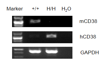
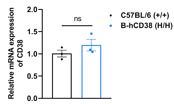
      mRNA Expression Analysis in CD38 Humanized Mice

      Species specific analysis of CD38 gene expression in wild-type C57BL/6 mice and homozygous humanized B-hCD38 mice by RT-PCR. Spleen RNA was isolated from wild-type C57BL/6 mice (+/+) and homozygous B-hCD38 mice (H/H), and then cDNA libraries were synthesized by reverse transcription, followed by PCR with mouse Cd38 primers and human CD38 primers.

      Species specific analysis of CD38 gene expression in wild-type C57BL/6 mice and homozygous humanized B-hCD38 mice by RT-qPCR. Splenocytes were collected from wild-type C57BL/6 mice and homozygous B-hCD38 mice (female, 10-week-old, n = 3). Values are expressed as mean ± SEM. Significance is determined by unpaired t-test. *P < 0.05, **P < 0.01, ***P < 0.001.

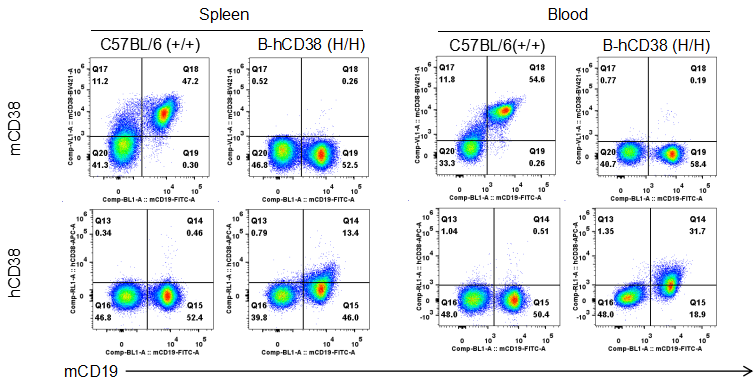
      Protein Expression Analysis in CD38 Humanized Mice

      Strain specific CD38 expression analysis in wild-type C57BL/6 mice and homozygous humanized B-hCD38 mice by flow cytometry. Spleen and blood were collected from wild-type C57BL/6 mice and homozygous B-hCD38 mice. Protein expression was analyzed with anti-mouse CD38 antibody (Biolegend, 102732) and anti-human CD38 antibody (Biolegend, 356606) by flow cytometry.

      B-hCD38 Mice: Anti-human CD38 Antibody Binding Assay

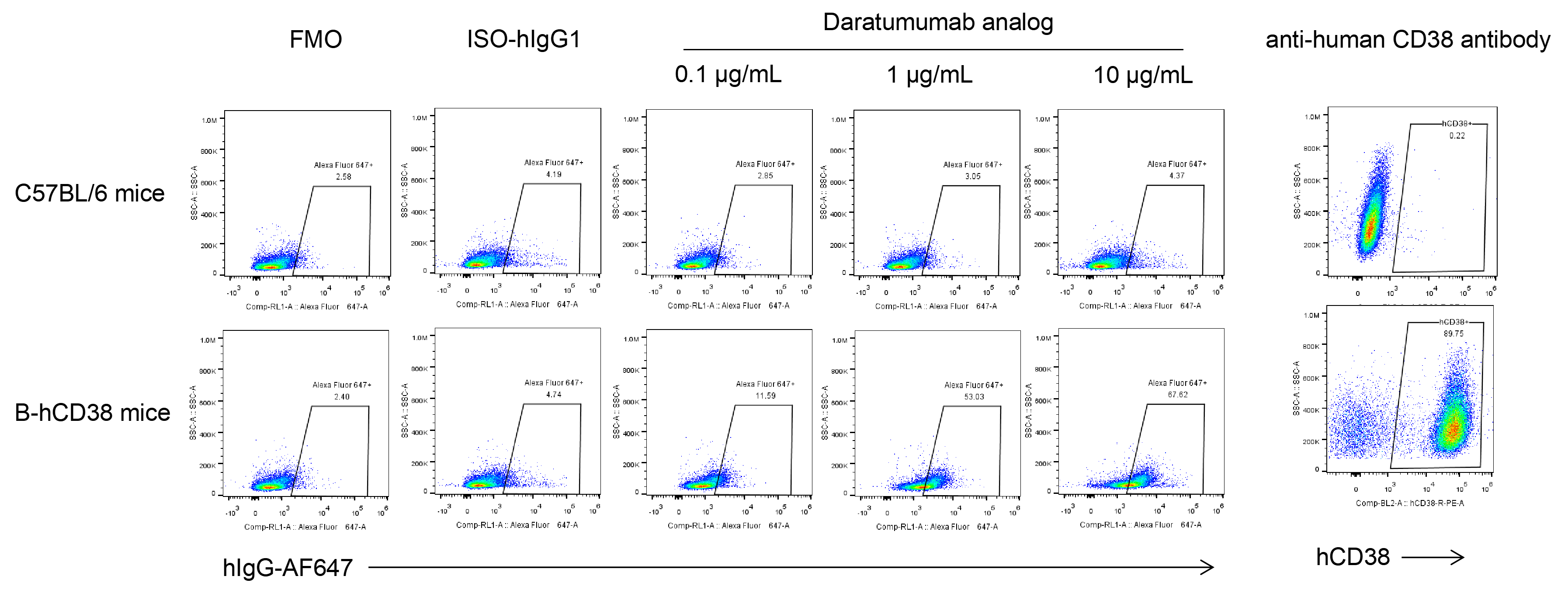
      Anti-human CD38 antibody binding assay in B-hCD38 mice. Splenocytes were collected from wild-type C57BL/6 mice and homozygous B-hCD38 mice and analyzed by flow cytometry with anti-hCD38 antibody Daratumumab analog (in-house) and anti-human CD38 antibody (Biolegend, 356606).

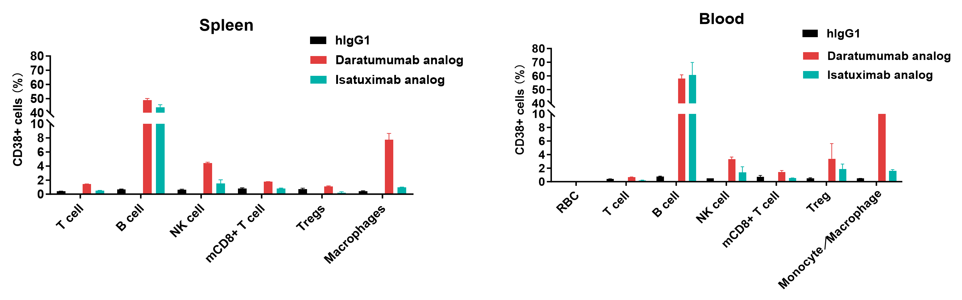
      Anti-human CD38 antibody binding assay B-hCD38 mice. Spleen and blood were collected from homozygous B-hCD38 mice and analyzed by flow cytometry with anti-hCD38 antibody Daratumumab analog (provided by the client) and Isatuximab analog (provided by the client).

      B-hCD38 Mice: In Vivo Efficacy of Anti-Human CD38 Antibody

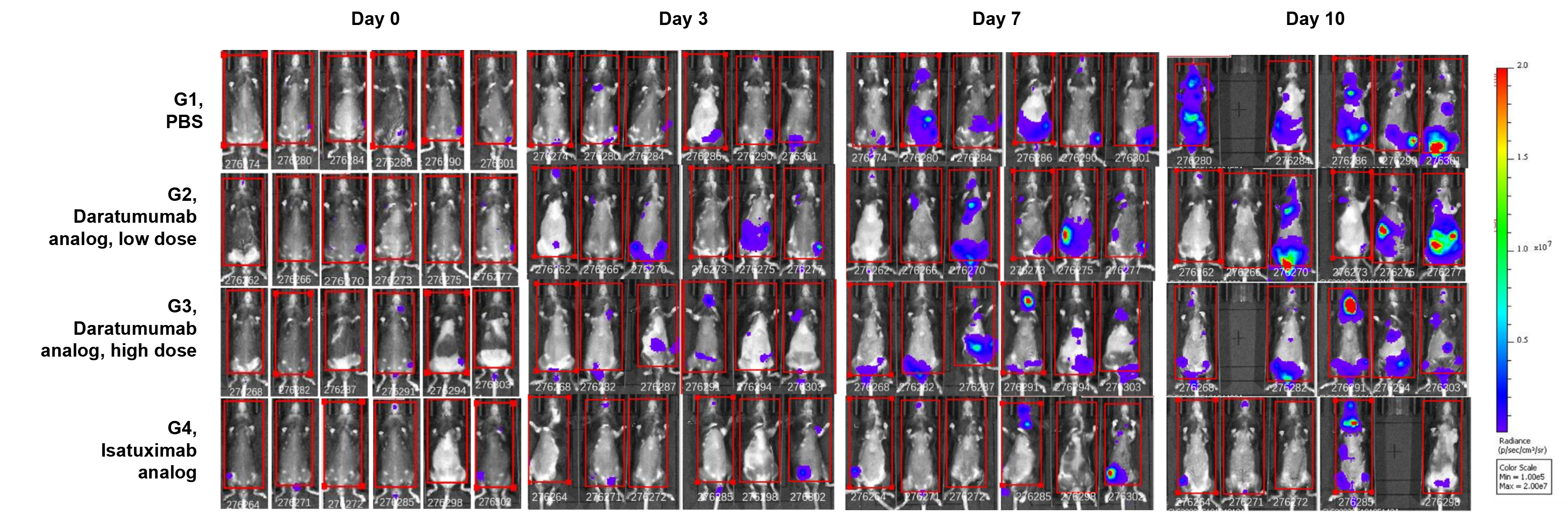
      Anti-tumor activity of anti-human CD38 antibody in B-hCD38 mice. (A) Anti-human CD38 antibody Daratumumab analog (in house) and Isatuximab analog (in house) inhibit B-hCD38-luc E.G7-OVA tumor growth in B-hCD38 mice. (B) Body weight changes during treatment. Values are expressed as mean ± SEM.

      In vivo luciferase images of B-hCD38-Luc E.G7-OVA cells. Murine T-cell lymphoma B-hCD38-luc E.G7-OVA cells were injected by tail vein into homozygous B-hCD38 mice (female, 6 week-old, n=6). Mice were grouped when total flux reached approximately 106 Ig, at which time they were treated with anti-human CD38 antibodies. Imaging was performed on day 0, Day3, day 7 and Day 10.

      B-hCD38 Mice: ADCC Functional Analysis of Anti-human CD38 Antibody

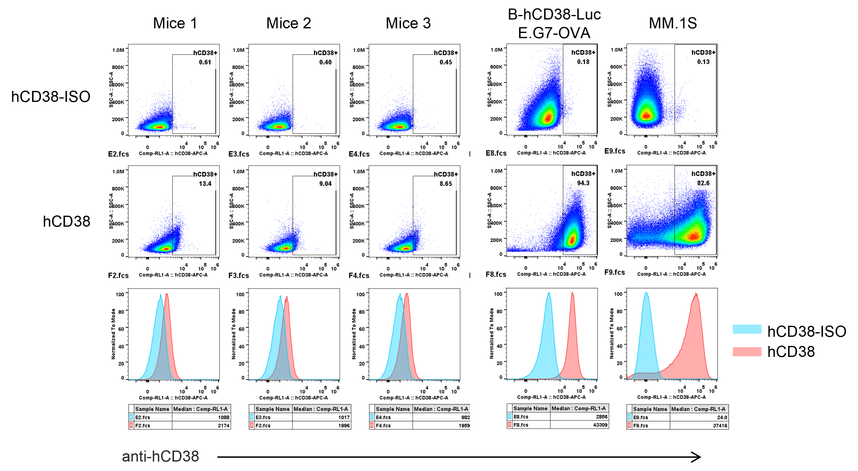
      The binding of anti-CD38 antibody Daratumumab analog (in-house) to mouse B cells of B-hCD38 mice, mouse T-cell lymphoma B-hCD38-Luc E.G7-OVA, and human multiple myeloma cells MM.1S. was assessed by FACS.

      Using B cells from the spleens of B-hCD38 mice, mouse T-cell lymphoma B-hCD38-Luc E.G7-OVA cells, or human multiple myeloma MM.1S cells as target cells, FcR-TANK cells were added at 5:1 ratio. Following the addition of varying concentrations of anti-hCD8 antibody and incubation at 37°C for 4 hours, specific lysis was assessed via flow cytometry. Panel A shows the results for three mice. Panel B shows the results for mouse T-cell lymphoma B-hCD38-Luc E.G7-OVA cells and human multiple myeloma MM.1S cells.

      B-hCD38 Mice: Analysis of Leukocyte Subpopulations

      Analysis of leukocyte subpopulations by flow cytometry in immune organs and blood. Splenocytes, peripheral blood, and lymph nodes were isolated from C57BL/6JNifdc mice and homozygous B-hCD38 mice (female, 8-week-old, n = 4). Single live cells were gated on the CD45⁺ population and analyzed by flow cytometry as indicated. Values are expressed as mean ± SEM.

      B-hCD38 Mice: Analysis of T Cell Subpopulations

      Analysis of T-cell subpopulations by flow cytometry in immune organs and blood. Splenocytes, peripheral blood, and lymph nodes were isolated from C57BL/6JNifdc mice and homozygous B-hCD38 mice (female, 8-week-old, n = 4). Single live cells were gated on the TCRβ⁺ T-cell population and analyzed by flow cytometry as indicated. Values are expressed as mean ± SEM.

      Frequently Asked Questions (FAQs) About CD38 Humanized Mice (B-hCD38)

      Q: What should be noted when using B-hCD38 mice (#110046)?

      A: In B-hCD38 mice (#110046), the extracellular domain of CD38 is only partially humanized rather than fully humanized. Therefore, it is necessary to verify the binding of anti-human CD38 antibodies to B-hCD38 mice (#110046) before starting in vivo experiments.


      Q: What is the difference between B-hCD38 mice (#110046) and B-hCD38 mice ad (#113475)?

      A: The targeting strategy for B-hCD38 mice (#110046) is humanization of a portion of the CD38 extracellular domain. B-hCD38 mice (#113475) were humanized with the full-length CD38 genome sequence, resulting in the expression of the full-length human CD38 protein.

      * When publishing results obtained using this animal model, please acknowledge the source as follows: The animal model [B-hCD38 mice] (Cat# 110046) was purchased from Biocytogen.