

C57BL/6-Cd3etm1(CD3E)Bcgen Cd3dtm1(CD3D)Bcgen Cd3gtm1(CD3G)Bcgen Cd28tm1(hCD28)Bcgen/Bcgen • 121285

CD3/CD28: A pivotal dual-receptor axis for complete T cell activation and its therapeutic engagement.

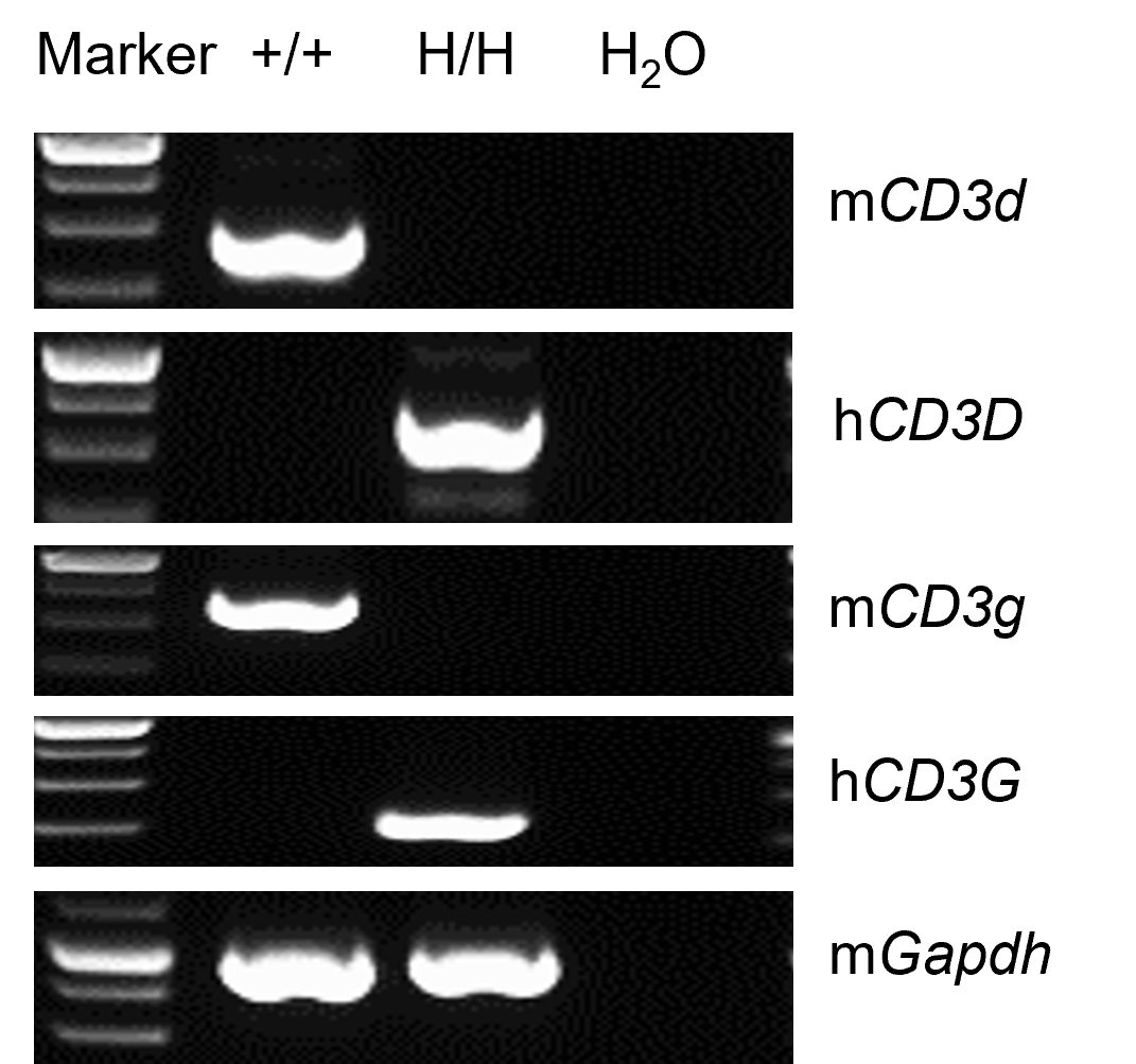
Mouse and human CD3D and CD3G analysis in thymus by RT-PCR and sequencing. Thymus RNA was isolated from wild-type C57BL/6 mice (+/+) and homozygous B-hCD3EDG/hCD28 mice (H/H, H/H), and then cDNA libraries were synthesized by reverse transcription, followed by PCR with mouse or human CD3D and CD3G primers.
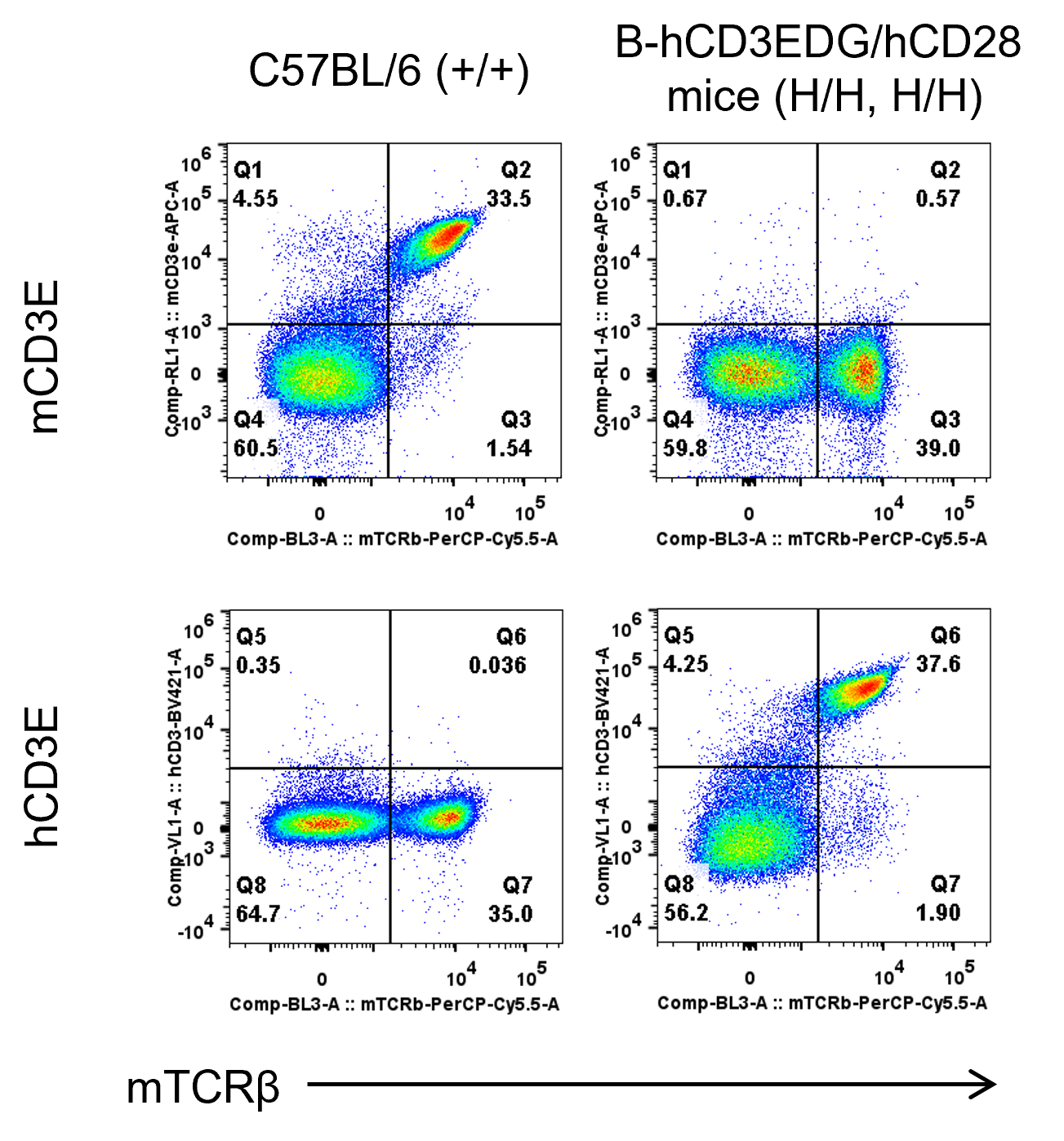
Mouse and human CD3E expression analysis in splenocytes. Splenocytes were collected from wild-type C57BL/6 mice and homozygous B-hCD3EDG/hCD28 mice (female, 8-week-old, n = 1). CD3E expression on T cells was analyzed by flow cytometry using species-specific anti-CD3E antibodies (anti-human CD3E antibody, BD Horizon, 562426; anti-mouse CD3E antibody, Biolegend, 100312 ).
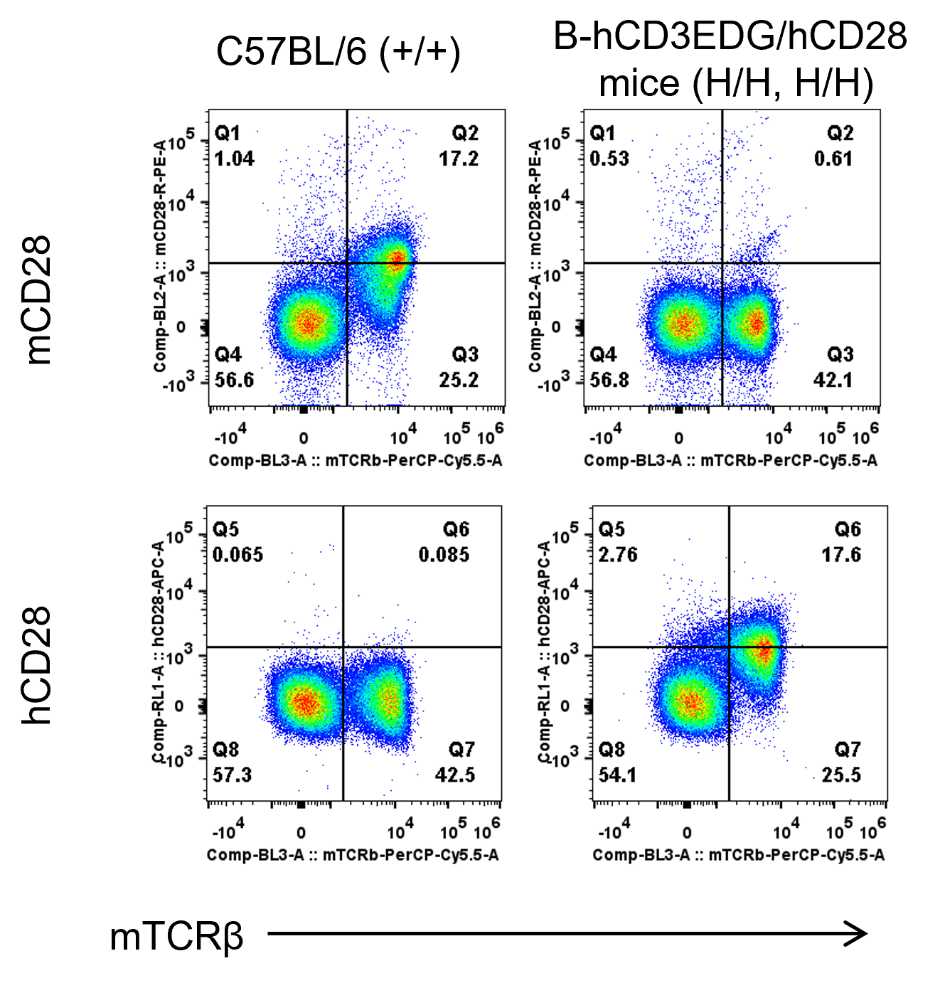
Mouse and human CD28 expression analysis in splenocytes. Splenocytes were collected from wild-type C57BL/6 mice and homozygous B-hCD3EDG/hCD28 mice (female, 8-week-old, n = 1). CD28 expression on T cells was analyzed by flow cytometry using species-specific anti-CD28 antibodies (anti-human CD28 antibody, Biolegend, 302912; anti-mouse CD28 antibody, Biolegend, 102105).
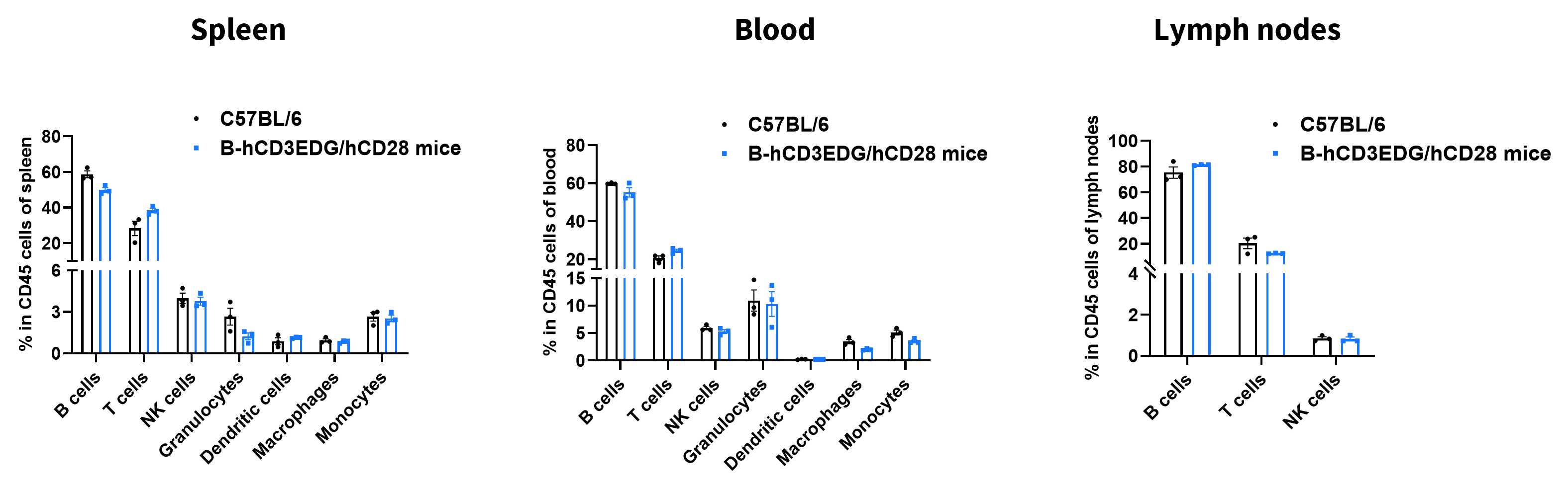
Analysis of leukocyte subpopulations by flow cytometry in immune organs and blood. Splenocytes, peripheral blood, and lymph nodes were isolated from female C57BL/6 and B-hCD3EDG/hCD28 mice (female, 8-week-old, n = 3). Single live cells were gated on the CD45⁺ population and analyzed by flow cytometry as indicated. Values are expressed as mean ± SEM.
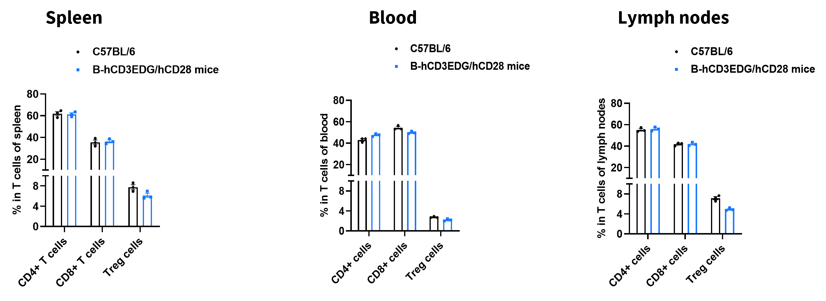
Analysis of T-cell subpopulations by flow cytometry in immune organs and blood. Splenocytes, peripheral blood, and lymph nodes were isolated from female C57BL/6 and B-hCD3EDG/hCD28 mice (female, 8-week-old, n = 3). Single live cells were gated on the TCR+ T-cell population and analyzed by flow cytometry as indicated. Values are expressed as mean ± SEM.
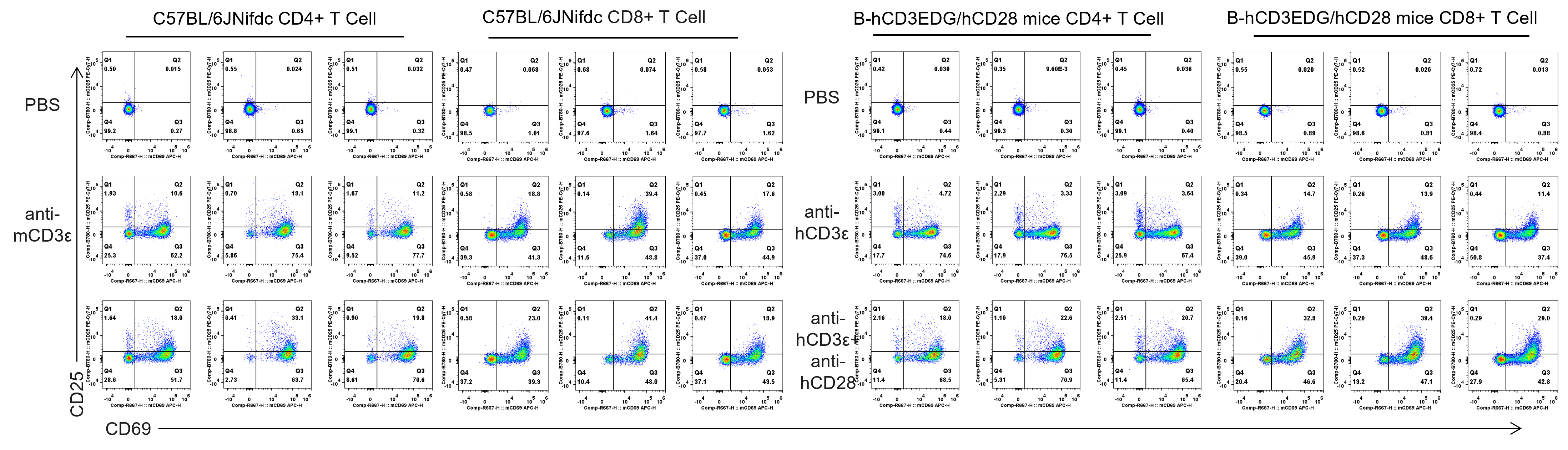
Analysis of T cell activation stimulated with anti-CD3 and anti-CD28 antibody in vitro. T cells were isolated from splenocytes of C57BL/6JNifdc and B-hCD3EDG/hCD28 mice (n=3, 7 week-old), and were incubated in the presence of anti-mCD3ε antibody (2ug/ml, BioXcell, BE0001-2) , anti-hCD3ε antibody (2ug/ml, BioXcell, BE0001-1), anti-mCD28 antibody (5ug/ml, BioXcell, BE0015-1) and anti-hCD28 antibody (5ug/ml, BioXcell, BE0248) for 24h, tested by flow cytometry (Anti-mCD25 antibody: Biolegend, 104514; Anti-mCD69 antibody: Biolegend, 102034).
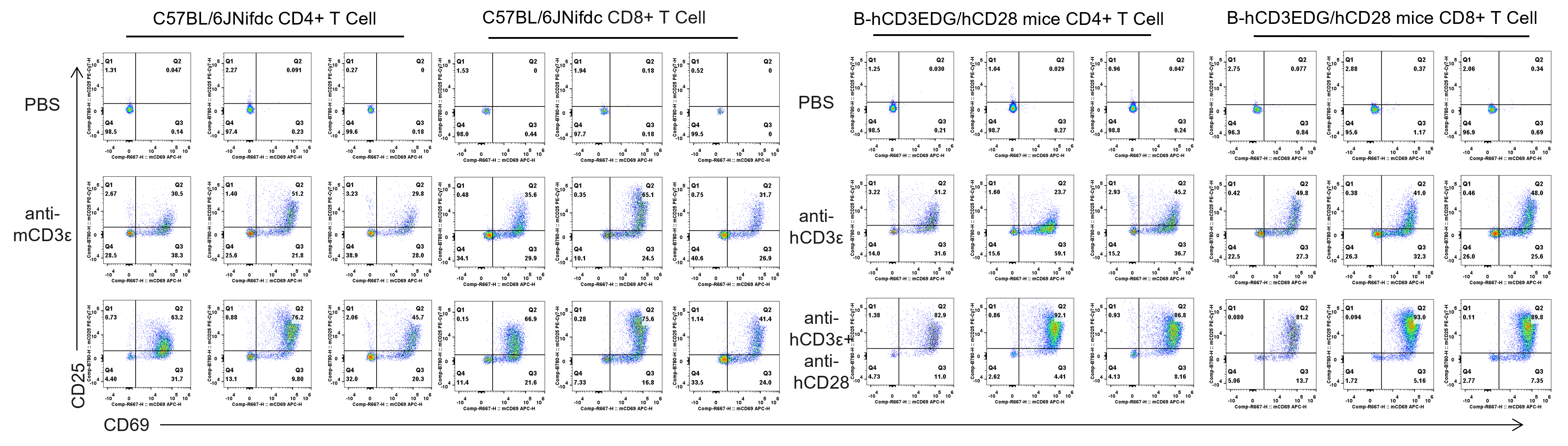
Analysis of T cell activation stimulated with anti-CD3 and anti-CD28 antibody in vitro. T cells were isolated from splenocytes of C57BL/6JNifdc and B-hCD3EDG/hCD28 mice (n=3, 7 week-old), and were incubated in the presence of anti-mCD3ε antibody (2ug/ml, BioXcell, BE0001-2) , anti-hCD3ε antibody (2ug/ml, BioXcell, BE0001-1), anti-mCD28 antibody (5ug/ml, BioXcell, BE0015-1) and anti-hCD28 antibody (5ug/ml, BioXcell, BE0248) for 48h, tested by flow cytometry (Anti-mCD25 antibody: Biolegend, 104514; Anti-mCD69 antibody: Biolegend, 102034).
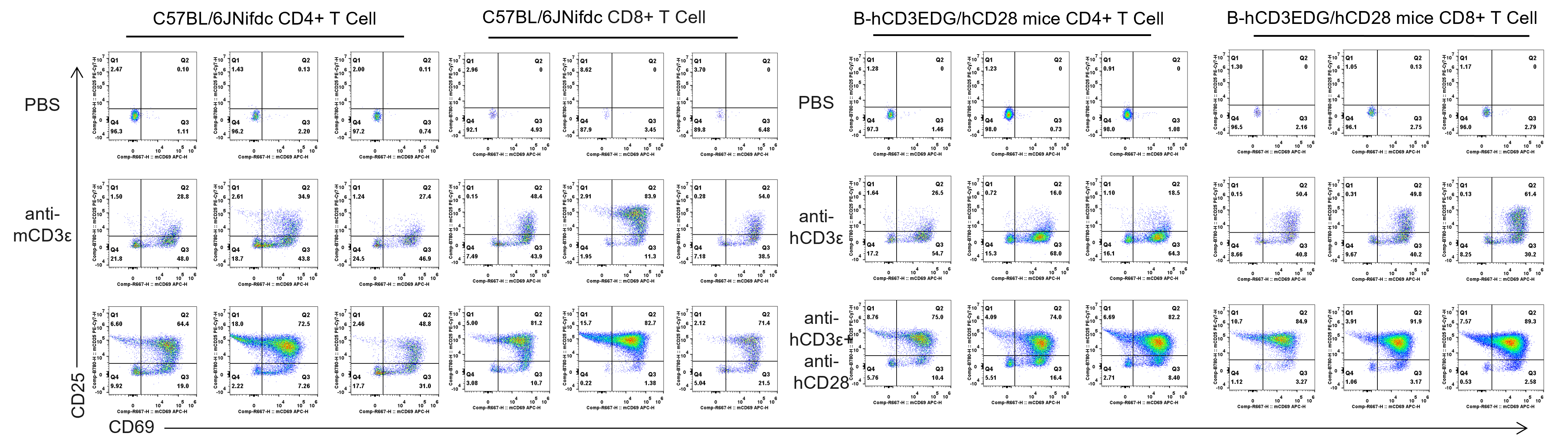
Analysis of T cell activation stimulated with anti-CD3 and anti-CD28 antibody in vitro. T cells were isolated from splenocytes of C57BL/6JNifdc and B-hCD3EDG/hCD28 mice (n=3, 7 week-old), and were incubated in the presence of anti-mCD3ε antibody (2ug/ml, BioXcell, BE0001-2) , anti-hCD3ε antibody (2ug/ml, BioXcell, BE0001-1), anti-mCD28 antibody (5ug/ml, BioXcell, BE0015-1) and anti-hCD28 antibody (5ug/ml, BioXcell, BE0248) for 72h, tested by flow cytometry (Anti-mCD25 antibody: Biolegend, 104514; Anti-mCD69 antibody: Biolegend, 102034).
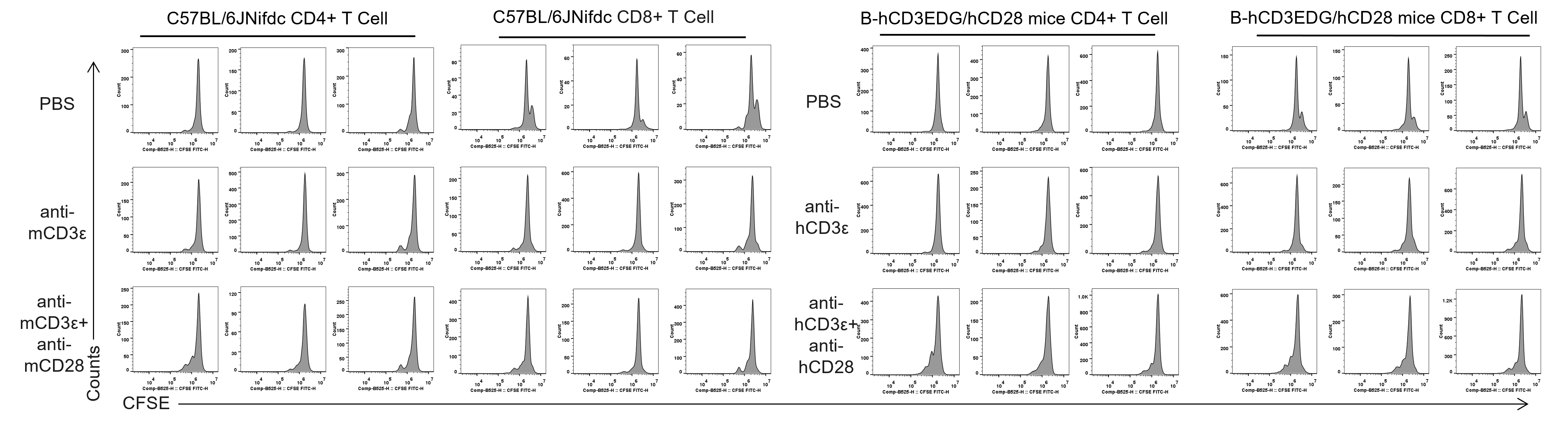
Analysis of T cell proliferaion stimulated with anti-CD3 and anti-CD28 antibody in vitro. T cells were isolated from splenocytes of C57BL/6JNifdc and B-hCD3EDG/hCD28 mice (n=3, 7 week-old), and were incubated in the presence of anti-mCD3ε antibody (2ug/ml, BioXcell, BE0001-2) , anti-hCD3ε antibody (2ug/ml, BioXcell, BE0001-1) , anti-mCD28 antibody (5ug/ml, BioXcell, BE0015-1) and anti-hCD28 antibody (5ug/ml, BioXcell, BE0248) for 48h, tested by flow cytometry (CFSE: Invitrogen, C34554).
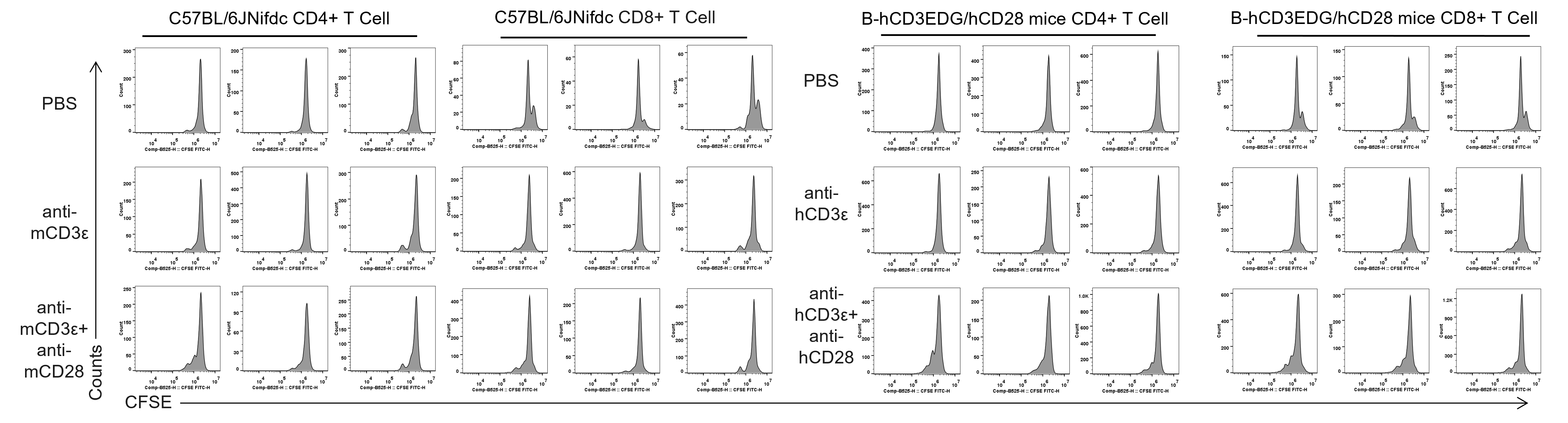
Analysis of T cell proliferaion stimulated with anti-CD3 and anti-CD28 antibody in vitro. T cells were isolated from splenocytes of C57BL/6JNifdc and B-hCD3EDG/hCD28 mice (n=3, 7 week-old), and were incubated in the presence of anti-mCD3ε antibody (2ug/ml, BioXcell, BE0001-2) , anti-hCD3ε antibody (2ug/ml, BioXcell, BE0001-1) , anti-mCD28 antibody (5ug/ml, BioXcell, BE0015-1) and anti-hCD28 antibody (5ug/ml, BioXcell, BE0248) for 72h, tested by flow cytometry (CFSE: Invitrogen, C34554).
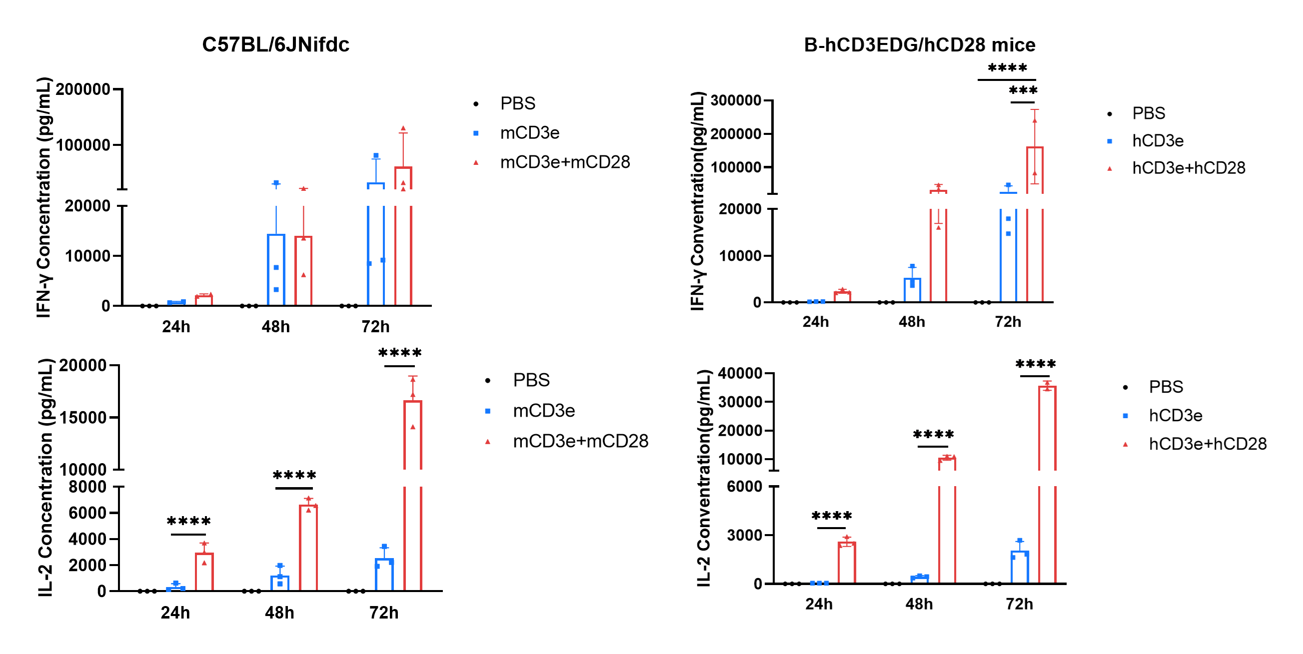
Analysis of T cell activation stimulated with anti-CD3 and anti-CD28 antibody in vitro. T cells were isolated from splenocytes of C57BL/6JNifdc and B-hCD3EDG/hCD28 mice (n=3, 7 week-old), and were incubated in the presence of anti-mCD3ε antibody (2ug/ml, BioXcell, BE0001-2) , anti-hCD3ε antibody (2ug/ml, BioXcell, BE0001-1), anti-mCD28 antibody (5ug/ml, BioXcell, BE0015-1) and anti-hCD28 antibody (5ug/ml, BioXcell, BE0248) for 24h, 48h and 72h. IFN-γ and IL-2 productions were then tested using the SLU Testing System (From SOLUTION). Values are expressed as mean ± SEM. Significance was determined by two-way ANOVA test. *P < 0.05, **P < 0.01, ***p < 0.001, ****p < 0.0001.
