Description
- Gene Information: PD-1 (PDCD1), PD-L1 (CD274), and HER2 (ERBB2) are clinically validated therapeutic targets involved in immune regulation and oncogenic signaling. Together, they represent key targets for cancer immunotherapy and targeted therapy.
- Protein Expression: PD-1 is primarily expressed on activated T cells, while PD-L1 is expressed on tumor cells and immune cells. HER2 is overexpressed in multiple solid tumors, especially breast and gastric cancers.
- Signaling Pathway: PD-1 binding to PD-L1 inhibits T-cell activation and promotes immune evasion. HER2 activates PI3K/AKT and MAPK signaling to drive tumor cell proliferation, survival, and metastasis.
- Model application : PD-1/PD-L1 blockade restores T-cell function, whereas HER2-targeted therapies directly eliminate HER2-positive tumor cells. Their combination has demonstrated enhanced anti-tumor efficacy and is widely explored in clinical development.
Targeting strategy
PD-1
- Exon 2 of the mouse PD-1 gene, which encode the IgV domain, was replaced with human PD-1 exon 2 in B-hPD-1/hPD-L1/hHER2 mice.
- The endogenous mouse promoter, 5′ UTR, 3′ UTR, and genomic regions encoding the signal peptide, non-IgV domain of the Extracellular domain, transmembrane and cytoplasmic domains are retained. The chimeric PD-1 expression is driven by the endogenous mouse PD-1 promoter, while endogenous mouse PD-1 transcription and translation are disrupted.
PD-L1
- Exon 3 of the mouse PD-L1 gene, which encode the IgV domain, was replaced with human PD-L1 exon 3 in B-hPD-1/hPD-L1/hHER2 mice.
- The endogenous mouse promoter, 5′ UTR, 3′ UTR, and genomic regions encoding the signal peptide, non-IgV domain of the Extracellular domain, transmembrane and cytoplasmic domains are retained. The chimeric PD-L1 expression is driven by the endogenous mouse PD-L1 promoter, while endogenous mouse PD-L1 transcription and translation are disrupted.
HER2
- The exons 2-17 of mouse Erbb2 gene that encode extracellular domain and transmembrane domain were replaced by human counterparts in B-hPD-1/hPD-L1/hHER2 mice.
- The genomic region of mouse Erbb2 gene that encodes cytoplasmic portion was retained. The promoter, 5’UTR and 3’UTR region of the Erbb2 gene were also retained. The chimeric ERBB2 expression was driven by endogenous mouse Erbb2 promoter, while mouse Erbb2 gene transcription and translation will be disrupted.
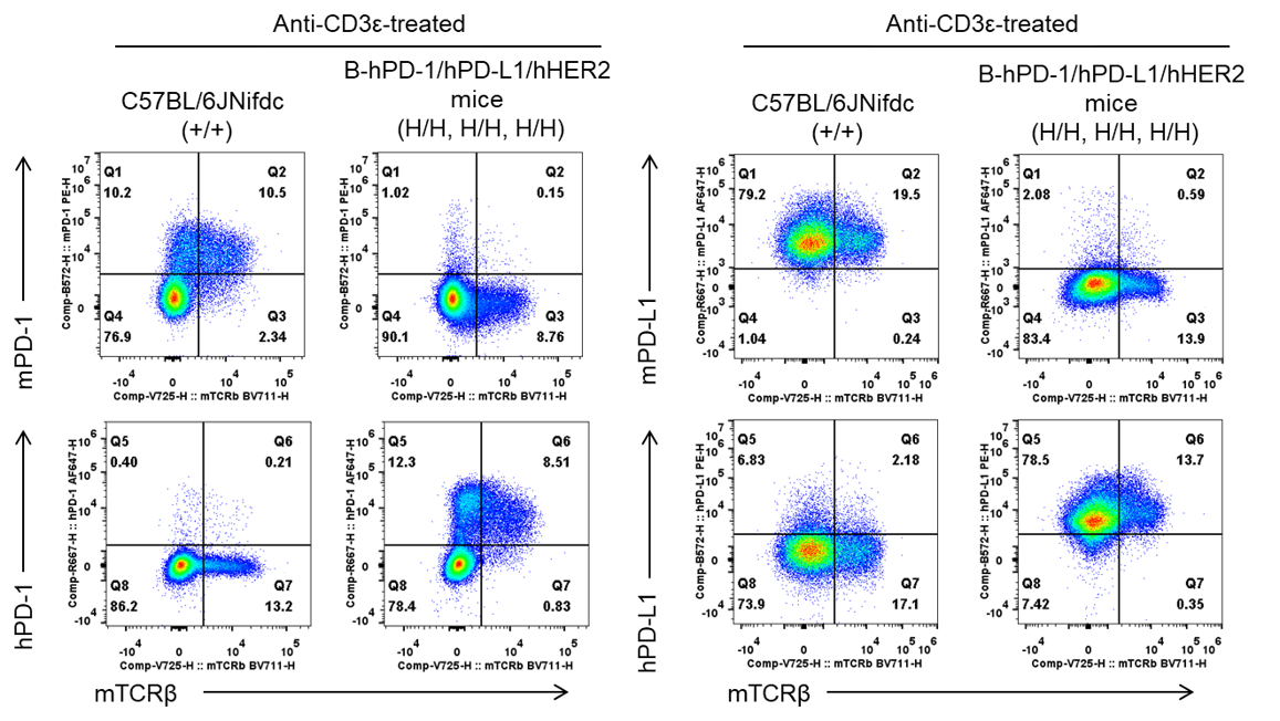
PD-1/PD-L1 Protein Expression Analysis
- Human PD-1 and PD-L1 are specifically detected in homozygous B-hPD-1/hPD-L1/hHER2 mice by flow cytometry.
Strain-specific analysis of PD-1 and PD-L1 protein expression in wild-type C57BL/6 mice and homozygous B-hPD-1/hPD-L1/hHER2 mice by flow cytometry. Splenocytes were isolated from wild-type C57BL/6 mice (+/+) and homozygous B-hPD-1/hPD-L1/hHER2 mice (H/H) and stimulated with anti-CD3ε antibody. Protein expression was analyzed with anti-human PD-1 antibody (Biolegend, 329908), anti-mouse PD-1 antibody (Biolegend, 109104), anti-human PD-L1 antibody (Biolegend, 329706) and anti-mouse PD-L1 antibody (Biolegend, 124312) by flow cytometry.
HER2 Protein Expression
- Human HER2 was detected in the uterus, breast, lung and colon of homozygous B-hPD-1/hPD-L1/hHER2 mice, but not in C57BL/6 mice.
Immunohistochemical (IHC) analysis of HER2 protein expression in wild-type mice and B-hPD-1/hPD-L1/hHER2 mice. Major tissues were collected from wild-type mice and homozygous B-hPD-1/hPD-L1/hHER2 mice and analyzed by IHC with anti-human HER2 antibody (HUABIO, HA721178).
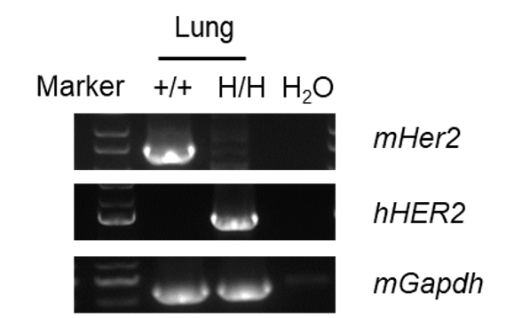
Expression by RT-PCR
- Mouse Her2 mRNA was detectable in wild-type C57BL/6 mice.
- Human HER2 mRNA was detectable only in homozygous B-hPD-1/hPD-L1/hHER2 mice but not in wild-type mice.
Strain specific analysis of HER2 mRNA expression in wild-type C57BL/6 mice and homozygous B-hPD-1/hPD-L1/hHER2 mice by RT-PCR. Lung RNA were isolated from wild-type C57BL/6 mice (+/+) and homozygous B-hPD-1/hPD-L1/hHER2 mice (H/H, H/H, H/H), then cDNA libraries were synthesized by reverse transcription, followed by PCR with mouse or human HER2 primers. Sequencing of the short-form PCR products confirmed that the amplified sequences were consistent with database reference sequences.
In vivo Efficacy of HER2 ADC DS8201 combined with Anti-hPD-1 Antibody Keytruda
- Anti-human HER2 ADC DS8201 with anti-hPD-1 antibody Keytruda therapy showed superior anti-tumor efficacy compared with monotherapy in B-hPD-1/hPD-L1/hHER2 mice, demonstrating that this model is a valuable platform for evaluating cancer immunotherapies and combination treatment strategies.
Establishment of a MC38 model in B-hPD-1/hPD-L1/hHER2 mice and In vivo Efficacy of HER2 ADC DS8201 combined with anti-hPD-1 Antibody Keytruda. Murine colon cancer B-hHER2 MC38 plus cells (Cat# 322353) were subcutaneously implanted into homozygous B-hPD-1/hPD-L1/hHER2 mice (female, 7-week-old, n=6). Mice were grouped when tumor volume reached approximately 250-300 mm3, at which time they were intravenously injected with anti-human HER2 ADC DS8201 (purchased from Daiichi Sankyo) or Intraperitoneal injected anti-hPD-1 antibody Keytruda (in-house).
CG: Complete tumor regression
- Anti-human HER2 ADC DS8201 with anti-hPD-1 antibody Keytruda therapy showed superior anti-tumor efficacy compared with monotherapy in B-hPD-1/hPD-L1/hHER2 mice, demonstrating that this model is a valuable platform for evaluating cancer immunotherapies and combination treatment strategies.
In vivo Efficacy of HER2 ADC DS8201 combined with Anti-hPD-1 antibody Keytruda-individual tumor growth curves.
* When publishing results obtained using this animal model, please acknowledge the source as follows: The animal model [B-hPD-1/hPD-L1/hHER2 mice] (Cat# 112936) was purchased from Biocytogen.