Description
SCF/KIT: A Critical Signaling Axis in Hematopoiesis, Mast Cell Biology, and Oncogenesis
- Gene Information: Stem Cell Factor (SCF / KITLG) is a protein-coding gene on chromosome 12q21.32 that encodes a cytokine ligand. Its receptor, KIT (c-Kit / CD117), is a type III receptor tyrosine kinase proto-oncogene located on chromosome 4q12.
- Protein Expression: SCF is expressed by bone marrow stromal cells, endothelial cells, fibroblasts, and keratinocytes as biologically active soluble or membrane-bound isoforms. KIT is highly expressed on hematopoietic stem cells (HSCs), mast cells, melanocytes, and interstitial cells of Cajal (ICCs).
- Signaling Pathway: SCF binding to the extracellular domain of KIT induces receptor homodimerization and intracellular tyrosine autophosphorylation. This activates downstream PI3K/Akt, MAPK/ERK, and JAK/STAT pathways to regulate cell survival, proliferation, and migration.
- Therapeutic Inhibition: Pathologic SCF/KIT signaling is targeted across two major clinical areas: Oncology: Small-molecule tyrosine kinase inhibitors (TKIs) like imatinib, sunitinib, and avapritinib block the ATP-binding site of mutated KIT, forcing tumor regression in GISTs and reducing mast cell burden in systemic mastocytosis. Autoimmunity & Allergy: In wild-type KIT diseases (e.g., Chronic Spontaneous Urticaria, asthma), biologics like barzolvolimab (anti-KIT mAb) or anti-SCF/anti-TSLP bispecific antibodies block ligand binding. This starves mast cells, inducing apoptosis to deplete tissue mast cells, lower serum tryptase, and suppress chronic inflammation.
Targeting strategy
KIT
- A chimeric CDS that encodes human KIT extracellular domain, mouse kit transmembrane and cytoplasmic domain, followed by mouse 3’UTR-STOP is inserted in exon 2 of mouse Kit gene.
- The chimeric KIT protein expression will be driven by endogenous mouse kit promoter, while mouse Kit gene transcription and translation will be disrupted.
KIT Protein Expression in Spleen
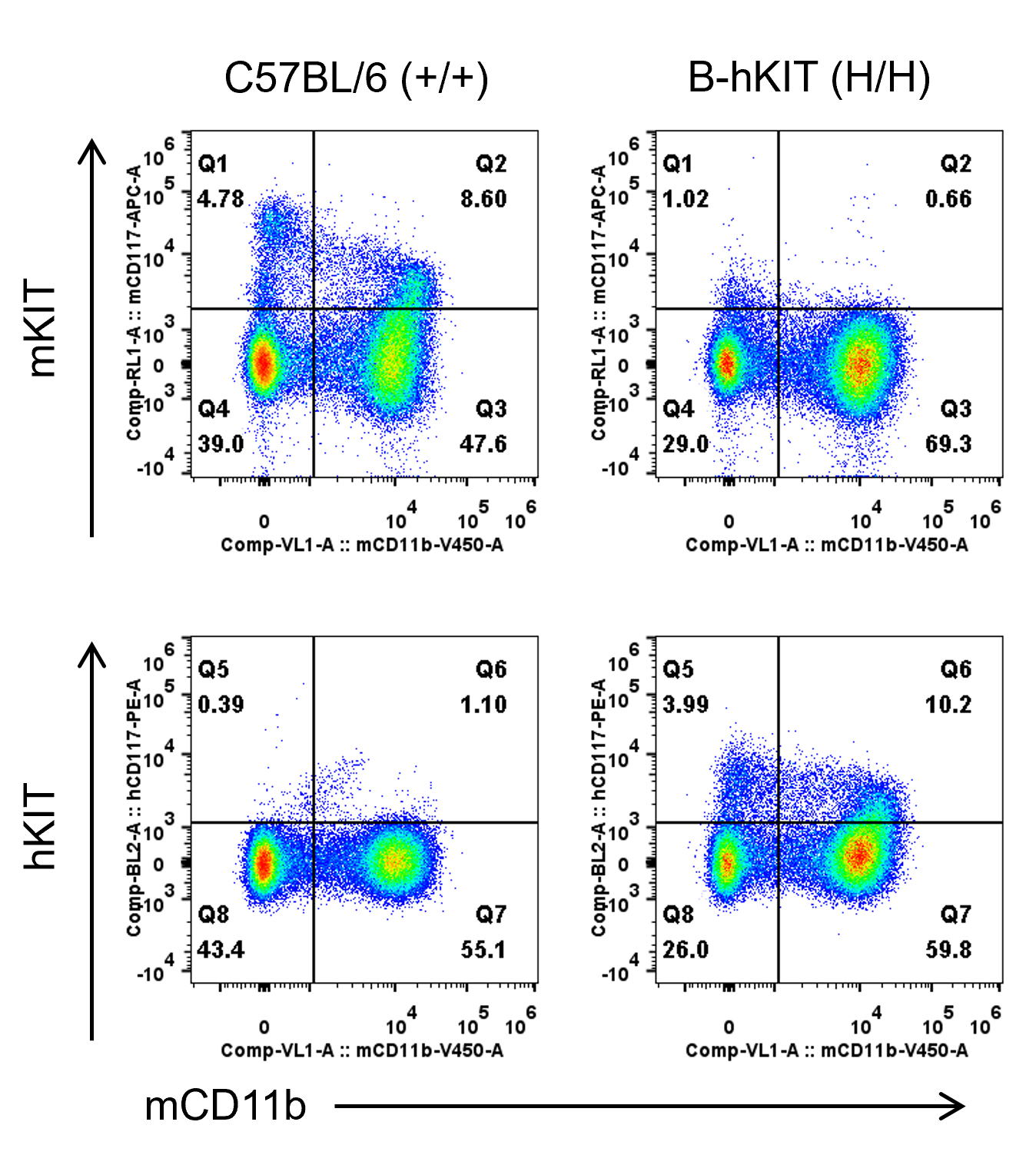
- Mouse KIT was detectable in wild-type mice.
- Human KIT was exclusively detectable in homozygous B-hKIT mice but not in wild-type mice.
Mouse and human KIT expression analysis in Bone marrow cells. Bone marrow were collected from wild-type C57BL/6 mice and homozygous B-hKIT mice. KIT expression was analyzed by flow cytometry using anti-mouse KIT antibody (Biolegend, 105811) and anti-human KIT antibody (Biolegend, 313203).
Compound 48/80 (C48/80) Induced Systemic Anaphylaxis Reaction
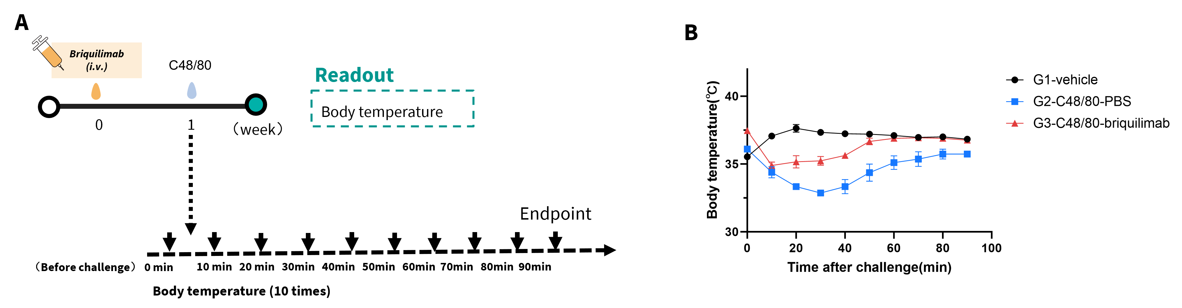
Effects of C48/80‑induced systemic anaphylaxis reaction. (A) Anti‑hKIT antibody briquilimab (25 mpk, commercially purchased) was injected before challenge at week 0. B‑hKIT mice (H/H) (n=3) were challenged with C48/80 at week 1. (B) Body temperature was recorded from 0 to 90 minutes after challenge with C48/80. briquilimab, by depleting mast cells, attenuated the decrease in body temperature induced by C48/80, demonstrating its inhibitory effect on the systemic anaphylaxis reaction in B-hKIT mice.
* When publishing results obtained using this animal model, please acknowledge the source as follows: The animal model [B-hKIT mice] (Cat# 112218) was purchased from Biocytogen.