

C57BL/6N-Slc3a2tm1(SLC3A2)Bcgen/Bcgen • 110983

Key Advantages:
Model Validation:
Applications:

Gene targeting strategy for CD98HC humanized mice. The exons 2-10 of mouse Cd98hc gene that encode extracellular domain were replaced by exons 4–12 of human CD98HC in CD98HC humanized mice. The genomic region of mouse Cd98hc gene that encodes cytoplasmic portion was retained. The promoter, 5' UTR and 3' UTR regions of the mouse gene were also retained. The chimeric CD98HC expression is driven by endogenous mouse Cd98hc promoter, while mouse Cd98hc gene transcription and translation will be disrupted.

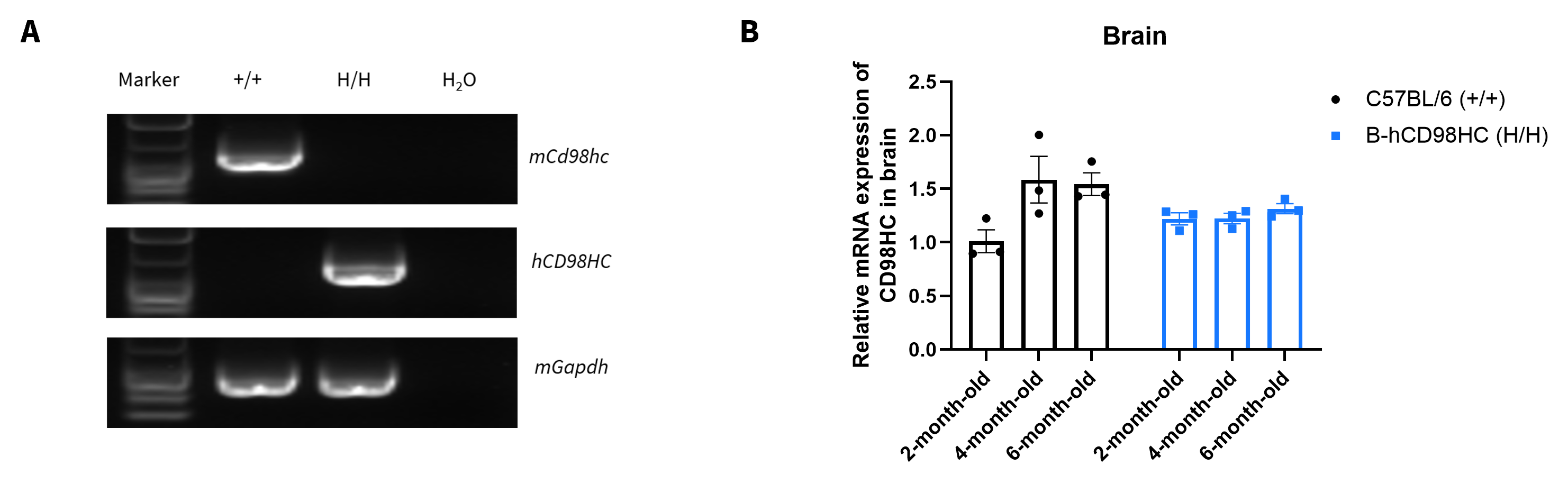
Strain-specific CD98HC mRNA expression analysis in wild-type C57BL/6 mice and homozygous CD98HC humanized mice by RT-PCR and qRT-PCR. (A) Total RNA was isolated from kidney tissues of wild-type C57BL/6 mice (+/+) and homozygous CD98HC humanized mice (H/H), then cDNA libraries were synthesized by reverse transcription, followed by PCR with mouse or human CD98HC primers. (B) Quantitative real-time PCR (qRT-PCR) analysis of relative CD98HC mRNA expression in the brain of male wild-type C57BL/6 mice and homozygous CD98HC humanized mice at 2, 4 and 6-months of age. Expression levels were normalized to CD98HC mRNA expression in the brain of 2-month-old C57BL/6 male mice. Values are expressed as mean ± SEM.
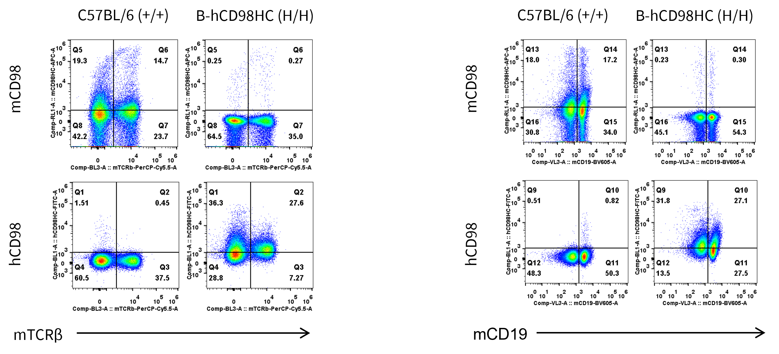
Strain specific CD98HC protein expression analysis in wild-type C57BL/6 mice and homozygous CD98HC humanized mice flow cytometry. Splenocytes were collected from wild-type C57BL/6 mice (+/+) and homozygous CD98HC humanized mice (H/H) (female, 7-week-old, n=1). Protein expression was analyzed with anti-mouse CD98HC antibody (Biolegend, 128211) and anti-human CD98HC antibody (Biolegend, 315603) by flow cytometry.
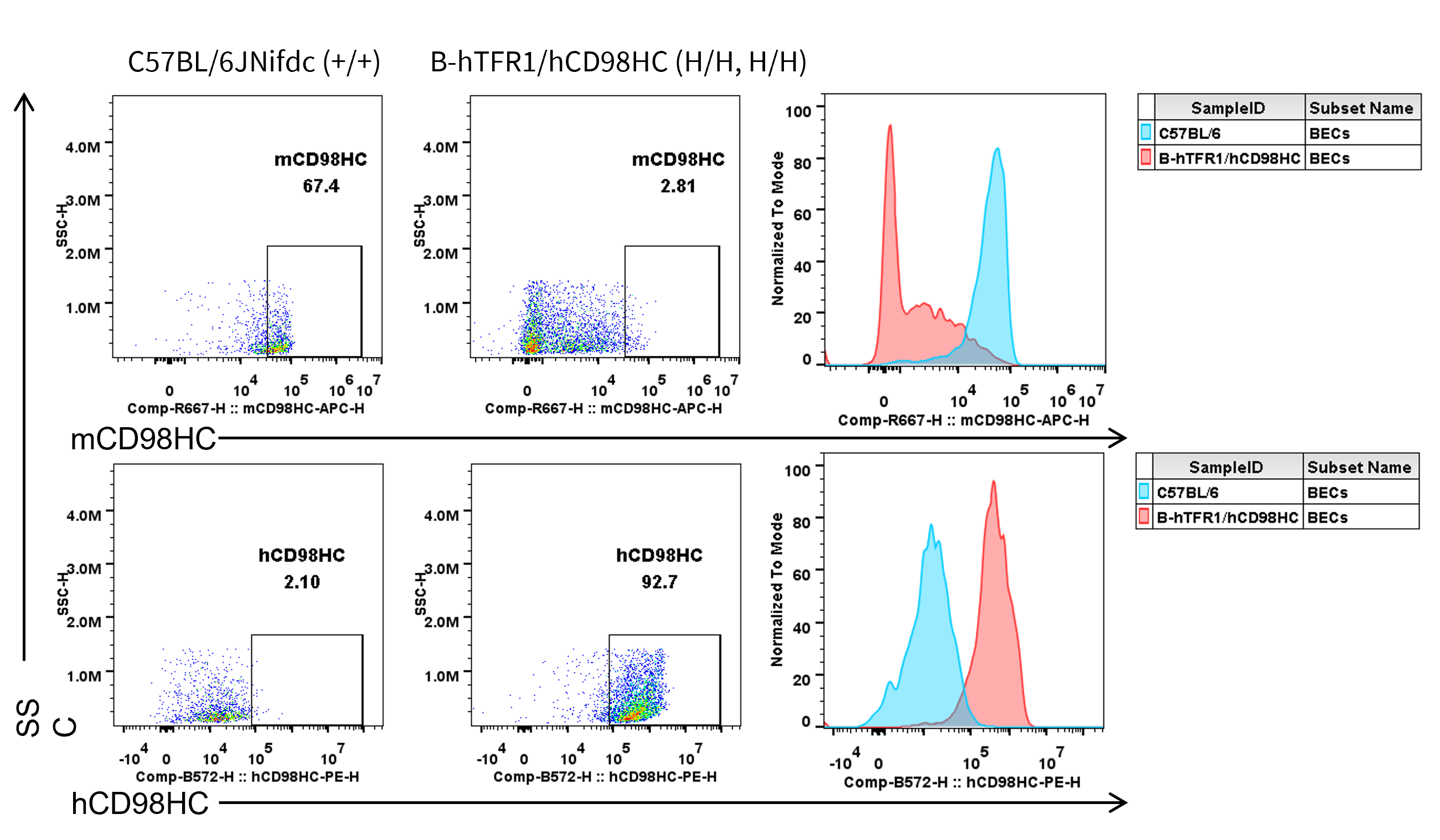
Strain specific CD98HC protein expression in brain cells from wild-type C57BL/6JNifdc and homozygous TFR1/CD98HC humanized mice by flow cytometry. Brain cells were collected from wild-type C57BL/6JNifdc (+/+) and homozygous TFR1/CD98HC humanized mice (H/H, H/H) and analyzed by flow cytometry with anti-mouse CD98HC antibody (Biolegend, 128211) and anti-human CD98HC antibody (231161-CD98BBBB-h1.L produced in-house).
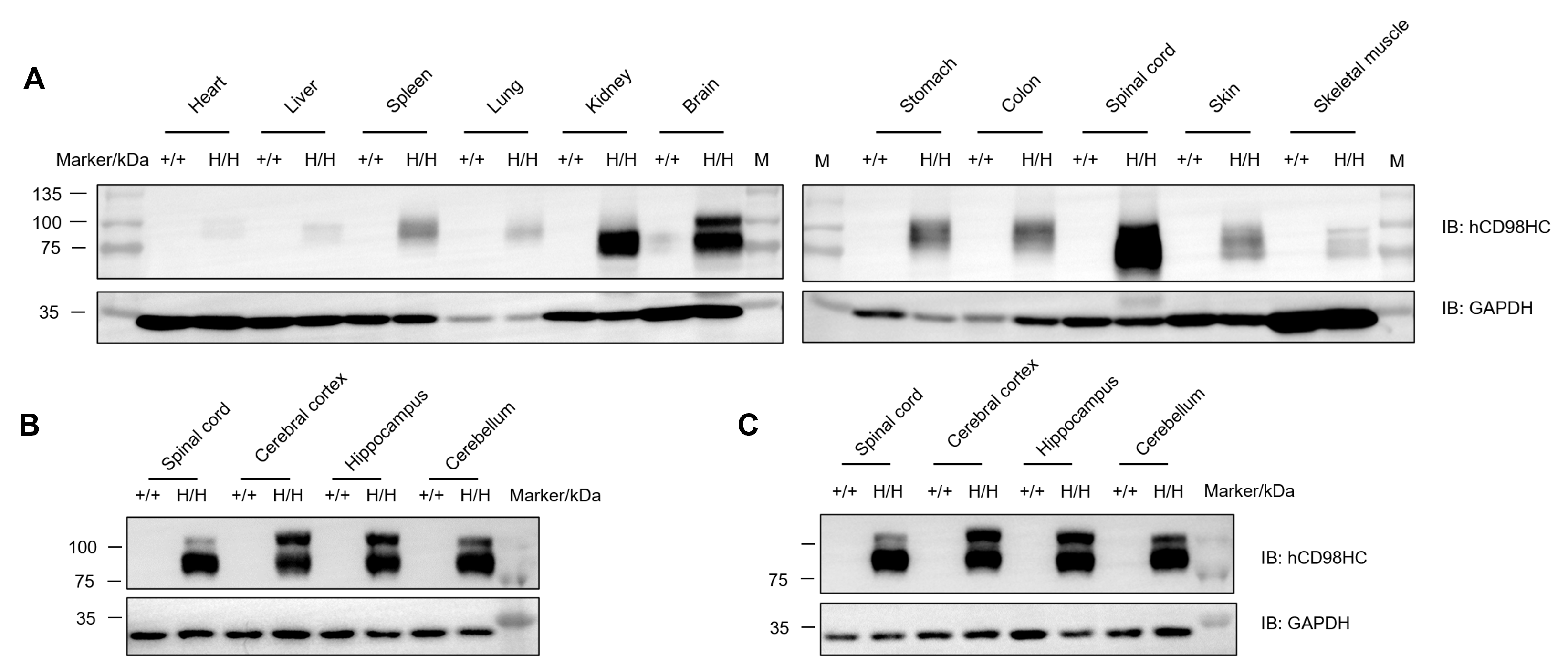
Western blot analysis of human CD98HC protein expression in homozygous CD98HC humanized mice.
Various tissue lysates were collected from wild-type C57BL/6 and homozygous CD98HC humanized mice (H/H), and then analyzed by western blot with species-specific anti-human CD98 antibody (Abcam, ab307587). (A) Male mice. (B) Male mice. (C) Female mice.
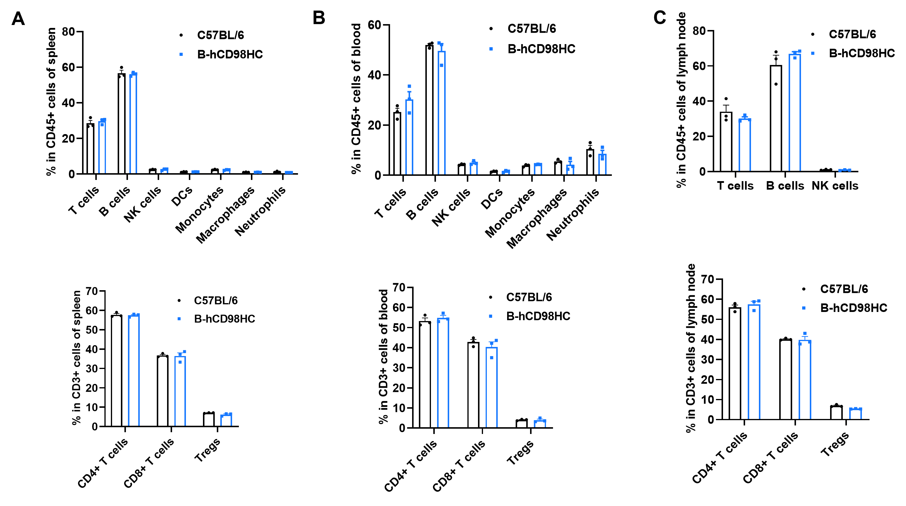
Humanization of CD98HC does not alter the frequency or distribution of immune cell populations in the spleen (A), peripheral blood (B), or lymph nodes (C) of CD98HC humanized mice.
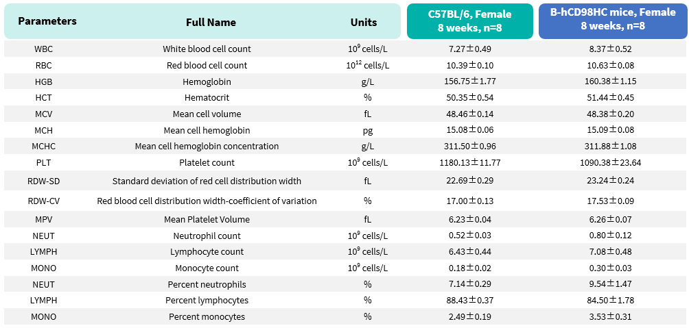
Complete blood count (CBC) of CD98HC humanized mice. Values are expressed as mean ± SD.
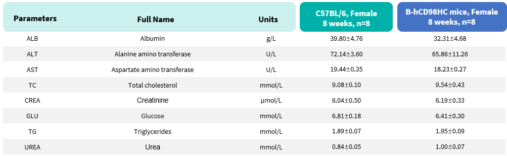
Biochemical test of CD98HC humanized mice. Values are expressed as mean ± SD.


In vivo pharmacokinetic (PK) evaluation of anti-human CD98HC antibody in CD98HC humanized mice. CD98HC humanized mice (n=2, female, 8-week-old) were injected with control IgG (10 mpk) and anti-human CD98HC antibody (CD98BBBB-h1.L analog, monovalent, produced in house, 13.3 mpk) via tail vein. Brain and serum were collected for in vivo pharmacokinetic (PK) evaluation. Brain and serum antibody concentration, as well as brain-to-serum ratio of anti-human CD98HC antibody were quantified. Data are presented as mean ± SEM.
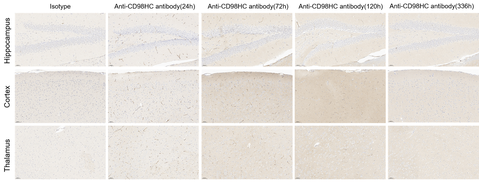
Immunohistochemical (IHC) staining of anti-CD98HC antibodies penetrates brain parenchyma of CD98HC humanized mice. CD98HC humanized mice were injected with control IgG (10 mpk) and anti-human CD98HC antibody (CD98BBBB-h1.L analog, monovalent, produced in house, 13.3 mpk) via tail vein. Brain comparts were collected for IHC staining after 120 h. Blue: nucleus; dark brown: antibodies.
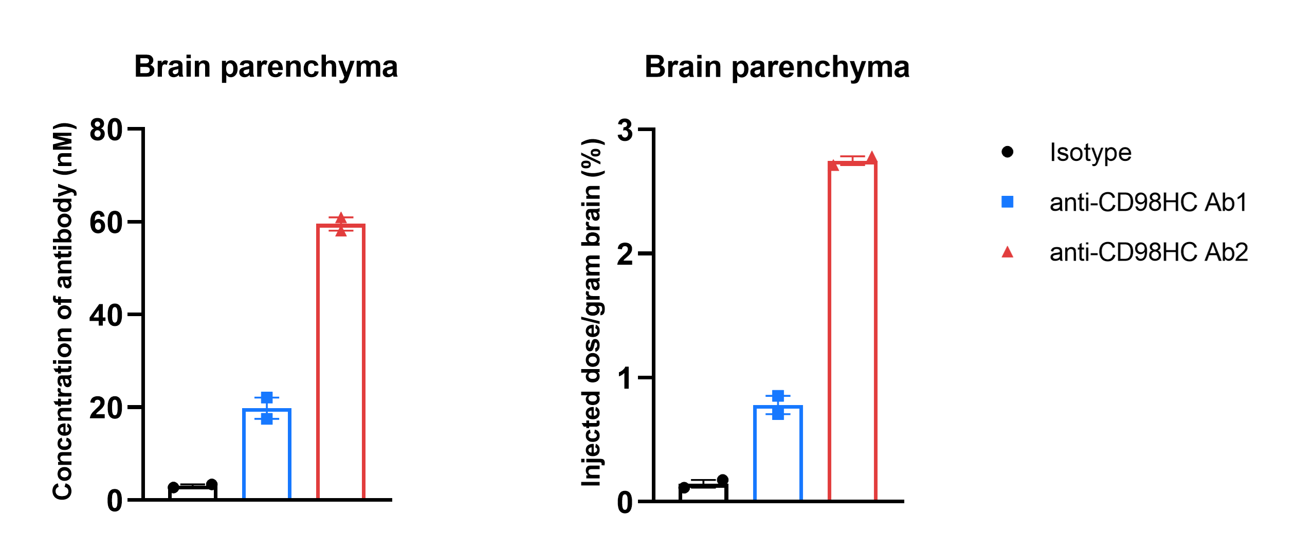
In vivo pharmacokinetic (PK) evaluation and comparison of different anti-human CD98HC antibodies in CD98HC humanized mice. CD98HC humanized mice (n=2, 8-week-old) were injected with Isotype IgG (20 mpk) and anti-human CD98HC antibodies (Ab1 and Ab2, produced in house according to patent) via tail vein. Brain tissues were collected for in vivo PK evaluation after dosing 5 days. Brain concentrations (A) and % of injection/gram brain (B) were quantified. As shown in panel, anti-CD98HC Ab2 exhibited higher brain exposure. Data are presented as mean ± SEM.
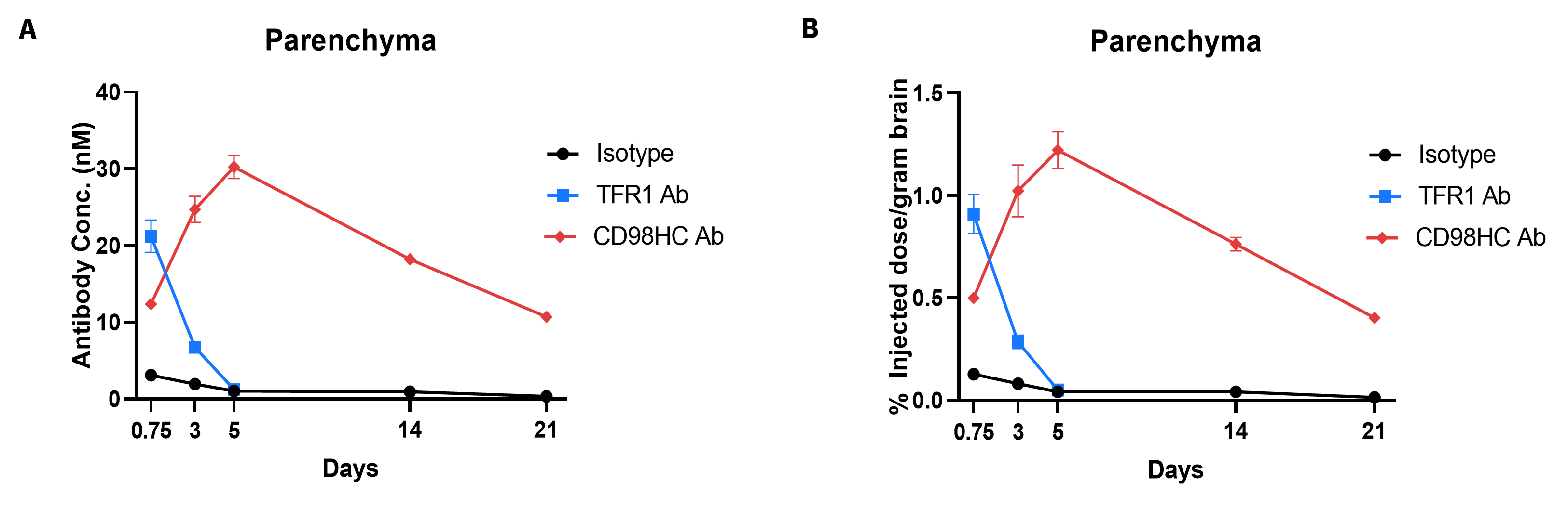
In vivo pharmacokinetic (PK) evaluation and comparison of anti-human TFR1 and anti-CD98HC antibodies in TFR1/CD98HC humanized mice.
TFR1/CD98HC humanized mice (n=2, female, 8-week-old) were injected with control IgG (10 mpk) anti-human TFR1 antibody (TFR1 Ab, JR-141 analog, monovalent, produced in house, 12.56 mpk) and anti-human CD98HC antibody (CD98HC Ab, CD98BBBB-h1.L analog, monovalent, produced in house, 13.3 mpk) via tail vein. Brain tissues were collected for in vivo PK evaluation after dosing 18 hours and 3, 5, 14, 21 days. Brain concentrations (A) and % of injection/gram brain (B) were quantified. As shown in panel, anti-human TFR1 antibody exhibited higher brain exposure at 24 h after dosing, while anti-human CD98HC antibody exhibited higher brain exposure at 72 h after dosing. The results confirmed that TFR1/CD98HC humanized mice enables uptake of an intravenously administered anti-human TFR1 antibody or anti-human CD98HC antibody, and these mice can be used for the comparison of penetration efficacy of shuttle molecules targeting TFR1 or CD98HC. Graphs represent mean ± SEM.

Q1: What are CD98HC humanized mice?
CD98HC humanized mice (B-hCD98HC) are engineered to replace murine Cd98 with human CD98 (SLC3A2), enabling expression of human CD98 heavy chain (CD98HC) in immune and CNS tissues.
Q2: Why are CD98HC humanized mice important?
CD98HC regulates integrin signaling and amino acid transport, and is highly expressed in proliferating cells. This model allows translational research in oncology, immunology, and CNS drug delivery.
Q3: Can CD98HC humanized mice be used for antibody validation?
Yes. Anti-human CD98HC antibodies can be tested for binding, pharmacokinetics, and brain penetration in vivo, making this model highly relevant for preclinical drug evaluation.
Q4: Do CD98HC humanized mice show normal immune function?
Yes. Flow cytometry confirmed that the distribution of leukocyte subsets in spleen, blood, and lymph nodes is comparable to wild-type controls, with no alterations in immune homeostasis.
Q5: What diseases or conditions can be studied using CD98HC humanized mice?
This model is particularly valuable for cancer, autoimmune diseases, and neurological disorders, and for testing the ability of therapeutic antibodies to cross the blood-brain barrier.
