CD36

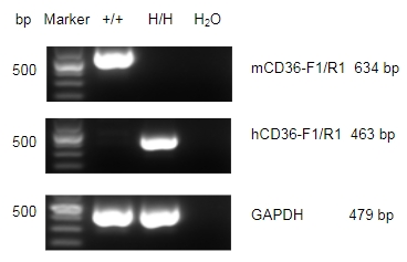
Strain-specific CD36 expression analysis in wild-type C57BL/6 mice and homozygous B-hCD36 mice. Lung RNA was isolated from wild-type C57BL/6 mice (+/+) and homozygous B-hCD36 mice (H/H) (female, 10-week-old, n=1), then cDNA libraries were synthesized by reverse transcription, followed by PCR with mouse or human CD36 primers.
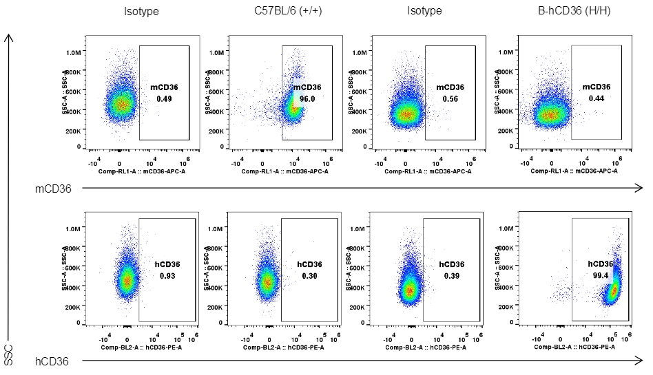
Strain specific CD36 expression analysis in homozygous B-hCD36 mice by flow cytometry. Peritoneal exudative macrophages were collected from wild-type mice (+/+) and homozygous B-hCD36 mice (H/H) (female, 7-week-old, n=1), and analyzed by flow cytometry with species-specific anti-CD36 antibody (mouse CD36: eBioscience, 17-0362-82; human CD36: BioLegend, 336205).
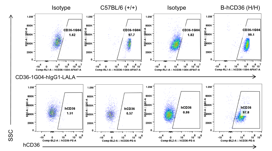
Strain specific CD36 expression analysis in homozygous B-hCD36 mice by flow cytometry. Peritoneal exudative macrophages were collected from wild-type C57BL/6 mice (+/+) and homozygous B-hCD36 mice (H/H), and analyzed by flow cytometry with anti-CD36 antibody.
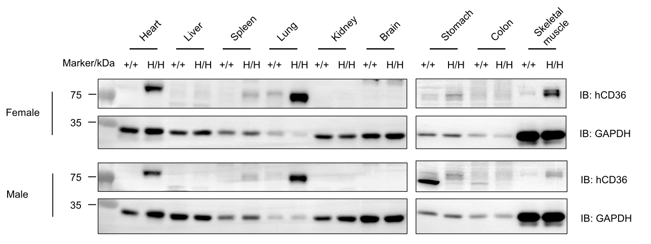
Western blot analysis of CD36 protein expression in homozygous B-hCD36 mice. Various tissue lysates were collected from wild-type C57BL/6N mice (+/+) and homozygous B-hCD36 mice (H/H) (6-week-old, female and male, n=1), and then analyzed by western blot with species-specific anti-CD36 antibody (abcam, ab133625). 40 μg total proteins were loaded for western blotting analysis.
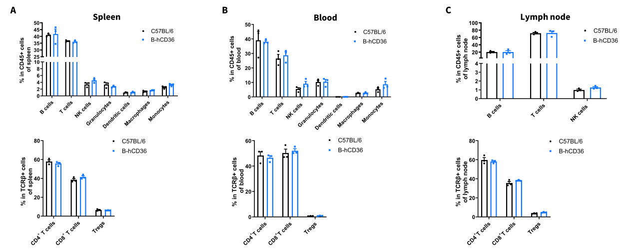
Analysis of leukocyte subpopulations by FACS. Splenocytes (A), blood cells (B) and lymph nodes cells (C) were isolated from female C57BL/6 and B-hCD36 mice (female, n=3, 8-week-old). Top, leukocyte subpopulations analysis. Bottom, T cell subpopulations analysis. Values are expressed as mean ± SEM.
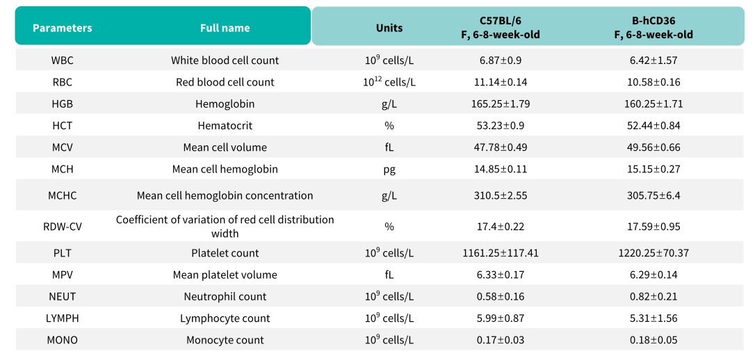
Complete blood count (CBC). Blood from female C57BL/6 and B-hCD36 mice (n=8, 6-8 week-old) was collected and analyzed for CBC. Values are expressed as mean ± SEM.
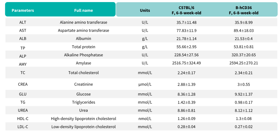
Blood chemistry tests of B-hCD36 mice. Serum from the C57BL/6 and B-hCD36 mice (n=8, 6-8 week-old) was collected and analyzed for levels of indicators. Values are expressed as mean ± SEM.

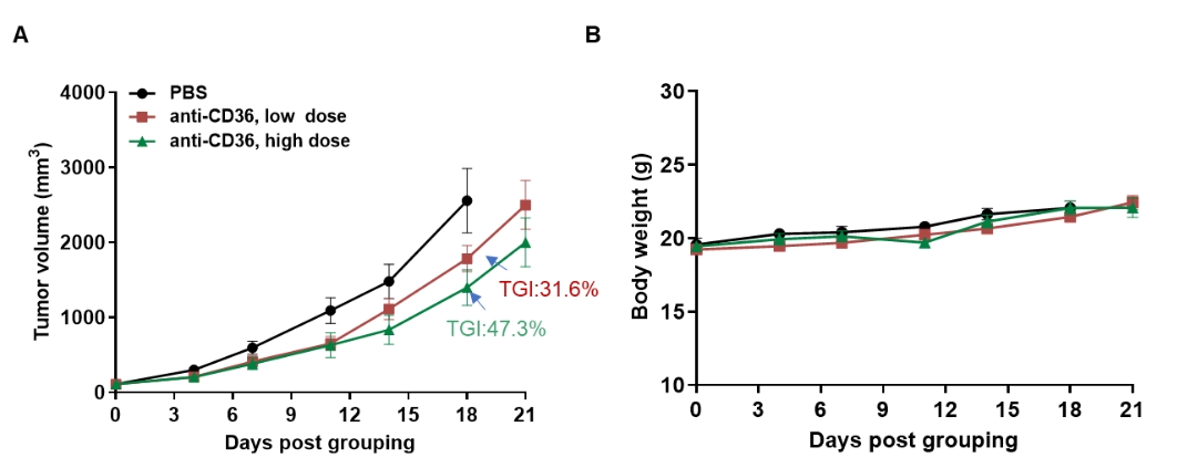
Antitumor activity of anti-CD36 antibodies in B-hCD36 mice. (A) Anti-CD36 antibodies (in house) inhibited MC38 tumor growth in B-hCD36 mice. Murine colon cancer MC38 cells were subcutaneously implanted into homozygous B-hCD36 mice (female, 8-week-old, n=6). Mice were grouped when tumor volume reached approximately 100 mm3, at which time they were treated with anti-CD36 antibodies. (B) Body weight changes during treatment. Values are expressed as mean ± SEM.
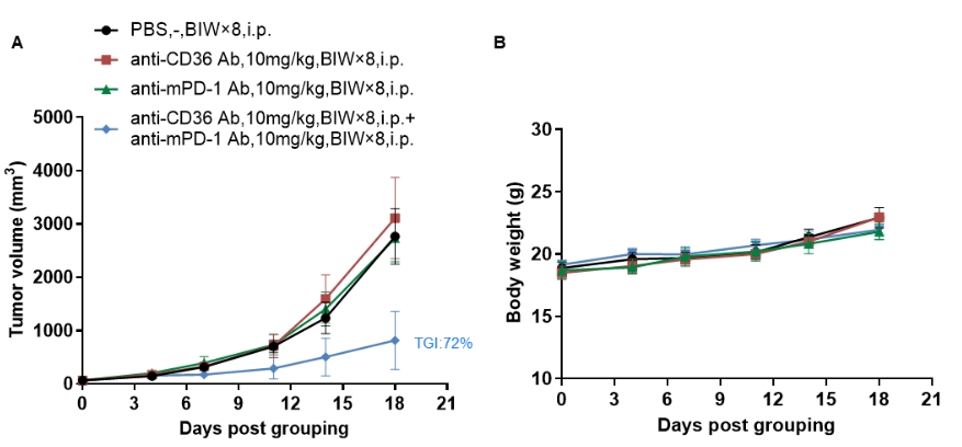
Anti-tumor activity of anti-mouse PD-1 antibody combined with anti-human CD36 antibody in B-hCD36 mice. (A) Anti-mPD-1 antibody combined with anti-hCD36 antibody inhibited MC38 tumor growth in B-hCD36 mice. Murine colon cancer MC38 cells were subcutaneously implanted into homozygous B-hCD36 mice (female, 7-week-old, n=5). Mice were grouped when tumor volume reached approximately 60 mm3, at which time they were treated with antibodies in panel A. (B) Body weight changes during treatment. Values are expressed as mean ± SEM. The data were obtained from the collaborator, and all antibodies used in the experiment were provided by the client.