Background:
Targeting strategy:
Verification:
Application:

Gene targeting strategy for B-hTFR1/hCD20 mice.
The exons 4-19 of mouse Tfr1 gene that encode extracellular domain are replaced by human counterparts in B-hTFR1/hCD20 mice. The genomic region of mouse Tfr1 gene that encodes cytoplasmic portion is retained. The promoter, 5’UTR and 3’UTR region of the mouse gene are also retained. The chimeric TFR1 expression is driven by endogenous mouse Tfr1 promoter, while mouse Tfr1 gene transcription and translation will be disrupted.
The exons 2-7 of mouse Cd20 gene that encode the whole molecule (ATG to STOP codon) are replaced by human counterparts in B-hTFR1/hCD20 mice.


Strain specific CD20 expression analysis in wild-type C57BL/6JNifdc and homozygous B-hTFR1/hCD20 mice by flow cytometry. Splenocytes were collected from wild-type C57BL/6JNifdc (+/+) and homozygous B-hTFR1/hCD20 mice (H/H, H/H) and analyzed by flow cytometry with anti-mouse CD20 antibody (Biolegend, 152107) and anti-human CD20 antibody (Biolegend, 302305). mCD20 was only detectable in wild-type C57BL/6JNifdc mice, and hCD20 was exclusively detectable in homozygous mice but not in wild-type mice.

Strain specific TFR1 expression analysis in wild-type C57BL/6JNifdc and homozygous B-hTFR1/hCD20 mice by flow cytometry. Bone marrow cells were collected from wild-type C57BL/6JNifdc (+/+) and homozygous B-hTFR1/hCD20 mice (H/H, H/H) and analyzed by flow cytometry with anti-mouse TFR1 antibody (Biolegend, 113808) and anti-human CD20 antibody (Biolegend, 334108). mTFR1 was only detectable in wild-type C57BL/6JNifdc mice, and hTFR1 was exclusively detectable in homozygous mice but not in wild-type mice.

Frequency of leukocyte subpopulations in spleen by flow cytometry. Splenocytes were isolated from wild-type C57BL/6JNifdc mice (male, n=3, 10-week-old) and homozygous B-hTFR1/hCD20 mice (male, n=3, 10-week-old). A. Flow cytometry analysis of the splenocytes was performed to assess the frequency of leukocyte subpopulations. B. Frequency of T cell subpopulations. Percentages of T cells, B cells, NK cells, DCs, neutrophils, monocytes, macrophages, CD4+ T cells, CD8+ T cells and Tregs in B-hTFR1/hCD20 mice were similar to those in C57BL/6JNifdc mice. The frequency of leukocyte subpopulations in lymph nodes and blood of B-hTFR1/hCD20 mice were also comparable to wild-type C57BL/6JNifdc mice (Data not shown). Values are expressed as mean ± SEM. Significance was determined by two-way ANOVA test. *P < 0.05, **P < 0.01, ***p < 0.001.
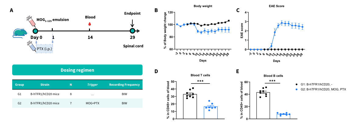
Construction of MOG1-125-induced experimental autoimmune encephalomyelitis (EAE) mouse model using B-hTFR1/hCD20 mice. (A) Experimental scheme. B-hTFR1/hCD20 mice (n=7-8, female, 8-week-old) were subcutaneously injected with MOG1-125 emulsion in the neck and buttock regions on day 0. Pertussis toxin (PTX) was administered intraperitoneally at 2 and 24 hours after MOG immunization (G2). Body weight (B) and clinical scores (C) were recorded every other day. (D-E) Blood were collected on day 14. Levels of T, B cells were evaluated by flow cytometry. Values are expressed as mean ± SEM. Two-way ANOVA with Dunnett’s test. *P
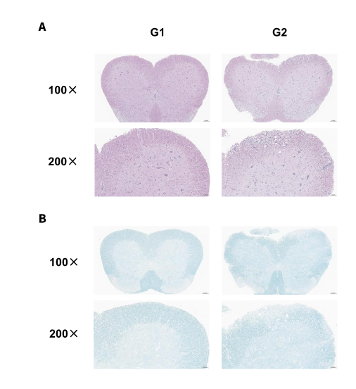
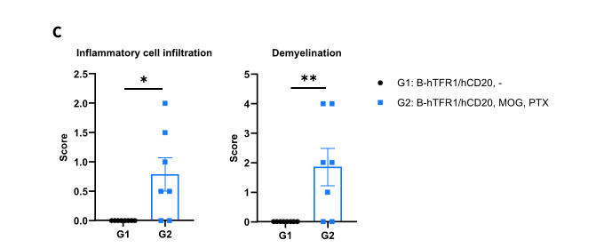
Construction of MOG1-125-induced experimental autoimmune encephalomyelitis (EAE) mouse model using B-hTFR1/hCD20 mice. Spinal cords were removed from B-hTFR1/hCD20 mice on day 29 and stained with Hematoxylin and eosin (H&E) (A) or Luxol fast blue (LFB) (B). Representative sections are shown, as well as the score of inflammatory cells and demyelination of spinal cord (C). MOG1-125-induced experimental autoimmune encephalomyelitis (EAE) mouse model was successful constructed using B-hTFR1/hCD20 mice, according to EAE score, percentage of blood B cells, and infiltration of inflammatory cells and demyelination of spinal cord. Values are expressed as mean ± SEM, Versus G1, *P