Description
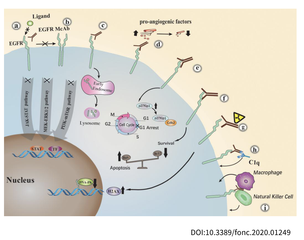
EGFR: A Key Driver of Cell Growth and Cancer Progression
- Gene Information: EGFR encodes a transmembrane receptor tyrosine kinase belonging to the ErbB receptor family. EGFR plays a central role in regulating cell proliferation, survival, differentiation, and tissue repair and is frequently dysregulated in cancer.
- Protein Expression: EGFR is widely expressed in epithelial tissues, including the skin, lung, gastrointestinal tract, liver, and kidney. It is also detected in various epithelial-derived tumors, where overexpression, amplification, or activating mutations can drive tumor growth and disease progression.
- Signaling Pathway: EGFR is activated by ligands such as EGF and TGF-α, inducing receptor dimerization and activation of downstream pathways including RAS/RAF/MEK/ERK, PI3K/Akt/mTOR, JAK/STAT, and PLCγ/PKC. These signaling cascades promote cell proliferation, survival, migration, angiogenesis, and tumor progression.
- Therapeutic Inhibition: Current EGFR-targeted therapies include monoclonal antibodies and tyrosine kinase inhibitors (TKIs). Emerging approaches such as bispecific antibodies, ADCs, and combination therapies are being developed to overcome resistance and enhance anti-tumor efficacy.
FcRn MOA: pH-Dependent Binding of IgG to FcRn
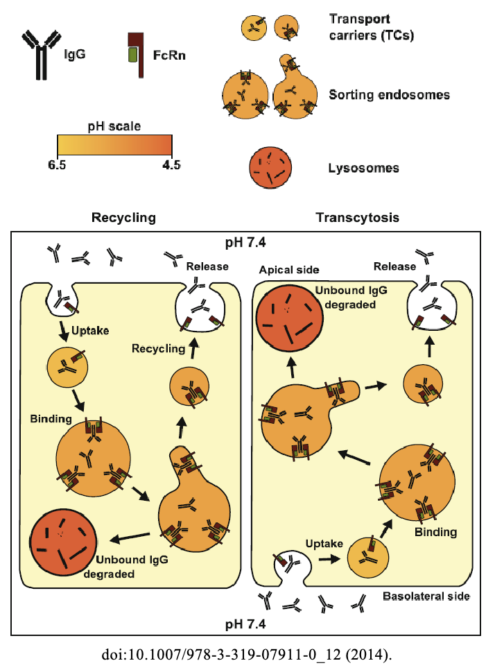
- IgG Binding to FcRn at Acidic pH: In acidic environments like as endosomes (pH 6.0-6.5), IgG binds strongly to FcRn. Histidine residues on the IgG Fc region become protonated, allowing a high-affinity interaction that recruits IgG into the salvage and recycling pathways.
- Intracellular Trafficking: Following endocytosis, the IgG-FcRn complexes are diverted away from lysosomal degradation, avoiding destructive pathways. They are instead sorted into recycling endosomes or transcytosis carriers.
- IgG Dissociation at Neutral pH: Upon reaching the plasma membrane, the environment shifts to near-neutral or physiological pH (~7.4). The loss of histidine protonation causes rapid dissociation, releasing free IgG back into the extracellular fluid or circulation.
- Therapeutic Application: Enhancing or inhibiting this pH-dependent IgG-FcRn interaction can modulate antibody dynamics, enabling the design of longer-lived therapeutic IgGs or the use of FcRn antagonists to rapidly clear pathogenic autoantibodies in autoimmune diseases.
Why Humanized FcRn? Species-Specific Differences in IgG Binding
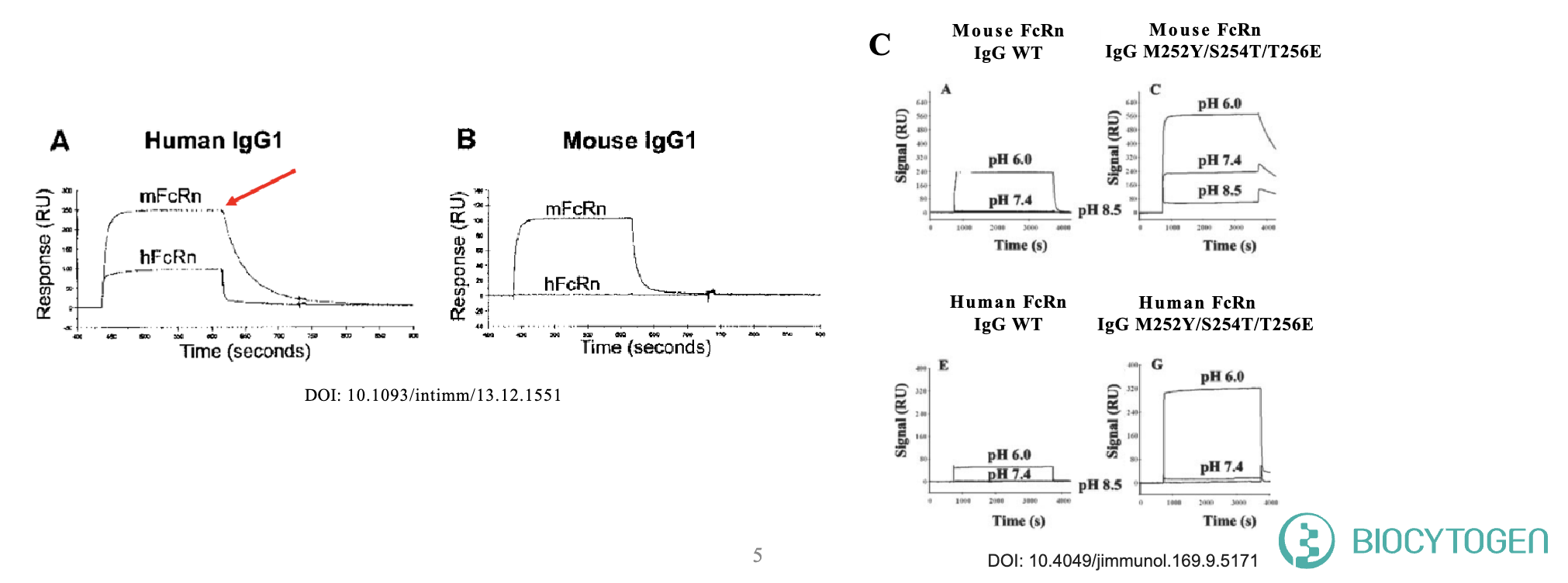
Different IgG Binding Sites Between Mouse and Human FcRn:
- Human IgG binds to mouse FcRn through different amino acid residues, leading to pharmacokinetic (PK) data that do not align with human results.
Different Binding Affinity to IgG variants Between Mouse and Human FcRn:
- Some human IgG variants (M252Y/S254T/T256E) bind more strongly to murine FcRn at pH 7.4 compared to WT IgG, but not to human FcRn.
- This increased affinity hinders efficient serum release, offsetting the benefits of enhanced binding at pH 6.0, resulting in reduced serum concentrations in mice.
Targeting strategy
B-hFcRn/hEGFR Mice: Gene Targeting Strategy
EGFR
- Exons 2- 17 of the mouse Egfr gene that encode the extracellular domain are replaced by human counterparts in B-hFcRn/hEGFR mice.
- The genomic region of the mouse Egfr gene that encodes the signal peptide, transmembrane domain and cytoplasmic portion is retained. The promoter, 5’UTR and 3’UTR regions of the mouse gene are also retained. The chimeric EGFR expression is driven by the endogenous mouse Egfr promoter, while mouse Egfr transcription and translation are disrupted.
FCRN
- The CDS that encodes full-length human FCGRT, followed by the mouse 3’UTR-STOP, is inserted right after the mouse Fcgrt ATG to replace exons 2-4 of the mouse Fcgrt gene.
- The FcRn protein expression will be driven by the endogenous mouse Fcgrt promoter, while the mouse Fcgrt gene transcription and translation will be disrupted.
B-hFcRn/hEGFR Mice: EGFR mRNA Expression by RT-PCR
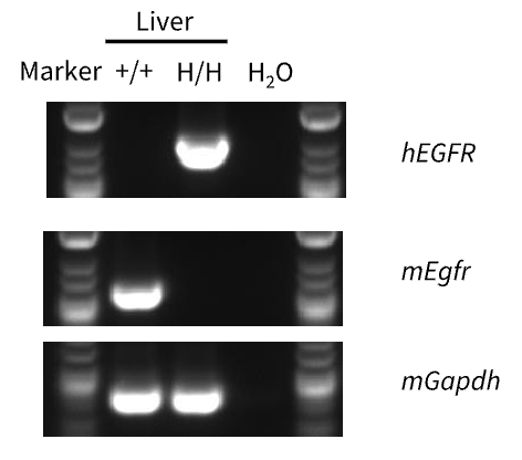
- Mouse EGFR mRNA was only detectable in wild-type C57BL/6JNifdc mice.
- Human EGFR mRNA was exclusively detectable in homozygous B-hFcRn/hEGFR mice but not in wild-type mice.
Strain-specific analysis of EGFR mRNA expression in wild-type C57BL/6JNifdc mice and homozygous B-hFcRn/hEGFR mice by RT-PCR. Liver tissue was isolated from wild-type C57BL/6JNifdc mice (+/+) and homozygous B-hFcRn/hEGFR mice (H/H); cDNA libraries were synthesized by reverse transcription, followed by PCR with mouse or human EGFR primers.
B-hFcRn/hEGFR Mice: EGFR Protein Expression Analysis
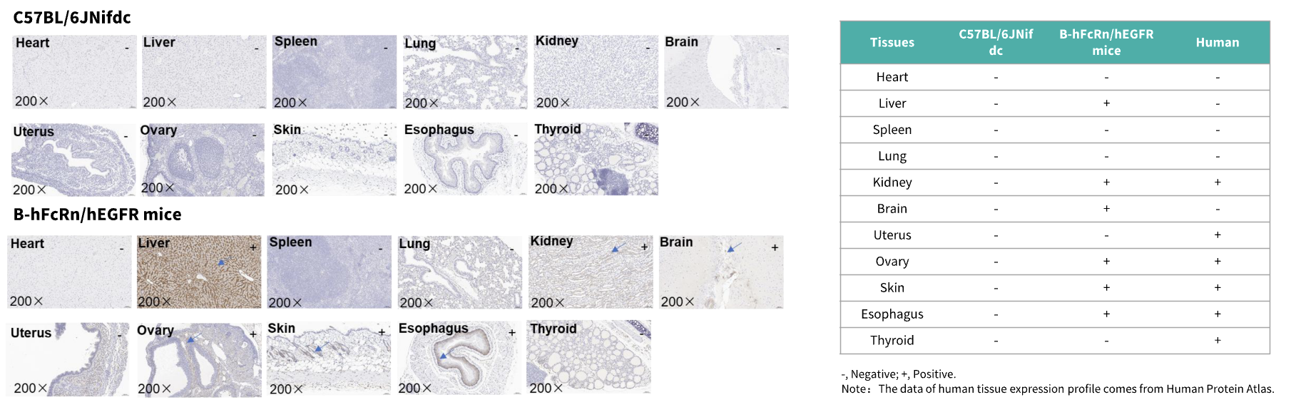
- Human EGFR was detected in the liver, kidney, brain, ovary, skin, and esophagus of B-hFcRn/hEGFR mice, but not in the heart, spleen, lung, uterus, or thyroid tissues.
- Because the antibody could not bind to mouse EGFR, no signal was detected in these tissues in wild-type mice.
Immunohistochemical (IHC) analysis of EGFR expression in B-hFcRn/hEGFR mice. Eleven tissues were collected from C57BL/6JNifdc and homozygous B-hFcRn/hEGFR mice and analysed by IHC with an anti-hEGFR antibody (Invitrogen, MA5-49312). The arrow indicates tissue cells with positive EGFR staining (brown). “+” indicates that the tissue is positive and “-” indicates that the tissue is negative.
B-hFcRn/hEGFR Mice: FcRn Protein Expression Analysis
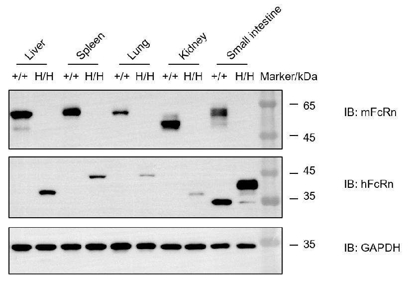
Mouse FcRn was only detected in wild-type C57BL/6JNifdc mice. Human FcRn was only detected in B-hFcRn mice.
Western blot analysis of FcRn protein expression in homozygous B-hFcRn/hEGFR mice. Various tissue lysates were collected from wild-type C57BL/6JNifdc mice (+/+) and homozygous B-hFcRn/hEGFR mice (H/H), and then analyzed by western blot with species-specific anti-mFcRn antibody (RD, AF6775) and anti-hFcRn antibody (NOVUS, NBP1-89128). 40 μg of total protein was loaded for Western blotting analysis.
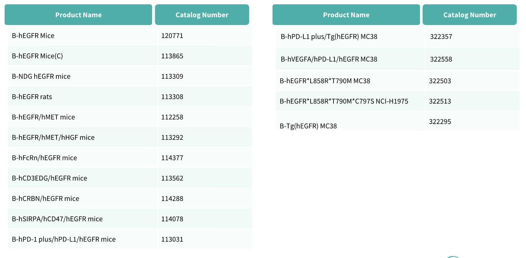
B-hFcRn/hEGFR Mice: Related Product List
* When publishing results obtained using this animal model, please acknowledge the source as follows: The animal model [B-hFcRn/hEGFR mice] (Cat# 114377) was purchased from Biocytogen.