Description
NECTIN1 MOA: a Cell Adhesion Molecule that Directs Junctional Assembly and Cell Polarit
Nectin-1 (CD111): Gene Architecture, Expression Profile, Downstream Signaling, and Therapeutic Landscape
- Gene Information: Encoded by the NECTIN1 gene (formerly PVRL1, CD111, or HVEC) located on chromosome 11q23.3. Transcript produces three main alternatively spliced variants: Nectin-1α, Nectin-1β, Nectin-1γ.
- Protein Expression: Broadly expressed across diverse tissues with high abundance in nervous system, epithelia and endothelia. Integrates directly into adherens junctions (AJs) and tight junctions (TJs) at the cell membrane.
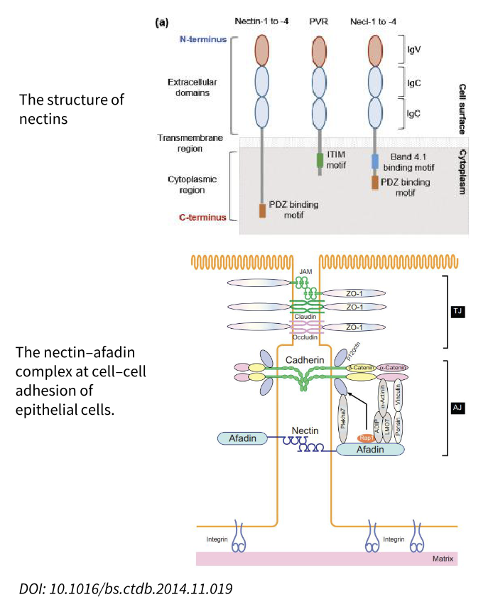
- Signaling Pathway: a.Junction Assembly: Trans-dimerizes (homophilic or with Nectin-3) and binds afadin via its cytoplasmic tail, recruiting Rap1 and catenins to guide cadherin clustering into mature adherens junctions. B.Cytoskeleton & Polarity: Activates Cdc42 and Rac1 to drive actin remodeling, while recruiting the Par complex to establish apico-basal polarity. C. Growth Control: Signals through PI3K-Akt and MAPK cascades for cell survival, and regulates the Hippo pathway by suppressing YAP/TAZ nuclear translocation upon cell–cell contact.
- Therapeutic Strategy: Antiviral Blockade: Serves as the primary entry receptor for HSV-1/2 via glycoprotein D (gD); neutralizing mAbs and soluble Nectin-1-Fc decoys prevent viral entry and infection. Oncology Applications: Binds CD96 and TIGIT on T/NK cells; blocking this axis relieves immunosuppression in the tumor microenvironment. Targeted by antibody-drug conjugates (ADCs) in overexpressing tumors, or conversely exploited as an entry portal by oncolytic herpesviruses (e.g., T-VEC, G207) for selective tumor lysis.
NECTIN1 MOA: A Primary Entry Receptor for alpha-Herpesviruses
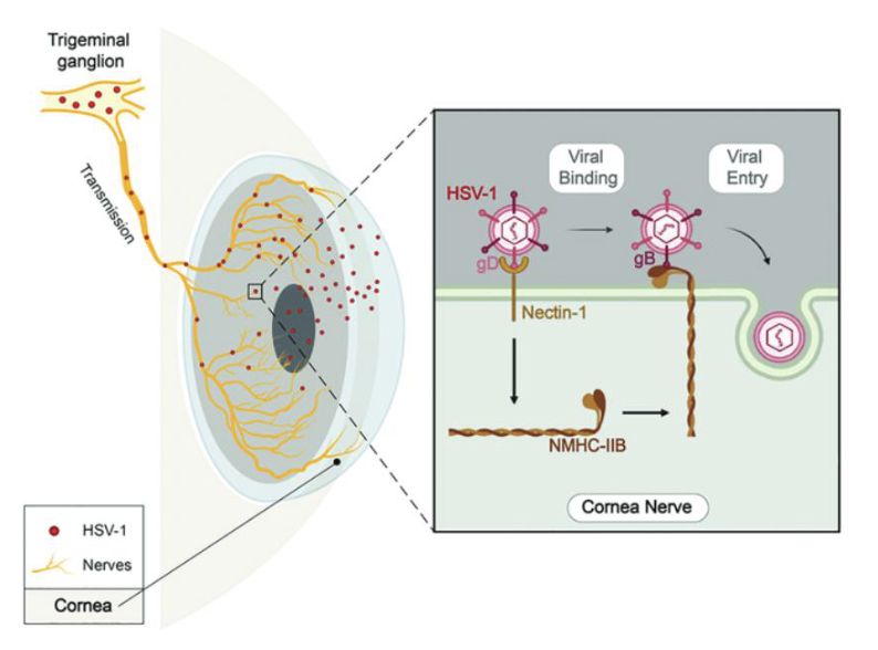
Schematic diagram of Nectin-1 and NMHC-IIB mediating HSV-1 entry into corneal nerves.
When HSV-1 infects the cornea, it enters the corneal nerves, travels along the ophthalmic branch of the trigeminal ganglion (TG), and ultimately establishes latency in the TG. During HSV-1 entry into corneal nerves, HSV-1 gD first binds to Nectin-1. This causes the virus to attach to the cell surface, and the expression of cell-surface NMHC-IIB is upregulated. Then, HSV-1 gB binds to NMHC-IIB, allowing the virus to fuse with the cell membrane, thereby completing its entry process.
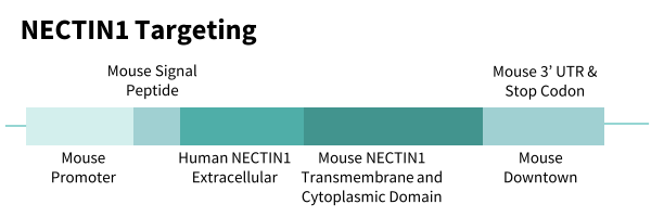
B-hNECTIN1 Mice: Gene Targeting Strategy
NECTIN1
- The exons 2-6 of mouse Nectin1 gene that encode the extracellular domain are replaced by human counterparts in B-hNECTIN1 mice. The genomic region of mouse Nectin1 gene that encodes transmembrane domain and cytoplasmic portion is retained. The promoter, 5’UTR and 3’UTR region of the mouse gene are also retained.
- The chimeric NECTIN1 expression is driven by endogenous mouse Nectin1 promoter, while mouse Nectin1 gene transcription and translation will be disrupted.
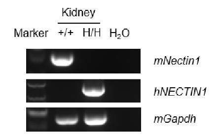
B-hNECTIN1 Mice: NECTIN1 Expression by RT-PCR
Human NECTIN1 is specifically and correctly expressed in kidney of B-hNECTIN1 mice.
Human NECTIN1 was detectable in B-hNECTIN1 mice by RT-PCR and sequencing. Kidney RNA was isolated from wild-type C57BL/6JNifdc mice (+/+) and homozygous B-hNECTIN1 mice (H/H), then cDNA libraries were synthesized by reverse transcription, followed by PCR with mouse or human NECTIN1 primers. Sequencing of PCR products confirmed that the amplified sequences were consistent with database reference sequences.
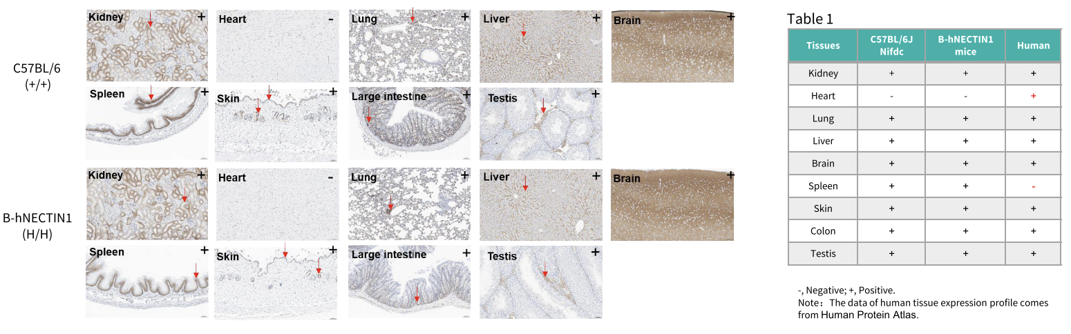
B-hNECTIN1 Mice: Immunohistochemical Analysis
- NECTIN1 expression was detected in the kidney, lung, liver, brain, spleen, skin, large intestine, and testis of homozygous B-hNECTIN1 mice, but not in the heart. The antibodies showed cross-reactivity with mouse NECTIN1 in C57BL/6JNifdc mice.
- The NECTIN1 expression profiles in B-hNECTIN1 mice and wild-type mice are summarized in Table 1.
Immunohistochemical analysis of organs in B-hNECTIN1 mice. Major organs were collected from wild-type C57BL/6JNifdc mice (male, 7 weeks old) and homozygous B-hNECTIN1 mice (male, 7 weeks old), and analyzed by IHC using anti-NECTIN1 antibody (Abcam, 323704). The arrow indicates tissue cells with positive NECTIN1 staining (brown). "+" and "-" indicate positive and negative tissues, respectively.
B-hNECTIN1 Mice: Related Product List
* When publishing results obtained using this animal model, please acknowledge the source as follows: The animal model [B-hNECTIN1 mice] (Cat# 114105) was purchased from Biocytogen.