HHLA2 Protein Expression by WB
SYNGAP1: A major genetic risk factor for intellectual disability
- Gene Information: SYNGAP1 (Synaptic Ras GTPase activating protein 1) is located on human chromosome 6, and it encodes a brain-enriched Ras GAP protein. Heterozygous loss-of-function variants in SYNGAP1 gene causes SYNGAP1-related intellectual disability, a neurodevelopmental disorder diseases.
- Protein Expression: SYNGAP1 protein is predominantly expressed in excitatory neurons of the central neurons system and enriched at postsynaptic densities.
- Signaling Pathway: SYNGAP1 acts as a Ras GTPase-activating protein to negatively regulate Ras. It suppresses postsynaptic Ras–ERK signaling, thereby restraining growth-associated processes including protein translation and postsynaptic AMPA receptor insertion under basal resting conditions. Loss of SYNGAP1 elevates Ras activity and increases neuronal excitability.
- Therapeutic Application: SYNGAP1 serves as a promising therapeutic target for intellectual disability. Potential strategies include gene replacement therapy, antisense oligonucleotide-mediated rescue, and small-molecule agents to restore synaptic function.
Targeting strategy
SYNGAP1
- The exons 1-19 of mouse Syngap1 gene that encode the whole molecule (ATG to STOP codon), including the promoter, 5’UTR and 3’UTR, are replaced by human counterparts in B-hSYNGAP1 mice.
- The human SYNGAP1 expression is driven by human SYNGAP1 promoter, while mouse Syngap1 gene transcription and translation will be disrupted.
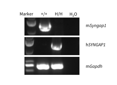
SYNGAP1 mRNA Expression by RT-PCR
Strain specific analysis of SYNGAP1 mRNA expression in wild-type mice and homozygous B-hSYNGAP1 mice by RT-PCR. Brain RNA was isolated from wild-type C57BL/6JNifdc mice (+/+) and homozygous B-hSYNGAP1 Mice (H/H), then cDNA libraries were synthesized by reverse transcription, followed by PCR with mouse and human SYNGAP1 primers. Mouse Syngap1 mRNA was only detectable in wild-type mice. Human SYNGAP1 mRNA was exclusively detectable in homozygous B-hSYNGAP1 mice.
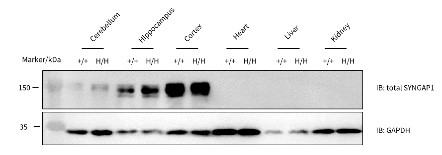
SYNGAP1 Protein Expression Analysis
Western blot analysis of SYNGAP1 protein expression in homozygous B-hSYNGAP1 mice. Various tissue lysates were collected from wild-type C57BL/6JNifdc mice (+/+) and homozygous B-hSYNGAP1 mice (H/H, male, 6-week-old), and then analyzed by western blot with anti-SYNGAP1 antibody (Cell signal, 62193S). 40 μg total proteins were loaded for western blot analysis. SYNGAP1 were detectable in hippocampus, cortex (high level) and cerebellum (low level) from both wild-type mice and homozygous B-hSYNGAP1 mice. This antibody cross-recognized both human and mouse SYNGAP1 protein.
* When publishing results obtained using this animal model, please acknowledge the source as follows: The animal model [B-hSYNGAP1 mice] (Cat# 114311) was purchased from Biocytogen.