Description
- Gene Information: CD3: Encoded by CD3E, D, G; part of the Ig superfamily. It forms the essential signaling backbone of the T cell receptor (TCR) complex. CDH3: encodes P cadherin, a classical type I cadherin and calcium dependent cell cell adhesion molecule. It is a single pass transmembrane glycoprotein with five extracellular cadherin repeats, a transmembrane domain, and a conserved cytoplasmic tail that interacts with catenins and the actin cytoskeleton.
- Protein Expression: CD3: Constitutive and universal marker for all mature T cells (CD4+, CD8+). Always present on the cell surface. CDH3: normally expressed in selected epithelial tissues, including basal/myoepithelial cell populations and specific regions of the mammary gland, skin, gastrointestinal, and genitourinary tracts. In many solid tumors, CDH3/P cadherin is aberrantly overexpressed.
- Signaling Pathway: CD3: Operates via ITAM phosphorylation and ZAP 70. It triggers the initial "on" switch, Ca2+ flux, and immediate cytotoxicity. CDH3: Functions primarily in calcium dependent cell cell adhesion through interactions with p120 catenin and β catenin. Dysregulated CDH3 expression can alter cell adhesion and activate or modulate signaling pathways, including PI3K/AKT, MAPK/ERK, Rho family GTPases, and Wnt/β catenin related pathways.
- Therapeutic Inhibition: CDH3 targeted therapies aim to selectively eliminate CDH3 positive tumor cells while minimizing damage to normal tissues. Potential strategies include monoclonal antibodies, CD3×CDH3 bispecific antibodies, antibody drug conjugates (ADCs), CAR T or other engineered cell therapies, and cancer vaccines.
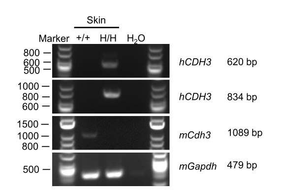
CDH3 mRNA Expression
Strain specific analysis of CDH3 mRNA expression in wild-type C57BL/6JNifdc mice and homozygous B-hCD3EDG/hCDH3 mice by RT-PCR. Skin tissue were isolated from wild-type C57BL/6 mice (+/+) and homozygous B-hCD3EDG/hCDH3 mice (H/H), then cDNA libraries were synthesized by reverse transcription, followed by PCR with mouse or human CDH3 primers.
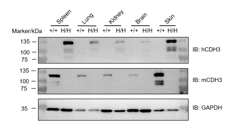
CDH3 Protein Expression
Western blot analysis of CDH3 protein expression in homozygous B-hCD3EDG/hCDH3 mice. Various tissue lysates were collected from wild-type C57BL/6JNifdc mice (+/+) and homozygous B-hCD3DG/hCDH3 mice (H/H; H/H), and then analyzed by western blot with species-specific anti-mCDH3 antibody (R&D, AF761-SP) and anti-hCDH3 antibody (R&D, MAB861-SP). 40 μg total proteins were loaded for western blotting analysis.
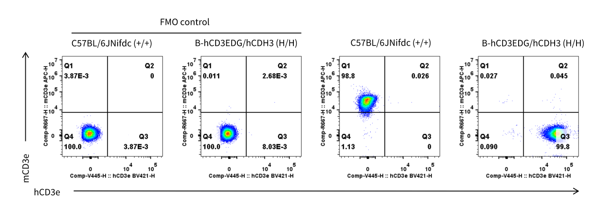
CD3e Protein Expression in Spleen
Mouse and human CD3e expression analysis in splenocytes. Splenocytes were collected from wild-type C57BL/6JNifdc mice (+/+) (Female, 6-week-old, n=1) and homozygous B-hCD3EDG/hCDH3 mice (H/H) (Female, 6-week-old, n=1). Protein expression was analyzed with anti-mouse CD3e antibody (Biolegend, 100312) and anti-human CD3e antibody (BD, 562426) by flow cytometry.
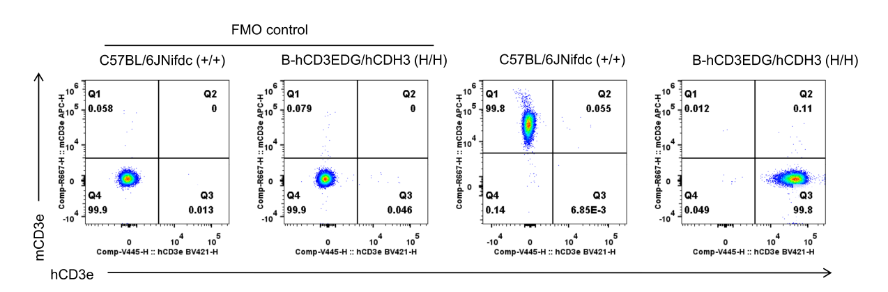
CD3e Protein Expression in Blood
Mouse and human CD3e expression analysis in blood T cells. Blood T cells were collected from wild-type C57BL/6JNifdc mice (+/+) (Female, 6-week-old, n=1) and homozygous B-hCD3EDG/hCDH3 mice (H/H) (Female, 6-week-old, n=1). Protein expression was analyzed with anti-mouse CD3e antibody (Biolegend, 100312) and anti-human CD3e antibody (BD, 562426) by flow cytometry.
* When publishing results obtained using this animal model, please acknowledge the source as follows: The animal model [B-hCD3EDG/hCDH3 mice] (Cat# 114436) was purchased from Biocytogen.