Gene Information: PD‑1 is encoded by the PDCD1 gene on chromosome 2q37.3, while PD‑L1 is encoded by CD274 on chromosome 9p24.1. Both are type I transmembrane proteins; PD‑1 belongs to the immunoglobulin superfamily and functions as an inhibitory receptor, whereas PD‑L1 is a member of the B7 family of co‑stimulatory molecules.
Protein Expression: PD-1 is primarily expressed on activated T cells, B cells, and NK cells, whereas PD-L1 is expressed on antigen-presenting cells and many tumor cells, with expression often induced by IFN-γ.
Signaling Pathway: Binding of PD-L1 to PD-1 recruits SHP2 phosphatase and suppresses TCR/CD28-mediated PI3K–AKT signaling, resulting in reduced T-cell proliferation, cytokine production, and cytotoxic activity.
Therapeutic Inhibition: Anti-PD-1 and anti-PD-L1 antibodies block this inhibitory pathway, restoring T-cell function and enhancing anti-tumor immune responses.
VEGFR2: A central regulator of angiogenesis and its therapeutic intervention
Gene Information: Vascular endothelial growth factor receptor 2 (VEGFR2), also known as KDR or FLK1, is a protein-coding gene located on chromosome 4q12. It encodes a receptor tyrosine kinase belonging to the VEGFR family.
Protein Expression: VEGFR2 is primarily expressed on vascular endothelial cells, lymphatic endothelial cells, and hematopoietic cells. It predominantly exists as a full-length transmembrane receptor, serving as the main transducer of angiogenic signals.
Signaling Pathway: VEGFR2 binds VEGFA, VEGFC, and VEGFD, utilizing NP1/2 as co-receptors. It possesses strong tyrosine kinase activity and plays a primary role in mediating vascular permeability, endothelial cell proliferation, migration, and survival..
Therapeutic Inhibition: By blocking VEGFR2 binding, dimerization, or kinase activity with monoclonal antibodies or tyrosine kinase inhibitors, downstream signaling and cell migration are inhibited, offering a major strategy for anti-angiogenic therapy.
Complete Human Pathway: Triple humanization of PD-1, PD-L1, and VEGFR2 preserves native human interactions, enabling precise in vivo evaluation of combination therapy.
IND-Enabling Data Package: Provides a robust platform for geng preclinical safety and efficacy data to support IND submissns for anti-PD-1/PD-L1, anti-VEGFR2, and bispecific therapeutics.
Versatile Disease Modeling: Enables robust in vivo modeling of PD-L1-driven immune suppression and VEGFR2-dependent pathological angiogenesis in syngeneic tumor models.
Target & Pathway Validation: Enables study of the PD-1/PD-L1 inhibitory axis and its crosstalk with VEGFR2-mediated angiogenesis signaling, facilitating the validation of dual-target therapeutic strategies.
Efficacy & Safety Evaluation: Supports efficacy and safety assessment of anti-PD-1/VEGFR2 or PD-L1/VEGFR2 bispecific antibodies and combination therapies in syngeneic tumor models.
Novel Therapeutic Screening: Support the discovery, screening, and optimization of new treatment strategies, leveraging a relevant humanized models for translational research.

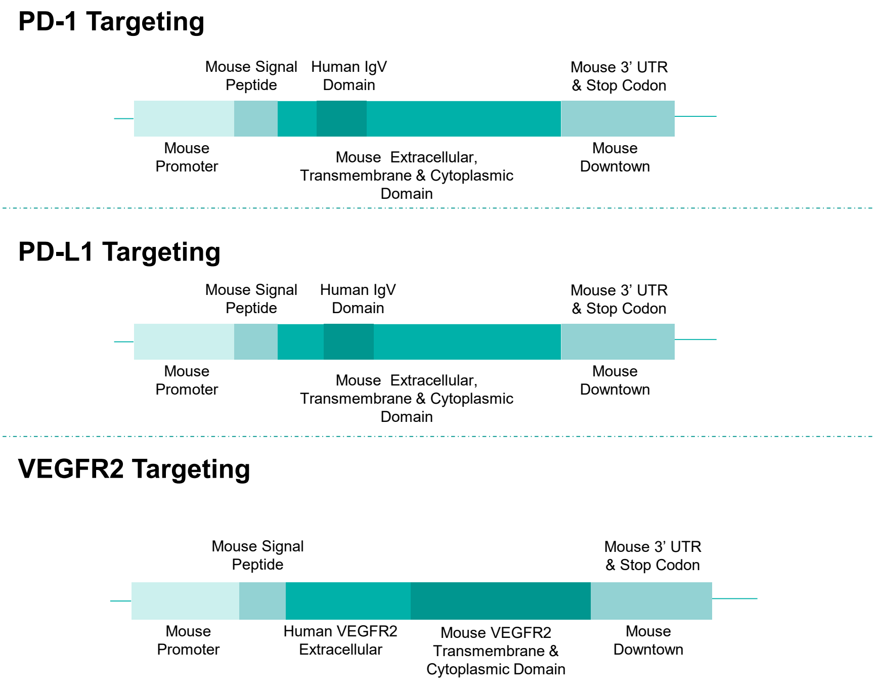
PD-1
The exon 2 of mouse Pdcd1 gene that encodes the IgV domain was replaced by human PDCD1 exon 2 in B-hPD-1/hPD-L1/hVEGFR2 mice.
PD-L1
The exon 3 of mouse Cd274 gene that encodes the IgV domain was replaced by human CD274 exon 3 in B-hPD-1/hPD-L1/hVEGFR2 mice. Sequences of other regions still belonged to mice.
VEGFR2
The exons 2-15 of mouse Kdr gene that encodes the extracellular region coding sequences were replaced by human KDR exons 2-15 in B-hPD-1/hPD-L1/hVEGFR2 mice.

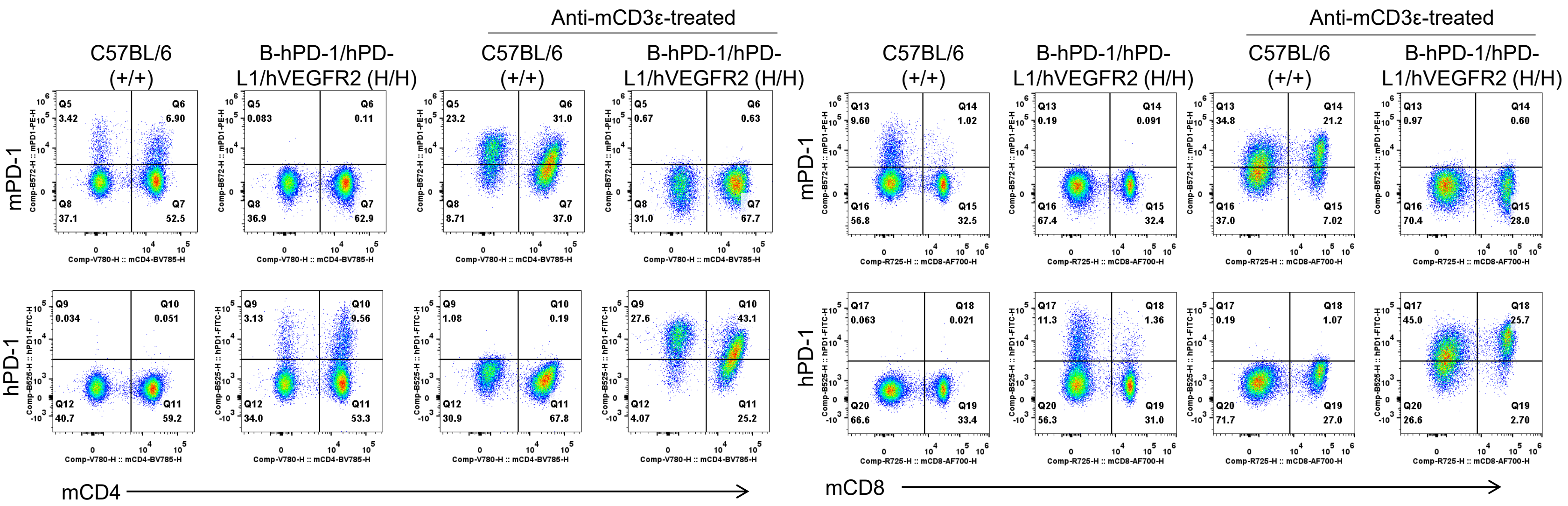
Mouse and human PD-1 expression analysis in splenocytes. Splenocytes were collected from wild-type C57BL/6 mice (+/+) and homozygous B-hPD-1/hPD-L1/hVEGFR2 mice (H/H) after stimulated with anti-CD3ε (7.5 μg, i.p.) in vivo or not. Protein expression was analyzed with anti-mouse PD-1 antibody (Biolegend, 109104) and anti-human PD-1 antibody (Biolegend, 329904) by flow cytometry.
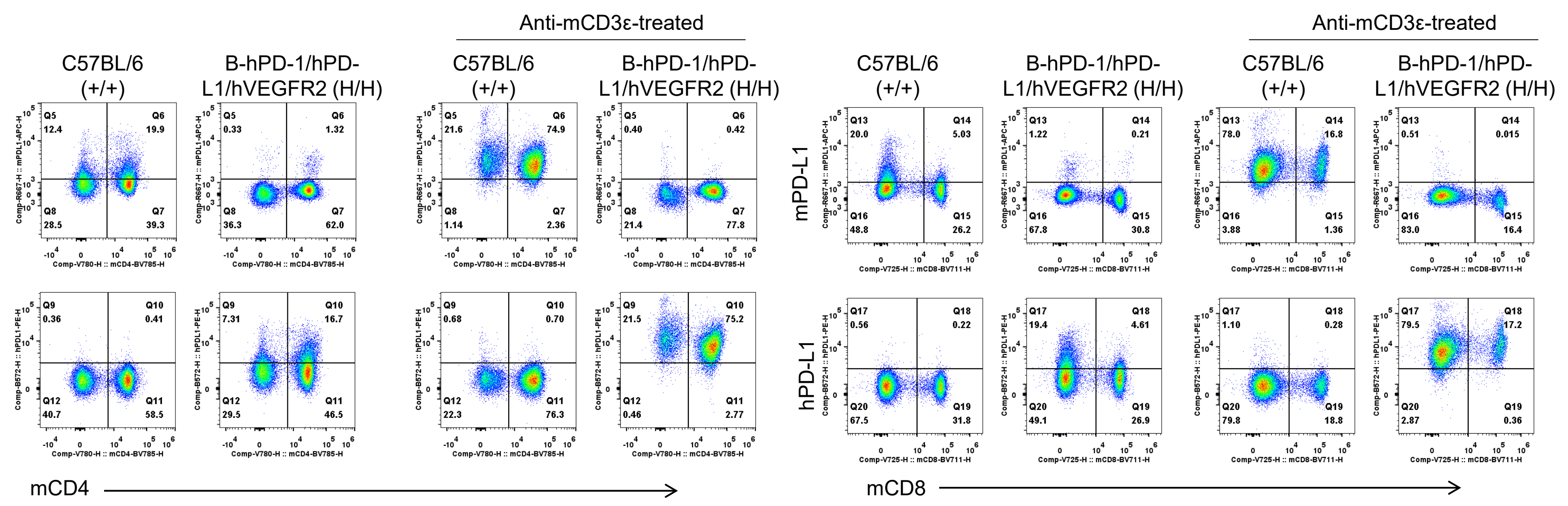
Mouse and human PD-L1 expression analysis in splenocytes. Splenocytes were collected from wild-type C57BL/6 mice (+/+) and homozygous B-hPD-1/hPD-L1/hVEGFR2 mice (H/H) after stimulated with anti-CD3ε (7.5 μg, i.p.) in vivo or not. Protein expression was analyzed with anti-mouse PD-L1 antibody (Biolegend, 124312) and anti-human PD-L1 antibody (Biolegend, 329706) by flow cytometry.
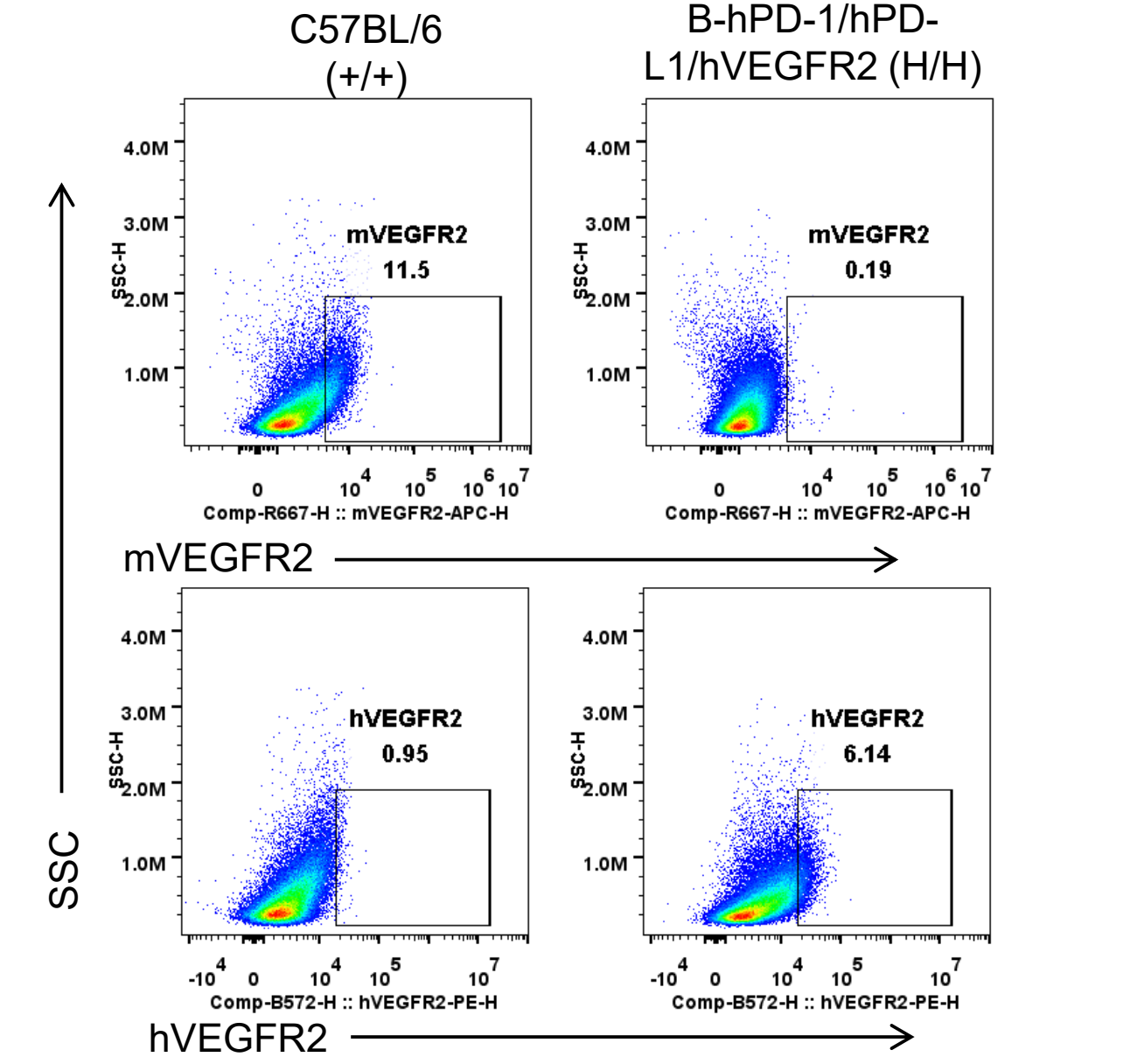
Mouse and human VEGFR2 expression analysis in splenocytes. Lung endothelial cells were collected from wild-type C57BL/6 mice (+/+) and homozygous B-hPD-1/hPD-L1/hVEGFR2 mice (H/H). Protein expression was analyzed with anti-mouse VEGFR2 antibody (Biolegend, 136406) and anti-human VEGFR2 antibody (Biolegend, 359904) by flow cytometry.

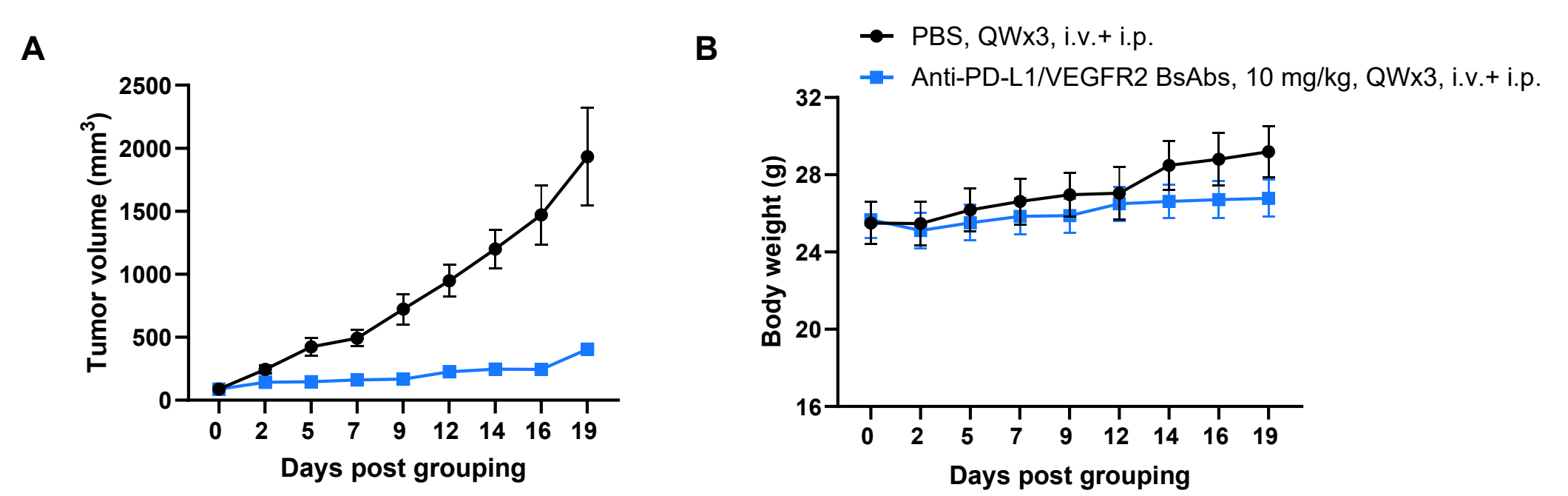
Antitumor activity of anti-PD-L1/VEGFR2 BsAbs in B-hPD-1/hPD-L1/hVEGFR2 mice. Murine colon cancer B-hVEGFA/hPD-L1 plus MC38 were subcutaneously implanted into homozygous B-hPD-1/hPD-L1/hVEGFR2 mice (male, 7-week-old, n=6). Mice were grouped when tumor volume reached approximately 80-120 mm3, at which time they were injected with anti-PD-L1/VEGFR2 bispecific antibody indicated in panel. (A) Tumor growth curves. (B) Body weight changes during treatment. As shown in panel A, anti-PD-L1/VEGFR2 BsAbs was efficacious in controlling tumor growth in B-hPD-1/hPD-L1/hVEGFR2 mice. The antibody provided by the client. Values are expressed as mean ± SEM.
The overage of this tumor model is 66%.