

PD-1: An immune-checkpoint receptor and therapeutic target for restoring T-cell anti-tumor activity

Gene targeting strategy for B-hPD-1 mice(C). The signal peptide and extracellular region of human PD-1 gene and the transmembrane, cytoplasmic region of mouse PD-1 gene were constructed into a chimeric CDS vector and inserted after the mouse 5’UTR sequences in B-hPD-1 mice(C).


Strain specific PD-1 expression analysis in wild-type BALB/c mice and homozygous B-hPD-1 mice(C) by flow cytometry. Splenocytes were collected from wild-type BALB/c mice and homozygous B-hPD-1 mice(C) stimulated with anti-CD3ε in vivo (7.5 μg/mice, stimulated for 24 hours, i.p.). Mouse PD-1 was only detectable in wild-type mice. Human PD-1 was only detectable in homozygous B-hPD-1 mice(C).
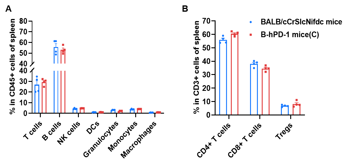
Frequency of leukocyte subpopulations in spleen by flow cytometry. Splenocytes were isolated from wild-type BALB/cCrSlcNifdc mice and homozygous B-hPD-1 mice(C) (female, 7-week-old, n=4). A. Flow cytometry analysis of the splenocytes was performed to assess the frequency of leukocyte subpopulations. B. Frequency of T cell subpopulations. Frequencies of T cells, B cells, NK cells, DCs, granulocytes, monocytes, macrophages, CD4+ T cells, CD8+ T cells and Tregs in B-hPD-1 mice(C) were similar to those in BALB/cCrSlcNifdc mice, demonstrating that humanization of PD-1 does not change the frequency or distribution of these cell types in spleen. Values are expressed as mean ± SEM.
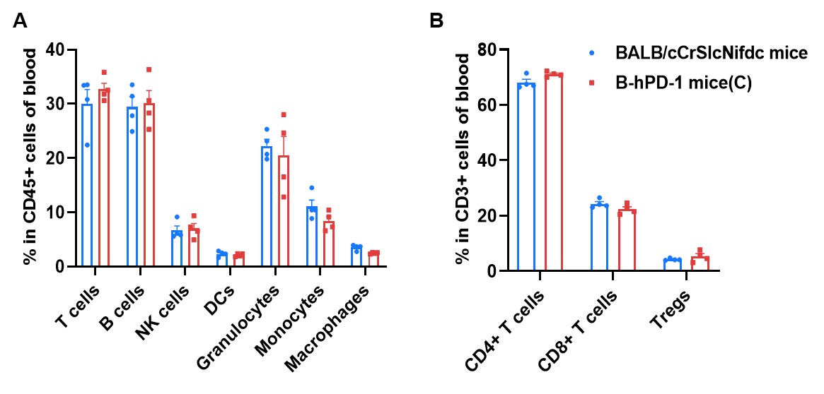
Frequency of leukocyte subpopulations in blood by flow cytometry. Blood cells were isolated from wild-type BALB/cCrSlcNifdc mice and homozygous B-hPD-1 mice(C) (female, 7-week-old, n=4). A. Flow cytometry analysis of the blood was performed to assess the frequency of leukocyte subpopulations. B. Frequency of T cell subpopulations. Frequencies of T cells, B cells, NK cells, DCs, granulocytes, monocytes, macrophages, CD4+ T cells, CD8+ T cells and Tregs in B-hPD-1 mice(C) were similar to those in BALB/cCrSlcNifdc mice, demonstrating that humanization of PD-1 does not change the frequency or distribution of these cell types in blood. Values are expressed as mean ± SEM.
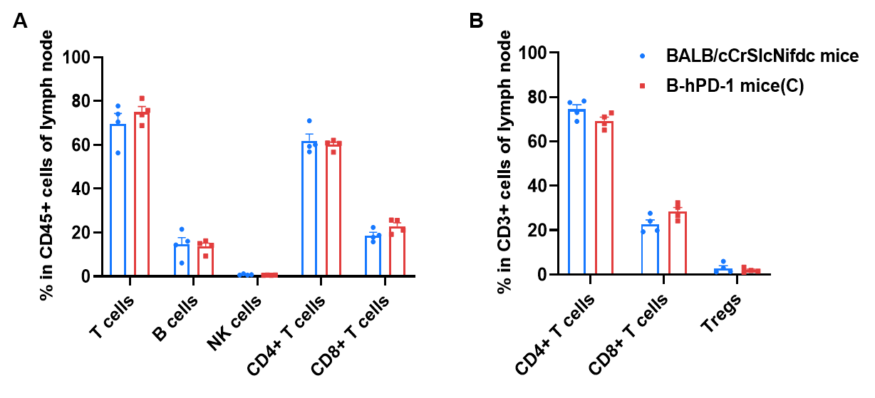
Frequency of leukocyte subpopulations in lymph nodes by flow cytometry. Lymph nodes were isolated from wild-type BALB/cCrSlcNifdc mice and homozygous B-hPD-1 mice(C) (female, 7-week-old, n=4). A. Flow cytometry analysis of the lymph node was performed to assess the frequency of leukocyte subpopulations. B. Frequency of T cell subpopulations. Frequencies of T cells, B cells, NK cells, CD4+ T cells, CD8+ T cells and Tregs in B-hPD-1 mice(C) were similar to those in BALB/cCrSlcNifdc mice, demonstrating that humanization of PD-1 does not change the frequency or distribution of these cell types in lymph nodes. Values are expressed as mean ± SEM.


Antitumor activity of anti-human PD-1 antibody pembrolizumab (in house) in B-hPD-1 mice(C). (A) Anti-human PD-1 antibody inhibited B-hPD-L1 CT26.WT tumor growth in B-hPD-1 mice(C). Murine colon cancer B-hPD-L1 CT26.WT cells were subcutaneously implanted into homozygous B-hPD-1 mice(C) (female, 9-week-old, n=6). Mice were grouped when tumor volume reached approximately 60-80 mm3, at which time they were treated with anti-human PD-1 antibody indicated in panel. (B) Body weight changes during treatment. As shown in panel A, anti-human PD-1 antibody was efficacious in controlling tumor growth in B-hPD-1 mice(C), demonstrating that the B-hPD-1 mice(C) provide a powerful preclinical model for in vivo evaluation of anti-human PD-1 antibodies. Values are expressed as mean ± SEM.