

Biological Features and Therapeutic Potential of CD3 and CEACAM5

CD3EDG
CEACAM5

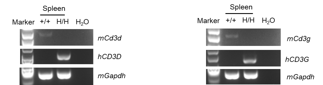
mRNA CD3D and CD3G were detectable in B-hCD3EDG/hCEACAM5 mice by RT-PCR and sequencing. Spleen tissues were isolated from wild-type C57BL/6JNifdc mice (+/+) and homozygous B-hCD3EDG/hCEACAM5 mice (H/H). Primers were designed to detect the mouse and human CD3D and CD3G. Sequencing of the PCR products confirmed that the amplified sequences were consistent with database reference sequences.
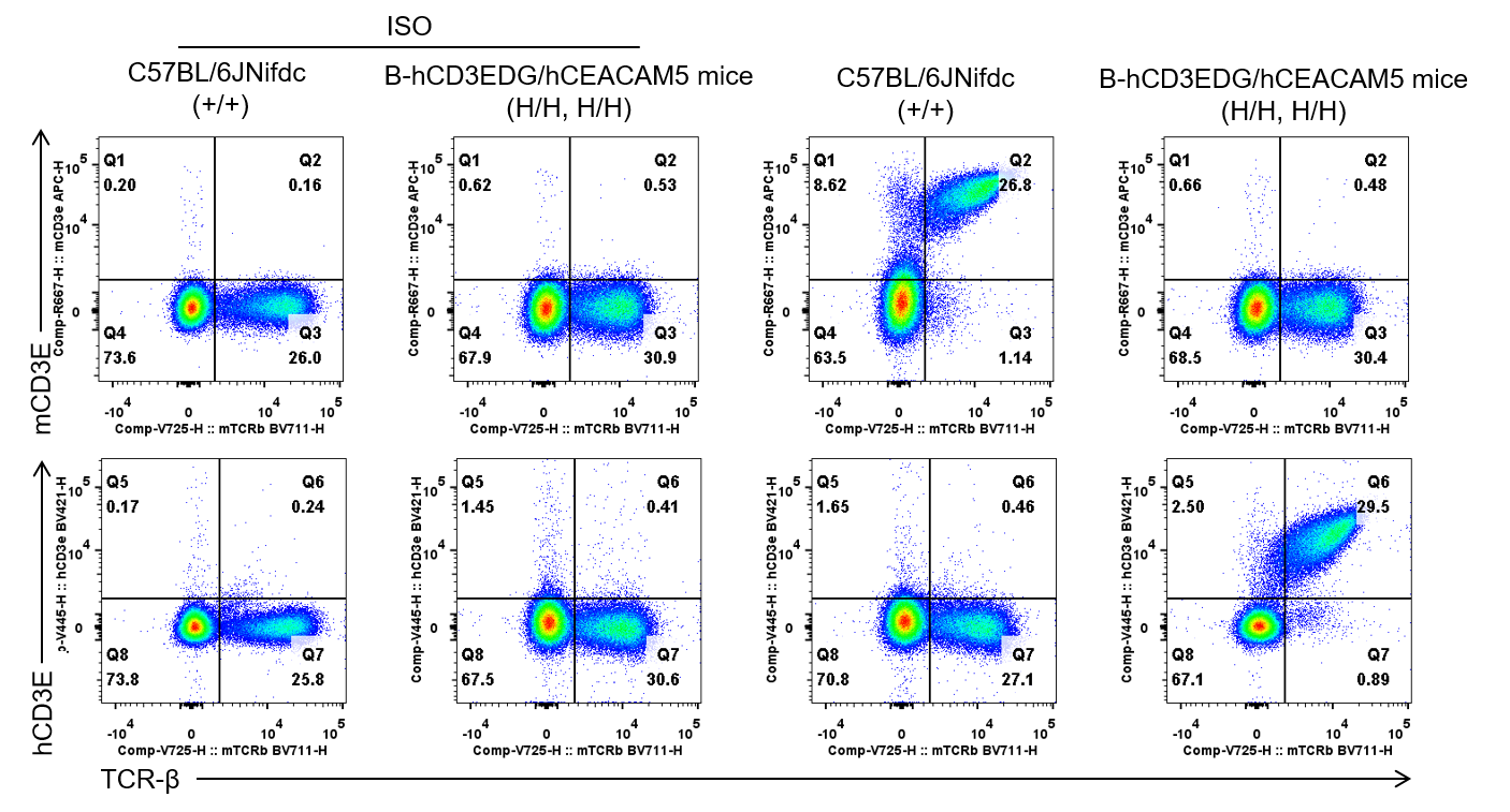
Mouse and human CD3E expression analysis in splenocytes. Splenocytes were collected from wild-type C57BL/6JNifdc mice and homozygous B-hCD3EDG/hCEACAM5 mice (Female, 5-week-old, n=1). CD3E expression on T cells was analyzed by flow cytometry using species-specific anti-CD3E antibodies (anti-human CD3E antibody, BD Horizon, 562426; anti-mouse CD3E antibody, Biolegend, 100312 ).
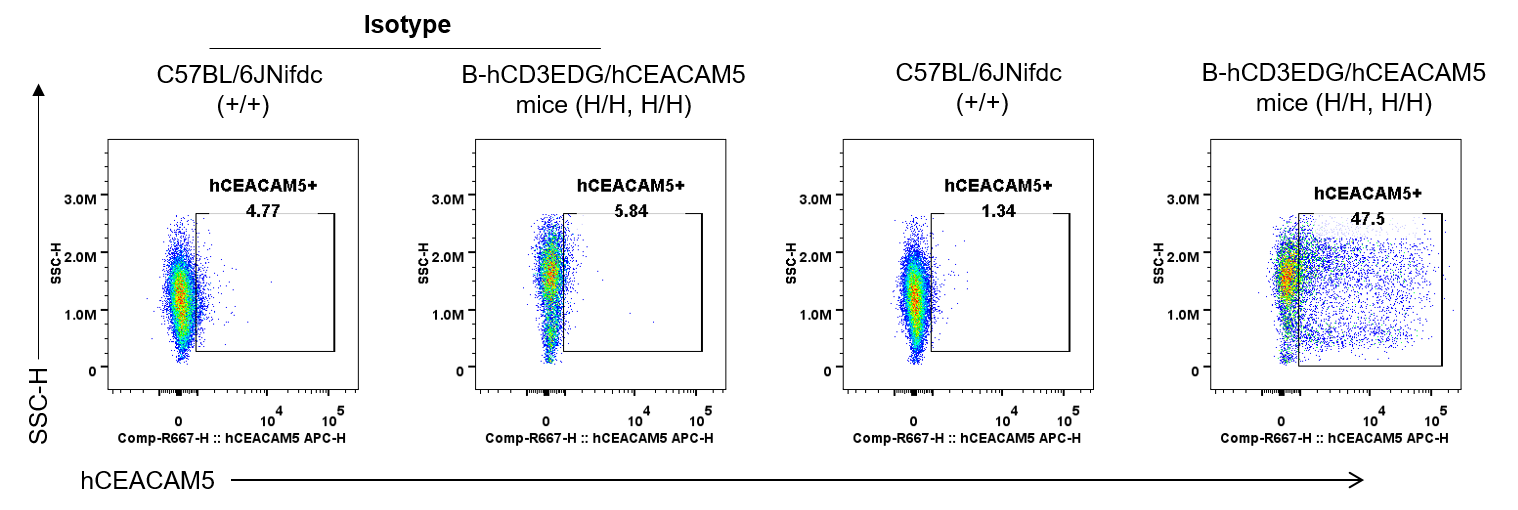
Human CEACAM5 expression analysis in colon. Colon was collected from wild-type C57BL/6JNifdc mice (+/+) and homozygous B-hCD3EDG/hCEACAM5 mice (H/H, H/H) and analyzed by flow cytometry with species-specific anti-human CD66a/c/e(CEACAM5) Antibody (Biolegend, 342308).
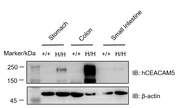
Western blot analysis of CEACAM5 protein expression in homozygous B-hCD3EDG/hCEACAM5 mice. Various tissue lysates were collected from wild-type C57BL/6JNifdc mice (+/+) and homozygous B-hCD3EDG/hCEACAM5 mice (H/H, H/H), and then analyzed by western blot with human-specific anti-CEACAM5 antibody (Invitrogen, PA5-88533). 40 μg total proteins were loaded for western blotting analysis.
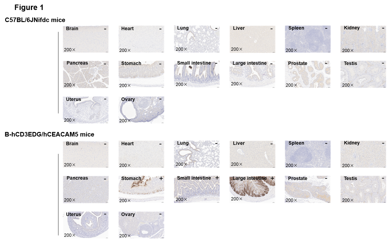
Immunohistochemical analysis of organs in B-hCD3EDG/hCEACAM5 mice. Major tissues were collected from wild-type mice and homozygous B-hCD3EDG/hCEACAM5 mice and analyzed by IHC with anti-human CEACAM5 antibody (Abcam, ab133633). The arrow indicates tissue cells with positive CEACAM5 staining (brown). "+" and "-" indicate positive and negative tissues, respectively.