CSF1 activates CSF1R signaling to promote the survival, proliferation, differentiation, and maintenance of monocytes and macrophages.

CSF1

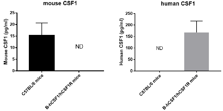
Strain specific CSF1 expression analysis in homozygous B-hCSF1/hCSF1R mice by ELISA. Splenic homogenate were collected from wild-type C57BL/6 mice (+/+) and homozygous B-hCSF1/hCSF1R mice (H/H) stimulated with LPS in vivo, and analyzed by ELISA with species-specific anti-mouse CSF1 ELISA kit (RayBio, ELH-MCSF) and anti-human CSF1 ELISA kit (RayBio, ELM-MCSF).
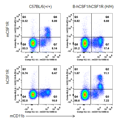
Strain specific CSF1R expression analysis in homozygous B-hCSF1/hCSF1R mice by flow cytometry. Blood were collected from wild-type C57BL/6 mice (+/+) and homozygous B-hCSF1/hCSF1R mice (H/H), and analyzed by flow cytometry with species-specific anti-mouse CSF1R antibody (Biolegend, 135509) and anti-human CSF1R antibody (Biolegend, 347303).

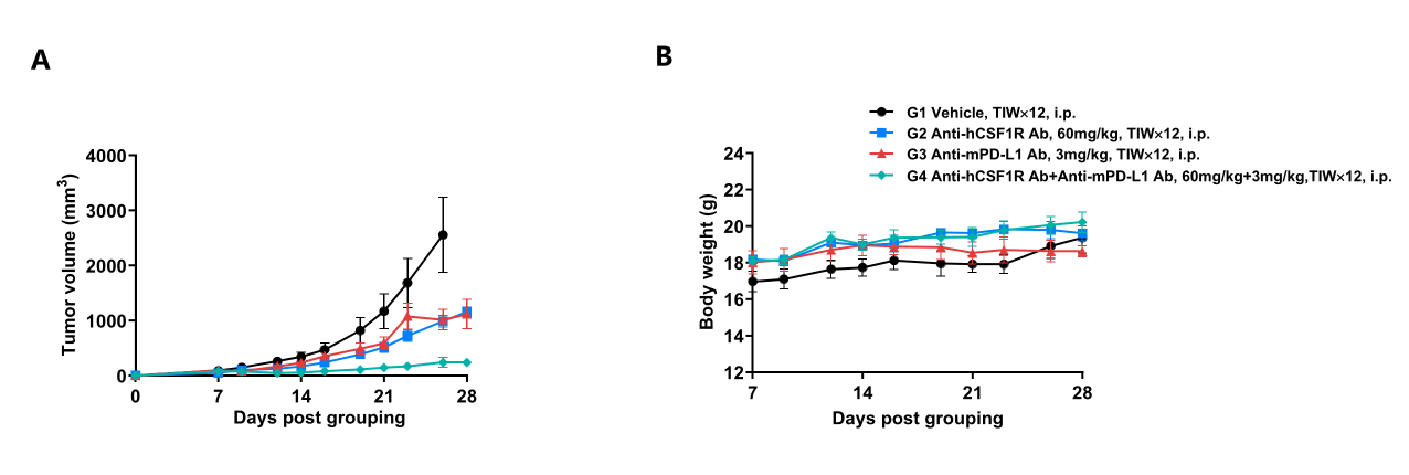
Antitumor activity of anti-mouse PD-L1 antibody combined with anti-human CSF1R antibody in B-hCSF1/hCSF1R mice. (A) Anti-mouse PD-L1 antibody combined with anti-human CSF1R antibody inhibited hCSF1-MC38 tumor growth in B-hCSF1/hCSF1R mice. Murine colon cancer hCSF1-MC38 cells (5Χ105) were subcutaneously implanted into homozygous B-hCSF1/hCSF1R mice (female, n=5). Mice were grouped on the first day, at which time they were treated with anti-mouse PD-L1 antibody and anti-human CSF1R antibody with doses and schedules indicated in panel A. (B) Body weight changes during treatment. Values are expressed as mean ± SEM.
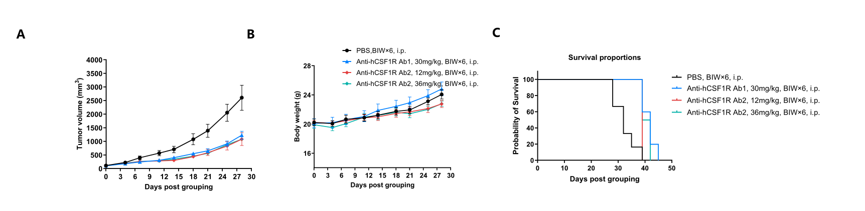
Antitumor activity of anti-human CSF1R antibodies in B-hCSF1/hCSF1R mice. (A) Anti-human CSF1R antibodies inhibited MC38 tumor growth in B-hCSF1/hCSF1R mice. Murine colon cancer MC38 cells were subcutaneously implanted into homozygous B-hCSF1/hCSF1R mice (female, 6-7-week-old, n=6). Mice were grouped when tumor volume reached approximately 100 mm3, at which time they were treated with two anti-human CSF1R antibodies with doses and schedules indicated in panel. (B) Body weight changes during treatment. (C) Mouse survival after grouping. Values are expressed as mean ± SEM. (Note: The client's antibodies may cross-reactivity between human and mouse)
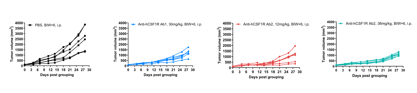
In vivo efficacy of anti-human CSF1R antibodies-individual tumor growth curves.
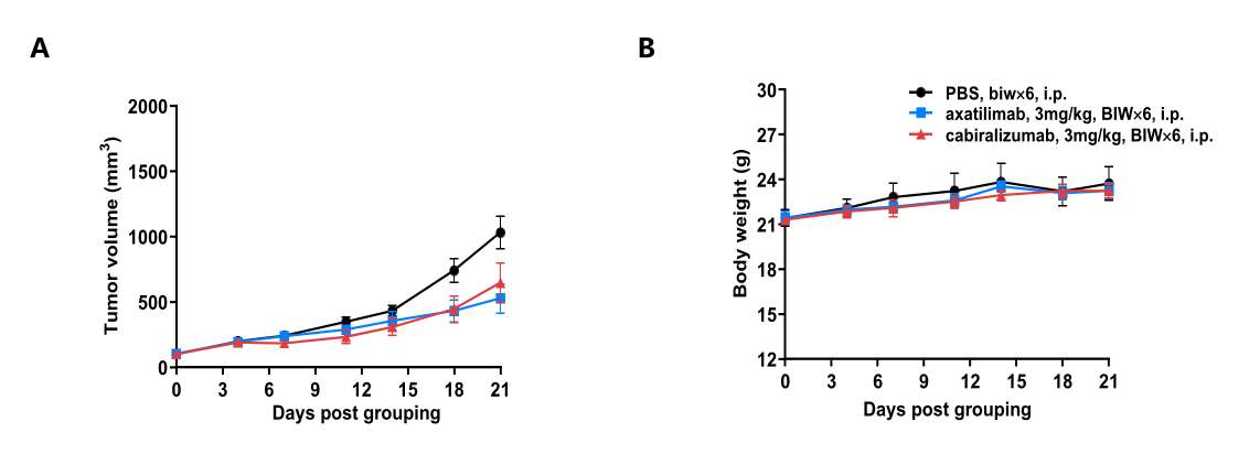
Antitumor activity of anti-human CSF1R antibodies in B-hCSF1/hCSF1R mice. (A) Axatilimab (in house) and cabiralizumab (in house) inhibited MC38 tumor growth in B-hCSF1/hCSF1R mice. Murine colon cancer MC38 cells were subcutaneously implanted into homozygous B-hCSF1/hCSF1R mice (female, 8-9-week-old, n=6). Mice were grouped when tumor volume reached approximately 100 mm3, at which time they were treated with axatilimab (in house) and cabiralizumab (in house) with doses and schedules indicated in panel. (B) Body weight changes during treatment. Values are expressed as mean ± SEM.

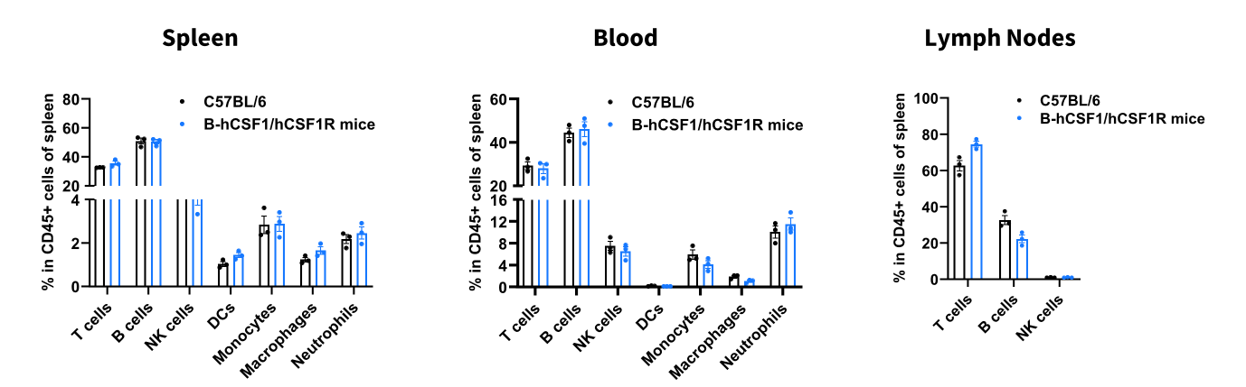
Analysis of leukocyte subpopulations by flow cytometry in immune organs and blood. Splenocytes, peripheral blood, and lymph nodes were isolated from female C57BL/6 and B-hCSF1/hCSF1R mice (female, 8-week-old, n=3). Single live cells were gated on the CD45⁺ population and analyzed by flow cytometry as indicated. Values are expressed as mean ± SEM.
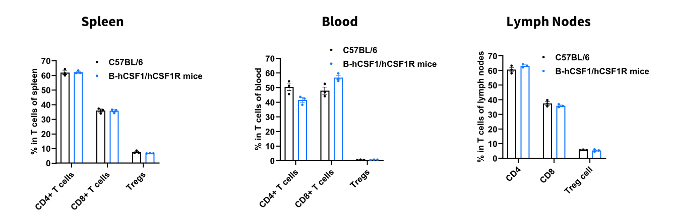
Analysis of T-cell subpopulations by flow cytometry in immune organs and blood. Splenocytes, peripheral blood, and lymph nodes were isolated from female C57BL/6 and B-hCSF1/hCSF1R mice (female, 8-week-old, n=3). Single live cells were gated on the CD3⁺ T-cell population and analyzed by flow cytometry as indicated. Values are expressed as mean ± SEM.